. 2010 Mar;31(9):2665-72.
doi: 10.1016/j.biomaterials.2009.12.005. Epub 2009 Dec 21.

The use of carboxymethylcellulose gel to increase non-viral gene transfer in mouse airways

Affiliations

The use of carboxymethylcellulose gel to increase non-viral gene transfer in mouse airways

Uta Griesenbach et al. Biomaterials. 2010 Mar.

Abstract

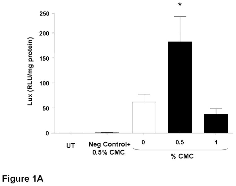
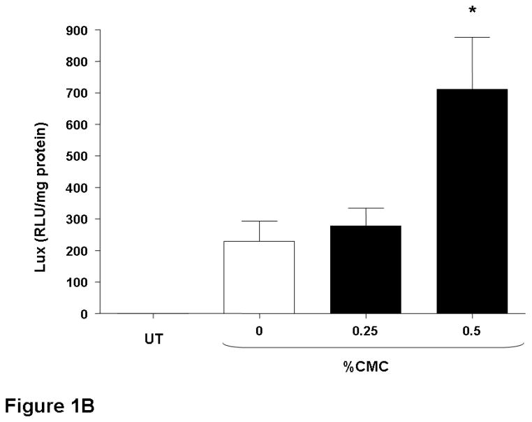
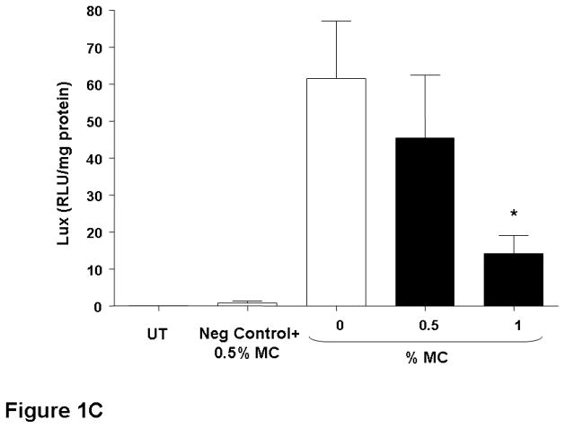
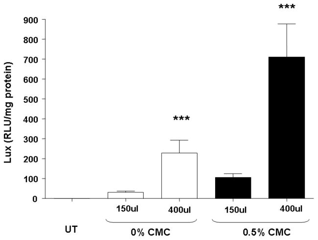
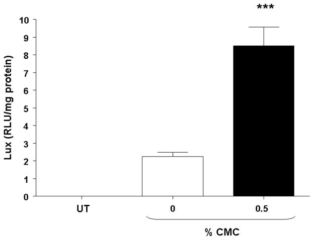
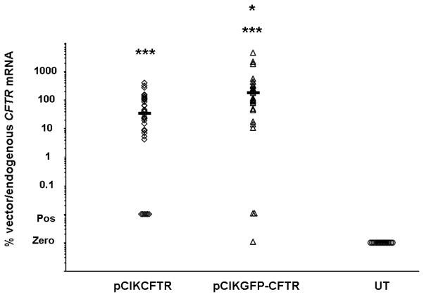
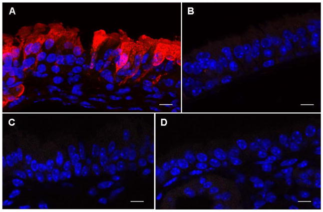
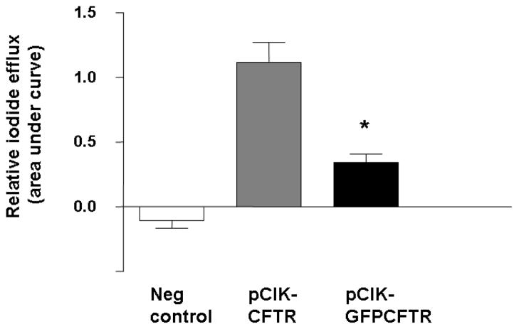
We have assessed whether viscoelastic gels known to inhibit mucociliary clearance can increase lipid-mediated gene transfer. Methylcellulose or carboxymethylcellulose (0.25-1.5%) was mixed with complexes of the cationic lipid GL67A and plasmids encoding luciferase and perfused onto the nasal epithelium of mice. Survival after perfusion with 1% CMC or 1% MC was 90 and 100%, respectively. In contrast 1.5% CMC was uniformly lethal likely due to the viscous solution blocking the airways. Perfusion with 0.5% CMC containing lipid/DNA complexes reproducibly increased gene expression by approximately 3-fold (n=16, p<0.05). Given this benefit, likely related to increased duration of contact, we also assessed the effect of prolonging contact time of the liposome/DNA complexes by delivering our standard 80 microg DNA dose over either approximately 22 or 60 min of perfusion. This independently increased gene transfer by 6-fold (n=8, p<0.05) and could be further enhanced by the addition of 0.5% CMC, leading to an overall 25-fold enhancement (n=8, p<0.001) in gene expression. As a result of these interventions CFTR transgene mRNA transgene levels were increased several logs above background. Interestingly, this did not lead to correction of the ion transport defects in the nasal epithelium of cystic fibrosis mice nor for immunohistochemical quantification of CFTR expression. To assess if 0.5% CMC also increased gene transfer in the mouse lung, we used whole body nebulisation chambers. CMC was nebulised for 1h immediately before, or simultaneously with GL67A/pCIKLux. The former did not increase gene transfer, whereas co-administration significantly increased gene transfer by 4-fold (p<0.0001, n=18). This study suggests that contact time of non-viral gene transfer agents is a key factor for gene delivery, and suggests two methods which may be translatable for use in man.


Figures

Figure 1
Figure 1. Addition of CMC increases gene transfer to mouse nasal epithelium
Mouse nasal epithelium was perfused with pCIKlux (80 μg/mouse) or pCIKempty (negative control) complexed to GL67A or remained untransfected (UT) (perfusion rate: 7 μl/min). Viscoelastic gels to final concentrations ranging from 0.25% to 1% were added to the lipid/DNA complexes (final volume 150 μl/mouse). 24 hr after transfection luciferase expression was quantified in tissue homogenate. (A and B) carboxymethylcellulose (CMC), (C) methylcellulose (MC). Bars represent group mean ±SEM (n=8/group). *=p<0.05 compared to 0% CMC.
Figure 2
Figure 2. Prolonged perfusion increases gene expression in mouse nasal epithelium
Mouse nasal epithelium was perfused with pCIKlux (80 μg/mouse) complexed to GL67A or remained untransfected (UT). Carboxymethylcellulose (CMC) to a final concentration of 0.5% was added to the lipid/DNA complexes. To increase contact time between epithelium and gene transfer agents the lipid/DNA complexes were either administered in 150 μl or 400 μl total volume and were then perfused onto the nasal epithelium (7 μl/min) for approximately 23 and 60 min, respectively. 24 hr after transfection luciferase expression was quantified in tissue homogenate. Bars represent group mean ±SEM (n=8/group). ***=p<0.005 compared to 150 μl group.
Figure 3
Figure 3. Addition of CMC increased gene transfer after nebulisation in mouse lung
Mouse lungs were transfected with pCIKlux complexed to GL67A for one hour using a nebulisation chamber or remained untransfected (UT). Carboxymethylcellulose (CMC) to a final concentration of 0.5% was added to the lipid/DNA complexes (25 mg DNA in 16 ml final volume). 24 hr after transfection luciferase expression was quantified in the lung homogenate. Bars represent group mean ±SEM (n=8/group). *=p<0.05 compared to 0% CMC
Figure 4
Figure 4. Expression of vector-specific CFTR mRNA in mouse nasal epithelium
The nasal epithelium of CF knockout mice was perfused with pCIKCFTR or pCIKGFPCFTR (80 μg DNA/mouse) complexed to GL67A including 0.5% CMC (final volume 150 μl/mouse) or remained untransfected (UT). 24 hr after transfection vector-specific and endogenous CFTR mRNA were quantified in nasal epithelium using real-time quantitative RT-PCR. Data are expressed as the ratio of vector-specific over endogenous CFTR mRNA. Each symbol represents one mouse (n=35/group). Horizontal bars indicates group median. In some samples mRNA was detectable, but not accurately quantifiable. These samples are labelled positive (pos). *=p<0.05 compared to pCIKCFTR, ***=p<0.001 compared to untransfected.
Figure 5
Figure 5. Detection of GFP-CFTR fusion protein in mouse nasal epithelium
The nasal epithelium of CF knockout mice was perfused with pCIKGFPCFTR complexed to GL67A plus 0.5% CMC. A recombinant Sendai virus (SeV) carrying GFP-CFTR was used as positive control and untransfected animals were negative controls. 24 hr after transfection nasal tissue was processed for immunohistochemistry and expression of GFP-CFTR fusion protein was visualized using an anti-GFP antibody and a secondary antibody conjugated to AlexaFluor 594. (A) SeVGFP-CFTR with anti-GFP primary antibody, (B) SeV-GFP-CFTR without anti-GFP primary antibody, (C) pCIKGFP-CFTR with anti-GFP primary antibody, (D) untransfected mouse with anti-GFP primary antibody. GFP-CFTR protein appears in red. DAPI stained nuclei appear in blue. Representative images are shown. n=10 mice/group. Scale bar=10 μm.
Figure 6
Figure 6. Assessment of CFTR function in a radio-tracer efflux assay
293T cells were transfected with either pCIKCFTR, pCIKGFP-CFTR or a negative control plasmid (pCIK-βgal) complexed to Lipofectamine 2000. 48 hr after transfection iodide efflux was quantified. Bars represent group mean ± SEM (n=6/group). *=p<0.05

References

    1. Hyde SC, Southern KW, Gileadi U, Fitzjohn EM, Mofford KA, Waddell BE, et al. Repeat administration of DNA/liposomes to the nasal epithelium of patients with cystic fibrosis. Gene Ther. 2000;7(13):1156–1165. - PubMed
    1. Alton EW, Stern M, Farley R, Jaffe A, Chadwick SL, Phillips J, et al. Cationic lipid-mediated CFTR gene transfer to the lungs and nose of patients with cystic fibrosis: a double-blind placebo-controlled trial. Lancet. 1999;353(9157):947–954. - PubMed
    1. Sorscher EJ, Logan JJ, Frizzell RA, Lyrene RK, Bebok Z, Dong JY, et al. Gene therapy for cystic fibrosis using cationic liposome mediated gene transfer: a phase I trial of safety and efficacy in the nasal airway. Hum Gene Ther. 1994;5(10):1259–1277. - PubMed
    1. Sinn PL, Shah AJ, Donovan MD, McCray PB., Jr Viscoelastic gel formulations enhance airway epithelial gene transfer with viral vectors. Am J Respir Cell Mol Biol. 2005;32(5):404–410. - PubMed
    1. Seiler MP, Luner P, Moninger TO, Karp PH, Keshavjee S, Zabner J. Thixotropic solutions enhance viral-mediated gene transfer to airway epithelia. Am J Respir Cell Mol Biol. 2002;27(2):133–140. - PubMed

Publication types

MeSH terms

Substances