

Effect of delivery of MMP inhibitors from PDMS as a model IOL material on PCO markers
- PMID: 20022368
- PMCID: PMC2972668
- DOI: 10.1016/j.biomaterials.2009.11.108
Effect of delivery of MMP inhibitors from PDMS as a model IOL material on PCO markers
Abstract
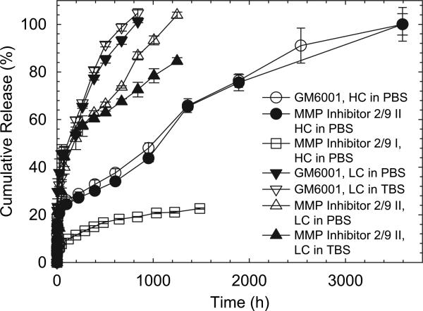
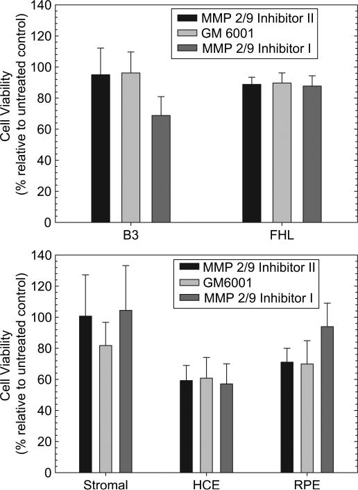
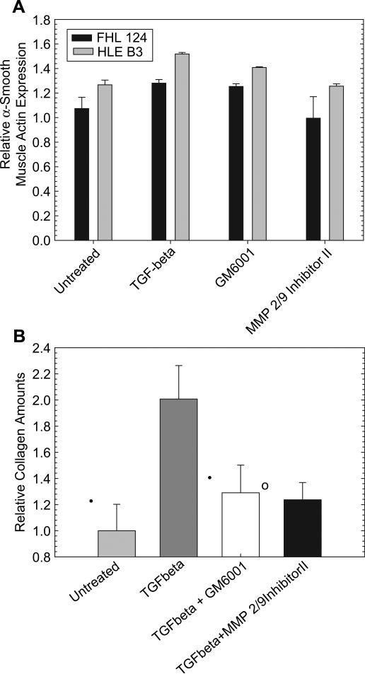
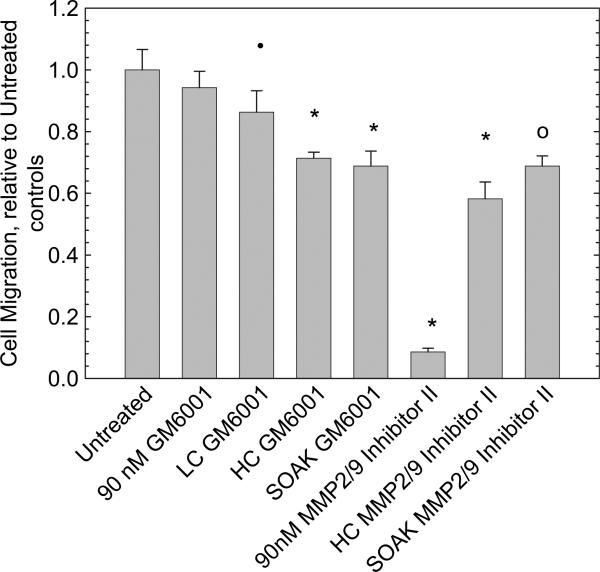
Posterior capsule opacification (PCO) or secondary cataract formation, following intraocular lens implantation, is a significant complication affecting an estimated 28% of cataract patients. Matrix metalloproteinases (MMPs) have been demonstrated to play a role in the formation of anterior subcapsular cataracts and it has been shown that the presence of MMP inhibitors (MMPI) decreases subcapsular cataract formation ex vivo. Since the mechanisms responsible for anterior subcapsular cataract formation and posterior capsule opacification are similar, it is reasonable to suggest that MMP inhibitors may also mitigate PCO. One of the most effective ways of delivering the inhibitors may be from the implanted intraocular lens (IOL) material itself. In the current work, delivery of three different MMP inhibitors from silicone rubber as a model IOL material was examined. Loading methods were developed which allowed continuous release of active MMPI for periods of over 5 months in some cases. Reduced migration rates were observed in human lens epithelial cells in vitro, suggesting that an effect on PCO may be possible. While further studies are necessary to tune the systems to achieve the desired rates of release, this work demonstrates that delivery of MMPI from silicone IOL materials has the potential to decrease the incidence of PCO.
Copyright (c) 2009 Elsevier Ltd. All rights reserved.
Figures


Similar articles
-
Sandwich theory: bioactivity-based explanation for posterior capsule opacification.J Cataract Refract Surg. 1997 Dec;23(10):1539-42. doi: 10.1016/s0886-3350(97)80026-0. J Cataract Refract Surg. 1997. PMID: 9456413
-
[Comparison of posterior capsule opacification in 2 types of implanted artificial intraocular lens].Cesk Slov Oftalmol. 2007 Jan;63(1):42-6. Cesk Slov Oftalmol. 2007. PMID: 17361627 Czech.
-
Posterior capsular opacification in pseudophakic eyes with a silicone or acrylic intraocular lens.Eur J Ophthalmol. 2002 May-Jun;12(3):212-8. doi: 10.1177/112067210201200307. Eur J Ophthalmol. 2002. PMID: 12113567 Clinical Trial.
-
Efficacy of different intraocular lens materials and optic edge designs in preventing posterior capsular opacification: a meta-analysis.Am J Ophthalmol. 2007 Mar;143(3):428-36. doi: 10.1016/j.ajo.2006.11.045. Epub 2006 Dec 28. Am J Ophthalmol. 2007. PMID: 17224119
-
Posterior capsule opacification.Curr Opin Ophthalmol. 2000 Feb;11(1):56-64. doi: 10.1097/00055735-200002000-00009. Curr Opin Ophthalmol. 2000. PMID: 10724829 Review.
Cited by
-
Sustained Release of a Peptide-Based Matrix Metalloproteinase-2 Inhibitor to Attenuate Adverse Cardiac Remodeling and Improve Cardiac Function Following Myocardial Infarction.Biomacromolecules. 2017 Sep 11;18(9):2820-2829. doi: 10.1021/acs.biomac.7b00760. Epub 2017 Aug 7. Biomacromolecules. 2017. PMID: 28731675 Free PMC article.
-
Sustained-release celecoxib from incubated acrylic intraocular lenses suppresses lens epithelial cell growth in an ex vivo model of posterior capsule opacity.J Ocul Pharmacol Ther. 2012 Aug;28(4):359-68. doi: 10.1089/jop.2011.0196. Epub 2012 Feb 28. J Ocul Pharmacol Ther. 2012. PMID: 22372691 Free PMC article.
-
RGD surface functionalization of the hydrophilic acrylic intraocular lens material to control posterior capsular opacification.PLoS One. 2014 Dec 11;9(12):e114973. doi: 10.1371/journal.pone.0114973. eCollection 2014. PLoS One. 2014. PMID: 25501012 Free PMC article.
-
Drug-Eluting Intraocular Lenses.Materials (Basel). 2011 Nov 1;4(11):1927-1940. doi: 10.3390/ma4111927. Materials (Basel). 2011. PMID: 28824115 Free PMC article. Review.
-
The Long Noncoding RNA H19 Promotes Fibrotic Processes in Lens Epithelial Cells.Invest Ophthalmol Vis Sci. 2023 Jun 1;64(7):21. doi: 10.1167/iovs.64.7.21. Invest Ophthalmol Vis Sci. 2023. PMID: 37310354 Free PMC article.
References
-
- Cheng JW, Wei RL, Cai JP, Xi GL, Zhu H, Li Y, et al. Efficacy of different intraocular lens materials and optic edge designs in preventing posterior capsular opacification: a meta-analysis. Am J Ophthalmol. 2007;143:428–36. - PubMed
-
- Hosal BM, Biglan AW. Risk factors for secondary membrane formation after removal of pediatric cataract. J Cataract Refract Surg. 2002;28:302–9. - PubMed
-
- Matsushima H, Iwamoto H, Mukai K, Katsuki Y, Nagata M, Senoo T. Preventing secondary cataract and anterior capsule contraction by modification of intraocular lenses. Expert Rev Med Dev. 2008;5:197–207. - PubMed
-
- Sacu S, Menapace R, Findl O, Georgopoulos M, Buehl W, Kriechbaum K, et al. Influence of optic edge design and anterior capsule polishing on posterior capsule fibrosis. J Cataract Refract Surg. 2004;30:658–62. - PubMed
-
- Buehl W, Findl O. Effect of intraocular lens design on posterior capsule opacification. J Cataract Refract Surg. 2008;34:1976–85. - PubMed
Publication types
MeSH terms
Substances
Grants and funding
LinkOut - more resources
Full Text Sources
