

Regulation of osteogenic differentiation of rat bone marrow stromal cells on 2D nanorod substrates
- PMID: 20022632
- PMCID: PMC4748726
- DOI: 10.1016/j.biomaterials.2009.11.041
Regulation of osteogenic differentiation of rat bone marrow stromal cells on 2D nanorod substrates
Abstract
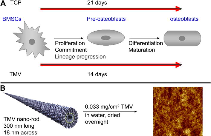
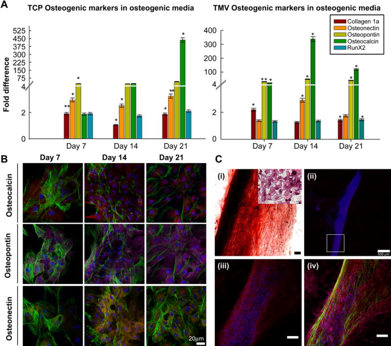
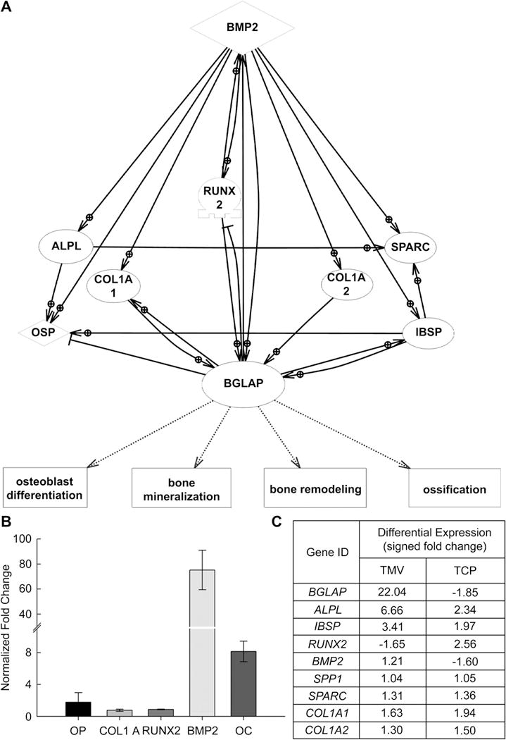
Bone marrow stromal cells (BMSCs) possess multi-lineage differentiation potential and can be induced to undergo differentiation into various cell types with the correct combination of chemical and environmental factors. Although, they have shown great prospects in therapeutic and medical applications, less is known about their behavior on nanosurfaces mimicking the extra cellular matrix (ECM). In this report we have employed 2D substrates coated with tobacco mosaic virus (TMV) nanorods to study the differentiation process of BMSCs into osteoblast like cells. TMV is a rod-shaped plant virus with an average length of 300 nm and diameter of 18 nm. The osteogenic differentiation of BMSCs on TMV was studied over time points of 7, 14 and 21 days. We examined the temporal gene expression changes during these time points by real-time quantitative PCR (RT-qPCR) analysis. As expected, osteo-specific genes (osteocalcin, osteopontin and osteonectin) were upregulated and showed a maximum change in expression on TMV at 14 days which was 7 days earlier than on tissue culture plastic (TCP). Based on the genes expression profile generated by RT-qPCR experiments, we proposed that the early interaction of cells with TMV triggers on signaling pathways which regulate speedy expression of osteocalcin in turn, resulting in early mineralization of the cells. To further investigate these regulating factors we studied global changes in gene expression (DNA microarray analyses) during osteogenic differentiation on the nanosubstrate. Multitudes of genes were affected by culturing cells on nanorod substrate, which corroborated our initial PCR findings. Microarray analysis further revealed additional targets influenced by the presence of nanorods on the surface, of which, the expression of bone morphogenetic protein 2 (BMP2) was of particular interests. Further investigation into the temporal change of BMP2, revealed that it acts as a major promoter in signaling the early regulation of osteocalcin on TMV coated substrates.
(c) 2009 Elsevier Ltd. All rights reserved.
Figures
Similar articles
-
The synergistic effects of multivalent ligand display and nanotopography on osteogenic differentiation of rat bone marrow stem cells.Biomaterials. 2010 Aug;31(22):5813-24. doi: 10.1016/j.biomaterials.2010.04.017. Epub 2010 May 10. Biomaterials. 2010. PMID: 20452665
-
Bone morphogenetic protein receptor in the osteogenic differentiation of rat bone marrow stromal cells.Yonsei Med J. 2010 Sep;51(5):740-5. doi: 10.3349/ymj.2010.51.5.740. Yonsei Med J. 2010. PMID: 20635450 Free PMC article.
-
Cyclic acetal hydroxyapatite composites and endogenous osteogenic gene expression of rat marrow stromal cells.J Tissue Eng Regen Med. 2010 Aug;4(6):422-36. doi: 10.1002/term.252. J Tissue Eng Regen Med. 2010. PMID: 20047194
-
Using Plant Virus Based Nanorods to Modulate the Differentiation of Human Bone Marrow Stem Cells.J Biomed Nanotechnol. 2019 Feb 1;15(2):363-372. doi: 10.1166/jbn.2019.2691. J Biomed Nanotechnol. 2019. PMID: 30596558
-
Real-time quantitative RT-PCR analysis of human bone marrow stromal cells during osteogenic differentiation in vitro.J Cell Biochem. 2002;85(4):737-46. doi: 10.1002/jcb.10174. J Cell Biochem. 2002. PMID: 11968014
Cited by
-
Genetically Engineered Flagella Form Collagen-like Ordered Structures for Inducing Stem Cell Differentiation.iScience. 2019 Jul 26;17:277-287. doi: 10.1016/j.isci.2019.06.036. Epub 2019 Jul 3. iScience. 2019. PMID: 31323474 Free PMC article.
-
Three dimensional extrusion printing induces polymer molecule alignment and cell organization within engineered cartilage.J Biomed Mater Res A. 2018 Aug;106(8):2190-2199. doi: 10.1002/jbm.a.36426. Epub 2018 Apr 30. J Biomed Mater Res A. 2018. PMID: 29659132 Free PMC article.
-
Fibroblast growth factor-4 maintains cellular viability while enhancing osteogenic differentiation of stem cell spheroids in part by regulating RUNX2 and BGLAP expression.Exp Ther Med. 2020 Sep;20(3):2013-2020. doi: 10.3892/etm.2020.8951. Epub 2020 Jun 26. Exp Ther Med. 2020. PMID: 32782511 Free PMC article.
-
Synthesis of photo-reactive poly (vinyl alcohol) and construction of scaffold-free cartilage like pellets in vitro.Regen Biomater. 2018 Jun;5(3):159-166. doi: 10.1093/rb/rby009. Epub 2018 May 3. Regen Biomater. 2018. PMID: 29942648 Free PMC article.
-
Polydopamine-modified poly(l-lactic acid) nanofiber scaffolds immobilized with an osteogenic growth peptide for bone tissue regeneration.RSC Adv. 2019 Apr 15;9(21):11722-11736. doi: 10.1039/c8ra08828d. eCollection 2019 Apr 12. RSC Adv. 2019. PMID: 35516986 Free PMC article.
References
-
- Mackay AM, Beck SC, Pittenger MF. Human mesenchymal stem cells progress to hypertrophic chondrocytes under specific conditions in vitro. Mol Biol Cell. 1998;9:173a–173a. - PubMed
-
- Mackay AM, Beck SC, Murphy JM, Barry FP, Chichester CO, Pittenger MF. Chondrogenic differentiation of cultured human mesenchymal stem cells from marrow. Tissue Eng. 1998;4(4):415–428. - PubMed
-
- Ferrari G. Skeletal muscle regeneration by bone marrow-derived myogenic progenitors. Science. 1998;281(5379):923–923. - PubMed
-
- Calvert JW, Marra KG, Cook L, Kumta PN, DiMilla PA, Weiss LE. Characterization of osteoblast-like behavior of cultured bone marrow stromal cells on various polymer surfaces. J Biomed Mater Res. 2000;52(2):279–284. - PubMed
-
- Benayahu D, Kletter Y, Zipori D, Wientroub S. Bone-Marrow Derived Stromal Cell-Line Expressing Osteoblastic Phenotype Invitro and Osteogenic Capacity Invivo. J Cell Physiol. 1989;140(1):1–7. - PubMed
Publication types
MeSH terms
Substances
Grants and funding
LinkOut - more resources
Full Text Sources
Other Literature Sources
Molecular Biology Databases
Research Materials
