Review
. 2010 Apr;35(4):189-98.
doi: 10.1016/j.tibs.2009.11.005. Epub 2009 Dec 21.

How antibodies fold

Affiliations
Review

How antibodies fold

Matthias J Feige et al. Trends Biochem Sci. 2010 Apr.

Abstract

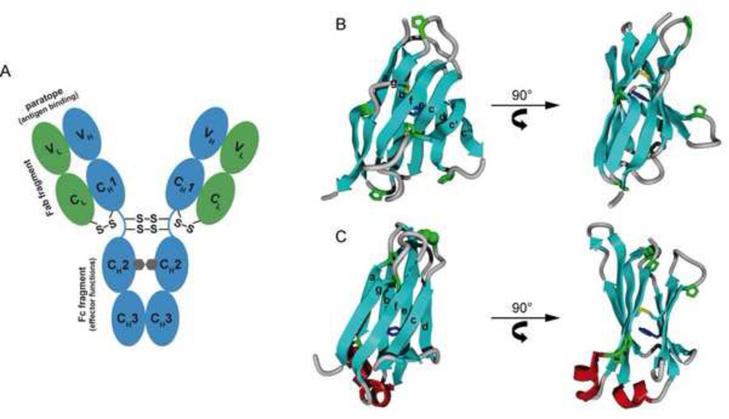
B cells use unconventional strategies for the production of a seemingly unlimited number of antibodies from a very limited amount of DNA. These methods dramatically increase the likelihood of producing proteins that cannot fold or assemble appropriately. B cells are therefore particularly dependent on 'quality control' mechanisms to oversee antibody production. Recent in vitro experiments demonstrate that Ig domains have evolved diverse folding strategies ranging from robust spontaneous folding to intrinsically disordered domains that require assembly with their partner domains to fold; in vivo experiments reveal that these different folding characteristics form the basis for cellular checkpoints in Ig transport. Taken together, these reports provide a detailed understanding of how B cells monitor and ensure the functional fidelity of Ig proteins.


Figures

Figure 1
Figure 1. overall antibody structure and domain architecture
(A) Domain arrangement of an IgG antibody molecule. The light chains are shown in green, the heavy chains in blue. The oligosaccharides between the CH2 domains are depicted as grey hexagons. Interchain disulfide bridges and important functional elements of the antibody (antigen binding paratope, Fab fragment, Fc fragment) are indicated. Domain architecture of the light chain variable (VL) (B) and constant (CL) domains (C). The strand nomenclature is indicated. The intrachain disulfide bridge (yellow) and the proximal conserved tryptophan residue (blue) are shown. The proline residues of the two domains are shown in green with the highly conserved cis-proline residue between strands b and c of CL highlighted in a CPK representation. Small helices (red) connect strands a and b and strands e and f of the CL domain.
Figure 2
Figure 2. three pathways of antibody domain folding
(i) CL and CH2 fold via a highly structured on-pathway intermediate that is trapped by the trans state of a proline residue in the loop connecting strands b and c (highlighted in yellow). In the intermediate, the core β-sheet structure and the two short helices connecting strands a and b and strands e and f are fully formed (shown in red). (ii) The obligate dimer CH3 folds via two intermediates, both most likely similar in structure to those of the CL and CH2 domains. In a first, rapidly formed intermediate, a critical proline residue (highlighted in yellow) must isomerize to its native cis state, leading to a second intermediate which can dimerize and thereby complete folding. (iii) CH1 is intrinsically disordered in isolation. Upon association with CL, it forms a loosely folded intermediate. In this complex, isomerization of the conserved proline residue between strands b and c (highlighted in yellow) limits the complete folding to the native state and formation of the interchain disulfide bridge between CH1 and CL.
Figure 3
Figure 3. Immunoglobulin quality control checkpoints at various stages in B cell development
After HC gene rearrangements, preB cells produce IgM HCs (μ HCs) (blue) bound to BiP (red). If their association with the surrogate LC, which is assembled from the VpreB (deep purple) and λ5 (light purple) proteins, induces BiP release and folding of the CH1 domain, and if the other Ig domains fold properly, the HC can traffic to the plasma membrane and engage signalling molecules (HC membrane anchor shown in yellow). If there is a failure in any of these steps, the μ HCs become substrates for ER associated degradation (ERAD) and are retrotranslocated to the cytosol for degradation by the 26S proteasome. Once conventional LCs (green) are produced in the B cell, they assemble with μ HCs, displace BiP from the CH1 domain, and induce its folding. As the ability of all domains of the HC to fold properly upon assembly was tested at the preB cell stage, quality control at this stage monitors the pairing and folding of the V domains. Plasma cell differentiation leads to the synthesis of extremely high levels of antibodies. Because the ability of the specific HC and LC combination to assemble and fold properly was verified at the B cell stage of development, quality control at this point involves monitoring the completeness of Ig assembly, focusing on the LC-induced release of BiP from the CH1 domain and its concomitant folding. There is a shift to production of the secretory form of μ HC in plasma cells, which possess a terminal cysteine that is involved in assembly with J chain and pentamer formation. Thiol-mediated retention mechanisms monitor the redox state of this cysteine and prevent IgM monomers from being secreted.
Figure 4
Figure 4. A comprehensive view of IgG folding and assembly
Folding, formation of disulfide bridges and glycosylation of the HC (blue) and LC (green) begins cotranslationally in the ER. The molecular chaperone BiP (red) interacts with most of the domains transiently before folding is completed. All constant domains except CH1 and most variable domains fold autonomously, populating an on-pathway intermediate on the way to the native state. CL is known to fold particularly fast in the cell. Once CH3 is folded, it induces HC dimerization which will be solidified by disulfide bridges in the hinge region. CH1 remains unfolded, unoxidized and stably bound to BiP until the LC displaces BiP and CL induces folding of the CH1 domain. Once the important CH1 prolines are in the correct isomerization state and CH1 is folded, a disulfide bridge between the LC and the HC forms rendering the IgG molecules ready for secretion. Most of these steps are likely to hold for other Ig classes. Chaperones and folding catalysts, such as Grp94, protein disulfide isomerase (PDI) and the peptidyl-prolyl isomerase CyclophilinB contribute to the individual steps in immunoglobulin biogenesis.

Comment in

References

    1. Murphy KM, et al. Janeway's Immunobiology. Garland Science; 2008.
    1. Maki R, et al. Exon shuffling generates an immunoglobulin heavy chain gene. Proc. Natl. Acad. Sci. U. S. A. 1980;77:2138–2142. - PMC - PubMed
    1. Early P, et al. An immunoglobulin heavy chain variable region gene is generated from three segments of DNA: VH D and JH. Cell. 1980;19:981–992. - PubMed
    1. Calame K, et al. Mouse Cmu heavy chain immunoglobulin gene segment contains three intervening sequences separating domains. Nature. 1980;284:452–455. - PubMed
    1. Alt FW, et al. Development of the primary antibody repertoire. Science. 1987;238:1079–1087. - PubMed

Publication types

MeSH terms

LinkOut - more resources