. 2010 Mar 5;285(10):7035-44.
doi: 10.1074/jbc.M109.096032. Epub 2009 Dec 18.

Her4 and Her2/neu tyrosine kinase domains dimerize and activate in a reconstituted in vitro system

Affiliations

Her4 and Her2/neu tyrosine kinase domains dimerize and activate in a reconstituted in vitro system

John Monsey et al. J Biol Chem. .

Abstract

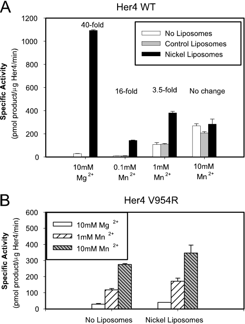
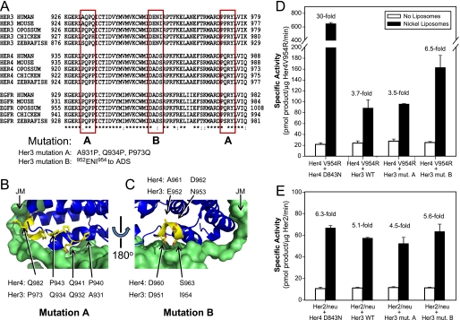
Her4 (ErbB-4) and Her2/neu (ErbB-2) are receptor-tyrosine kinases belonging to the epidermal growth factor receptor (EGFR) family. Crystal structures of EGFR and Her4 kinase domains demonstrate kinase dimerization and activation through an allosteric mechanism. The kinase domains form an asymmetric dimer, where the C-lobe surface of one monomer contacts the N-lobe of the other monomer. EGFR kinase dimerization and activation in vitro was previously reported using a nickel-chelating lipid-liposome system, and we now apply this system to all other members of the EGFR family. Polyhistidine-tagged Her4, Her2/neu, and Her3 kinase domains are bound to these nickel-liposomes and are brought to high local concentration, mimicking what happens to full-length receptors in vivo following ligand binding. Addition of nickel-liposomes to Her4 kinase domain results in 40-fold activation in kinase activity and marked enhancement of C-terminal tail autophosphorylation. Activation of Her4 shows a sigmoidal dependence on kinase concentration, consistent with a cooperative process requiring kinase dimerization. Her2/neu kinase activity is also activated by nickel-liposomes, and is increased further by heterodimerization with Her3 or Her4. The ability of Her3 and Her4 to heterodimerize and activate other family members is studied in vitro. Her3 kinase domain readily activates Her2/neu but is a poor activator of Her4, which differs from the prediction made by the asymmetric dimer model. Mutation of Her3 residues (952)ENI(954) to the corresponding sequence in Her4 enhanced the ability of Her3 to activate Her4, demonstrating that sequence differences on the C-lobe surface influence the heterodimerization and activation of ErbB kinase domains.


Figures

FIGURE 1.
FIGURE 1.
Activation of the Her4 kinase domain by nickel-liposomes. A, Her4 kinase domain protein containing the N-terminal polyhistidine tag or Her4 having the His6 tag removed by TEV protease treatment was incubated with liposomes for 15–20 min on ice. The kinase domain-liposome complex was incubated at 30 °C with a kinase reaction mixture containing 100 μm ATP, 1 μCi of [γ-32P]ATP, 10 mm MgCl2, and 100 μm biotin-GGMEDIYFEFMGGKKK peptide as in “Experimental Procedures.” The phosphorylated peptide was measured by liquid scintillating counting. B, His6-tagged Her4 kinase domain was incubated with liposomes in the presence of 0–100 mm imidazole, pH 7.5. Kinase assays were performed as above. C, Her4 kinase domain proteins were incubated with liposomes, and kinase assays performed as above. Mixing of Her4 I712Q and V954R proteins at 1:1 molar ratio was done prior to addition of liposomes. Fold change is calculated relative to the activity with control liposomes. The Her4 amino acid numbering scheme used here is based on the Her4 JM-a/Cyt-1 isoform with its signal peptide included. Data are representative of two (A and B) or four (C) independent experiments. D, predicted effects of the Her4 mutations used in C on kinase dimerization and activation.
FIGURE 2.
FIGURE 2.
Binding and activation of Her4 on the nickel-liposomes. A, binding of His6-tagged Her4 kinase domain to nickel-liposomes was measured by incubating 0–17.3 μm Her4 with nickel-liposome (8 μm bulk concentration of accessible Ni-NTA-DOGS lipid) and then separating bound from free Her4 by ultracentrifugation at 120,000 × g. Data were fit to a single site binding equation, y = ax/(b + x). The data are representative of four experiments. B, cooperativity of Her4 kinase domain activation on nickel-liposomes. Her4 kinase domain concentration during binding was varied and kinase activity measured as in Fig. 1 and the “Experimental Procedures.” Data points represent mean ± S.D. of n = 4. Data were fit to the Hill equation of y = y0 + [axb/(cb + xb)], where b represents the Hill coefficient. The inset shows transformation of the data so that the slope = −Hill coefficient. Linear regression of this transformed data demonstrates a Hill coefficient of 2.1 ± 0.2. Data are representative of three experiments. Her4 kinase domain concentrations reported in A and B represent bulk solution concentrations of the kinase.
FIGURE 3.
FIGURE 3.
Effect of divalent cations, Mg2+ and Mn2+, on Her4 kinase domain activation. A, Her4 WT kinase domain was incubated in the presence or absence of the indicated liposomes and the kinase reaction conducted in the presence of the indicated concentration of Mg2+ or Mn2+. Fold change is calculated relative to the activity in the absence of liposomes. B, Her4 V954R kinase domain was incubated in the absence or presence of nickel-liposomes and the kinase reaction conducted in the presence of the indicated concentration of Mg2+ or Mn2+. Data are representative of three (A) or two (B) independent experiments.
FIGURE 4.
FIGURE 4.
Autophosphorylation of Her4 and Her2/neu. A, Her4 kd+tail was incubated with 100 μm ATP, 1 μCi of [γ-32P]ATP, and either with or without nickel-liposomes, as in the “Experimental Procedures.” 32P incorporation into Her4 kd+tail was visualized by phosphorimaging. B, upper panel, Her2/neu kinase domain autophosphorylation was measured as in A. B, lower panel, autophosphorylation of Her2/neu was conducted in a non-radioactive reaction. Phosphorylation at Tyr-877 was measured using a phosphospecific Ab to this site.
FIGURE 5.
FIGURE 5.
Heterodimerization and activation of Her2/neu kinase domain by Her3 and kinase dead Her4. A, activation of Her2/neu kinase domain by Her3. B, activation of Her2/neu kinase domain by kinase-dead Her4 (Her4 D843N). For both A and B, kinase domain proteins were mixed at 1:1 molar ratio immediately prior to nickel-liposome addition, and kinase assay was measured as in Fig. 1. Fold change is calculated relative to Her2/neu kinase activity in the absence of liposomes. Data are representative of three experiments.
FIGURE 6.
FIGURE 6.
Heterodimerization of Her4 with Her3. A, predicted effects of the Her4 mutations and of Her3 on Her4 kinase activity. The mutations shown here are in the Her4 kinase domain. The prediction made by the asymmetric dimer model in the case of Her4 V954R mixed with Her3 is highlighted because the model prediction is not borne out by the observed experimental results (below). B, Her4 and Her3 kinase domain proteins were mixed at 1:1 molar ratio immediately prior to nickel-liposome addition, and the kinase assay was measured as in Fig. 1. Specific activity was corrected for the amount of active Her4 kinase present in each pairwise mixture. Fold change is calculated relative to kinase activity in the absence of liposomes. C, kinase-dead Her4 (D843N) or Her3 WT was mixed with WT Her4 kinase domain at the indicated molar ratio, nickel-liposomes were added, and kinase assays were performed as in Fig. 1. Data are representative of three (B) or two (C) experiments.
FIGURE 7.
FIGURE 7.
Mutation of Her3 to enhance its ability to activate Her4. A, multisequence alignment of the intracellular portions of Her3, Her4, and EGFR across five species, using ClustalW2 (47). Shown by red boxes are three regions on the C-lobe dimerization surface of the kinase domain where contact residues are divergent across this family. To make these contact sites in Her3 more “Her4-like,” the following mutations were created. Her3 mutation A: A931P/Q943P/P973Q. Her3 mutation B: 952ENI954 to ADS. B, contacts made by residues in mutation A. The Her4 donor monomer is shown in blue, and the PQPP and PQRY sequences in it are highlighted in yellow (structure shown is PDB ID: 2R4B) (17). The Her4 acceptor monomer is shown as a green surface. This crystal structure includes the juxtamembrane (JM) segment, which is seen on the left side of the image and contacts Her4 residue Gln-982 through Tyr-984 in the donor monomer. C, contacts made by residues in mutation B. The Her4 asymmetric dimer is rotated 180° relative to B, and the DADS sequence in the donor monomer is highlighted in yellow. Panels B and C are generated with PyMol version 1.1 (48). D, Her4 V954R was mixed with either kinase-dead Her4, WT Her3 or mutant Her3 at 1:1 molar ratio immediately prior to nickel-liposome addition and kinase assay was measured as in Fig. 1. Specific activity was corrected for the amount of active Her4 kinase present in each pairwise mixture. Fold change is calculated relative to kinase activity in the absence of liposomes. E, mixing of Her2/neu with the specified Her4 or Her3 kinase domain proteins was performed as in D. Data in D and E are representative of two independent experiments each.

Similar articles

Cited by

References

    1. Carpenter G. (2003) Exp. Cell Res. 284, 66–77 - PubMed
    1. Citri A., Skaria K. B., Yarden Y. (2003) Exp. Cell Res. 284, 54–65 - PubMed
    1. Tzahar E., Waterman H., Chen X., Levkowitz G., Karunagaran D., Lavi S., Ratzkin B. J., Yarden Y. (1996) Mol. Cell Biol. 16, 5276–5287 - PMC - PubMed
    1. Graus-Porta D., Beerli R. R., Daly J. M., Hynes N. E. (1997) EMBO J. 16, 1647–1655 - PMC - PubMed
    1. Karunagaran D., Tzahar E., Beerli R. R., Chen X., Graus-Porta D., Ratzkin B. J., Seger R., Hynes N. E., Yarden Y. (1996) EMBO J. 15, 254–264 - PMC - PubMed

Publication types

MeSH terms