. 2010 Mar 12;285(11):7866-79.
doi: 10.1074/jbc.M109.096222. Epub 2009 Dec 18.

mTOR Ser-2481 autophosphorylation monitors mTORC-specific catalytic activity and clarifies rapamycin mechanism of action

Affiliations

mTOR Ser-2481 autophosphorylation monitors mTORC-specific catalytic activity and clarifies rapamycin mechanism of action

Ghada A Soliman et al. J Biol Chem. .

Abstract

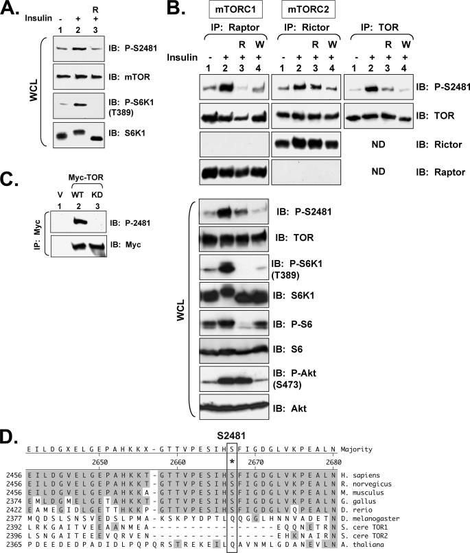
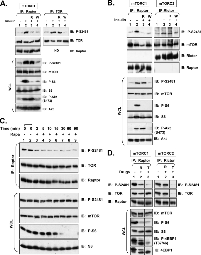
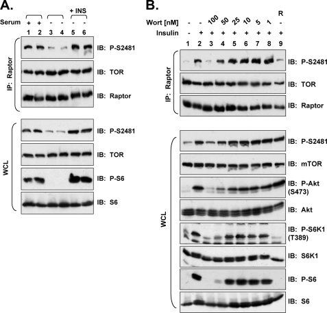
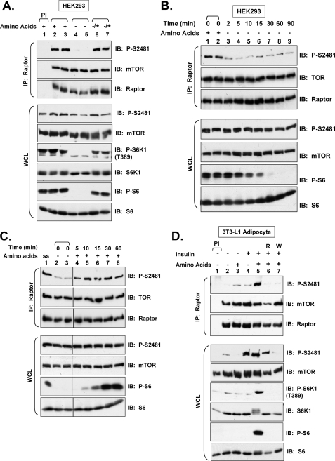
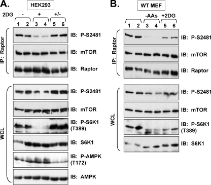
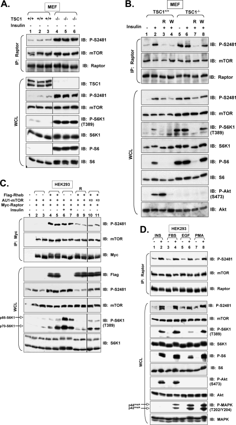
The mammalian target of rapamycin (mTOR) Ser/Thr kinase signals in at least two multiprotein complexes distinguished by their different partners and sensitivities to rapamycin. Acute rapamycin inhibits signaling by mTOR complex 1 (mTORC1) but not mTOR complex 2 (mTORC2), which both promote cell growth, proliferation, and survival. Although mTORC2 regulation remains poorly defined, diverse cellular mitogens activate mTORC1 signaling in a manner that requires sufficient levels of amino acids and cellular energy. Before the identification of distinct mTOR complexes, mTOR was reported to autophosphorylate on Ser-2481 in vivo in a rapamycin- and amino acid-insensitive manner. These results suggested that modulation of mTOR intrinsic catalytic activity does not universally underlie mTOR regulation. Here we re-examine the regulation of mTOR Ser-2481 autophosphorylation (Ser(P)-2481) in vivo by studying mTORC-specific Ser(P)-2481 in mTORC1 and mTORC2, with a primary focus on mTORC1. In contrast to previous work, we find that acute rapamycin and amino acid withdrawal markedly attenuate mTORC1-associated mTOR Ser(P)-2481 in cycling cells. Although insulin stimulates both mTORC1- and mTORC2-associated mTOR Ser(P)-2481 in a phosphatidylinositol 3-kinase-dependent manner, rapamycin acutely inhibits insulin-stimulated mTOR Ser(P)-2481 in mTORC1 but not mTORC2. By interrogating diverse mTORC1 regulatory input, we find that without exception mTORC1-activating signals promote, whereas mTORC1-inhibitory signals decrease mTORC1-associated mTOR Ser(P)-2481. These data suggest that mTORC1- and likely mTORC2-associated mTOR Ser-2481 autophosphorylation directly monitors intrinsic mTORC-specific catalytic activity and reveal that rapamycin inhibits mTORC1 signaling in vivo by reducing mTORC1 catalytic activity.


Figures

FIGURE 1.
FIGURE 1.
In 3T3-L1 adipocytes, insulin/PI3K signaling promotes rapamycin-sensitive and -resistant mTOR Ser-2481 autophosphorylation in mTORC1 and mTORC2, respectively. A, shown is regulation of total mTOR Ser(P)-2481 in whole cell lysates. Differentiated 3T3-L1 adipocytes were serum-deprived, pretreated with rapamycin (R), and stimulated with insulin (100 nm) for 30 min. After cell lysis, whole cell lysate was resolved on SDS-PAGE and immunoblotted with the indicated antibodies. B, shown is regulation of mTORC1- and mTORC2-associated mTOR Ser(P)-2481. 3T3-L1 adipocytes were serum-deprived, pretreated with rapamycin (R) or wortmannin (W), and stimulated with insulin. WCL was immunoprecipitated with anti-raptor antibodies (left panels) to immuno-isolate mTORC1, with anti-rictor antibodies (middle panels) to immuno-isolate mTORC2, and with anti-mTOR antibodies. Immunoprecipitates were immunoblotted with the indicated antibodies. WCL was also immunoblotted directly (lower panels) with the indicated antibodies to confirm the expected activation and/or inhibition of mTORC1 signaling. Note: the same four lysates (lanes 1–4) were used for raptor, rictor, and mTOR immunoprecipitation as well as for direct immunoblotting. ND, not done. C, shown is confirmation that Ser-2481 is a site of mTOR autophosphorylation. HEK293 cells were transfected with vector control (V), WT (1.0 μg), or KD (1.0 μg) Myc-mTOR plasmids. WCL was immunoprecipitated with Myc antibodies and immunoblotted as indicated. D, shown is alignment of mTOR Ser-2481 from various organisms using the algorithm ClustalW. The Caenorhabditis elegans sequence was omitted because of poor alignment resulting from large regions of non-homology.
FIGURE 2.
FIGURE 2.
In HEK293 cells, insulin/PI3K signaling promotes rapamycin-sensitive and -resistant mTOR Ser-2481 autophosphorylation in mTORC1 and mTORC2, respectively. A, shown is regulation of mTORC1-associated mTOR Ser(P)-2481. HEK293 cells were serum deprived, pretreated with rapamycin (R) or wortmannin (W), and stimulated with insulin. WCL was immunoprecipitated with anti-raptor antibodies (left panels) to immuno-isolate mTORC1 or anti-mTOR antibodies (right panels). Immunoprecipitates were immunoblotted with the indicated antibodies. WCL was also immunoblotted directly (lower panels) with the indicated antibodies to confirm the expected activation and/or inhibition of mTORC1 signaling. Note: the same four lysates (lanes 1–4) were used for raptor and mTOR immunoprecipitation as well as for WCL immunoblotting. ND, not done. B, shown is regulation of mTORC1- and mTORC2-associated mTOR Ser(P)-2481. Experiments were similar to A above, except WCL was immunoprecipitated with anti-raptor antibodies (left panels) to immuno-isolate mTORC1 or anti-rictor antibodies (right panels) to immuno-isolate mTORC2. C, rapamycin treatment of cycling cells mediates the rapid dephosphorylation of mTORC1-associated mTOR Ser-2481 in mTORC1. HEK293 cells cycling in DMEM/FBS were incubated in the absence (lanes 1 and 2) or presence of rapamycin for the indicated times (2–90 min) (lanes 3–9). WCL was immunoprecipitated with anti-raptor antibodies and immunoblotted with the indicated antibodies. WCL was also immunoblotted directly to confirm the expected inhibition of mTORC1 signaling (lower panels). D, Torin1 eliminates the residual mTORC1-associated mTOR Ser(P)-2481 and P-4EBP1 that remains upon rapamycin treatment. Raptor or rictor was immunoprecipitated from cycling HEK293 cells treated in the absence or presence of rapamycin or Torin1 for 1 h. Immunoprecipitates were immunoblotted for Ser(P)-2481 on raptor- or rictor-associated mTOR. WCLs were also immunoblotted to confirm the expected inhibition of mTORC1 and mTORC2 signaling by these drugs.
FIGURE 3.
FIGURE 3.
Serum withdrawal and inhibition of PI3K inhibit mTORC1-associated mTOR Ser(P)-2481 in HEK293 cells. A, withdrawal of serum growth factors mediates the dephosphorylation of mTORC1-associated mTOR Ser-2481. HEK293 cells were cultured in DMEM/FBS (lanes 1–2) or in media lacking serum growth factors for 20 h (lanes 3–6). Serum-deprived cells were then re-stimulated with insulin (INS) for 30 min (lanes 5 and 6). WCL was immunoprecipitated with anti-raptor antibodies and immunoblotted with the indicated antibodies. Note: 1 h before lysis the cycling cells (lanes 1 and 2) were re-fed with fresh media. WCL was also immunoblotted directly to confirm the expected regulation of mTORC1 signaling (lower panels). B, insulin-stimulated, mTORC1-associated mTOR Ser(P)-2481 requires PI3K. HEK293 cells were serum-deprived, pretreated in the absence (lane 2) or presence of various concentrations of wortmannin (100–1 nm) (lanes 3–8), or pretreated with rapamycin (R) (lane 9) for 30 min and then stimulated with insulin for 30 min (lanes 2–9). WCL was immunoprecipitated with anti-raptor antibodies and immunoblotted as indicated (upper panels). WCLs were also immunoblotted directly to confirm the expected activation and/or inhibition of PI3K and mTORC1 signaling by the various treatments (lower panels).
FIGURE 4.
FIGURE 4.
Sufficient levels of amino acids are required for mTORC1-associated mTOR Ser-2481 autophosphorylation. A, amino acid withdrawal mediates the dephosphorylation of mTORC1-associated mTOR Ser(P)-2481. Cycling HEK293 cells cultured in DMEM/FBS (lanes 1–3) were incubated in D-PBS/glucose containing dialyzed 10% FBS (D-PBS/Glc/FBS) for 60 min (lanes 4–7) to effect amino acid deprivation. Amino acid-deprived cells were then re-stimulated with DMEM/FBS for 30 min (lanes 6–7) as a source of amino acids. WCL was immunoprecipitated with anti-raptor antibodies and immunoblotted as indicated (upper panels). WCL was also immunoblotted directly to confirm the expected regulation of mTORC1 signaling (lower panels). B, experiments were similar to A above, except that amino acids were withdrawn for various amounts of time, 2–90 min. C, experiments were similar to A above, except that after amino acid deprivation for 60 min (lanes 2–8), amino acids were added back by refeeding with DMEM/FBS for 5–60 min. Note: ss indicates steady state, whereby cycling cells were cultured in DMEM/FBS before incubation in d-PBS/Glc/FBS. D, both amino acids and insulin are required for mTORC1-associated mTOR Ser(P)-2481. HEK293 cells were serum-deprived (∼20 h) and then amino acid-deprived via incubation in D-PBS/Glc (60 min). Factor-deprived cells were then pretreated with rapamycin (R) (lane 6) or wortmannin (W) (lane 7) and stimulated with amino acids alone (lane 3), insulin alone (lane 4), or both amino acids and insulin (lanes 5–7) for 30 min as indicated. Note: DMEM was used as source of amino acids. WCL was immunoprecipitated with pre-immune (PI) sera or with anti-raptor antibodies and immunoblotted as indicated. WCL was also immunoblotted directly to confirm the expected activation and/or inhibition of mTORC1 signaling by the various treatments.
FIGURE 5.
FIGURE 5.
Energy stress down-regulates mTORC1-associated mTOR Ser-2481 autophosphorylation. A, energy stress mediates mTORC1-associated mTOR Ser-2481 dephosphorylation. Cycling HEK293 cells were untreated (lanes 1 and 2) or treated with 2DG) (25 mm) for 15 min (lanes 3–6). After 2DG treatment, cells were re-fed with media lacking 2DG and incubated for an additional 30 min (lanes 5 and 6). WCL was immunoprecipitated with anti-raptor antibodies and immunoblotted as indicated. WCL was also immunoblotted directly to confirm the expected activation of AMPK and inhibition of mTORC1 signaling by 2DG. B, in MEFs, energy stress and amino acid withdrawal mediates mTORC1-associated mTOR Ser-2481 dephosphorylation. Cycling wild type MEFs (lanes 1 and 2) were incubated in D-PBS/Glc/FBS for 60 min to effect amino acid (AA) deprivation (lanes 3 and 4) or treated with 2DG (25 mm) for 15 min (lanes 5 and 6). WCL was immunoprecipitated with anti-raptor antibodies and immunoblotted as indicated. WCL was also immunoblotted directly to confirm the expected activation and/or inhibition of mTORC1 signaling by the various treatments.
FIGURE 6.
FIGURE 6.
TSC null status, Rheb overexpression, and PI3K/Akt-independent signaling promote mTORC1-associated mTOR Ser-2481 autophosphorylation. A, TSC suppresses mTORC1-associated mTOR Ser(P)-2481. Littermate-matched, 3T3 immortalized MEFs derived from TSC1+/+ or TSC1−/− mice were serum-deprived. Triplicate lysates were immunoprecipitated with anti-raptor antibodies and immunoblotted as indicated. WCL was also immunoblotted directly to confirm the absence of TSC1 and the expected activation of mTORC1 signaling. Note: we find that TSC1−/− fibroblasts express higher levels of total raptor protein when normalized for total protein content; thus, two-thirds of the immunoprecipitate from TSC1−/− cells was loaded relative to TSC1+/+ cells to normalize the amount of raptor between the two cell lines. WCL was loaded similarly. B, TSC1−/− cells exhibit constitutive, rapamycin-sensitive, and wortmannin-resistant mTORC1-associated mTOR Ser(P)-2481. Experiments were similar to A above, except that serum-deprived MEFs from TSC1+/+ or TSC1−/− animals were pretreated with rapamycin (R) or wortmannin (W) and then stimulated with or without insulin. C, Rheb promotes constitutive, mTORC1-associated mTOR Ser(P)-2481 in a rapamycin-sensitive manner that requires mTOR kinase activity. HEK293 cells were co-transfected with Myc-raptor (0.5 μg) and AU1-mTOR (2 μg) in the absence (lanes 1 and 3 and lanes 6 and 7) or presence (lanes 4 and 5 and lanes 8–11) of FLAG-Rheb (3 μg). Rheb-transfected cells were also treated with rapamycin (R) for 24 h (lanes 6-and 7) or were co-transfected with KD Myc-TOR (lanes 10 and 11) rather than wild type mTOR. Cells were serum-deprived and stimulated in the absence (lanes 1–5 and 8–11) or presence (lanes 6 and 7) of insulin. WCL was immunoprecipitated with Myc antibodies to pull down mTORC1 and immunoblotted with the indicated antibodies. WCL was also immunoblotted directly to confirm the expected activation and/or inhibition of mTORC1 signaling by the various treatments. D, EGF and PMA promote mTORC1-associated mTOR Ser(P)-2481 independently of PI3K. HEK293 cells were serum-deprived and stimulated with insulin (INS) (lane 2), 10% FBS (lane 4), EGF (lane 6), or PMA (lane 8) for 30 min. WCL was immunoprecipitated with anti-raptor antibodies and immunoblotted with the indicated antibodies. WCL was also immunoblotted directly to confirm the expected modulation of PI3K (P-Akt (S473)), MAPK (P-MAPK (T202/Y204)I, and mTORC1 (P-S6K1 (T389); P-S6) signaling by the various growth factors/mitogens. Note: p44mapk and p42mapk are also known as ERK1 and -2 (extracellular signal-regulated kinase 1 and 2), respectively.
FIGURE 7.
FIGURE 7.
Mechanism of mTOR Ser-2481 autophosphorylation. A, Rheb-GTP provided in vitro does not increase mTORC1-associated mTOR Ser(P)-2481 over basal levels. HEK293 cells on 10-cm plates were co-transfected with HA-raptor (2 μg) together with WT or KD Myc-mTOR alleles (8 μg) and serum-deprived. mTORC1 was immuno-isolated via anti-HA-raptor immunoprecipitation and preincubated with recombinant, GTPase-deficient GST-Rheb (Q64L) protein loaded with GTP. The level of HA-raptor-associated mTOR Ser(P)-2481 after immunoprecipitation but before the addition of GST-Rheb-GTP and initiation of in vitro kinase reactions is shown as Input. In vitro kinase reactions primed with Rheb-GTP were initiated with the addition of recombinant GST-4EBP1 and ATP (Assay). Myc-mTOR autophosphorylation was assessed by immunoblotting IVK reactions with Ser(P)-2481 antibodies, and substrate phosphorylation was assessed with P-Thr-37/46–4EBP1 (P-4EBP1 (T37/46)) antibodies. IVK reactions and WCLs were also immunoblotted with the antibodies indicated. B, mTOR Ser-2481 autophosphorylation occurs in cis rather than in trans. HEK293 cells were co-transfected with Myc-mTOR (1 μg) WT or KD in the absence or presence of FLAG-Rheb (4 μg). Myc-mTOR was immunoprecipitated from cycling cells and immunoblotted with the indicated antibodies. WCLs were also immunoblotted as shown.

References

    1. Dunlop E. A., Tee A. R. (2009) Cell. Signal. 21, 827–835 - PubMed
    1. Fingar D. C., Blenis J. (2004) Oncogene 23, 3151–3171 - PubMed
    1. Jacinto E., Lorberg A. (2008) Biochem. J. 410, 19–37 - PubMed
    1. Bhaskar P. T., Hay N. (2007) Dev. Cell 12, 487–502 - PubMed
    1. Guertin D. A., Sabatini D. M. (2009) Sci. Signal. 2, pe24. - PubMed

Publication types

MeSH terms