. 2010 Mar 5;285(10):7111-8.
doi: 10.1074/jbc.M109.052977. Epub 2009 Dec 17.

Deficiency in APOBEC2 leads to a shift in muscle fiber type, diminished body mass, and myopathy

Affiliations

Deficiency in APOBEC2 leads to a shift in muscle fiber type, diminished body mass, and myopathy

Yusuke Sato et al. J Biol Chem. .

Abstract

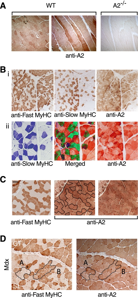
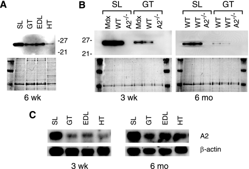
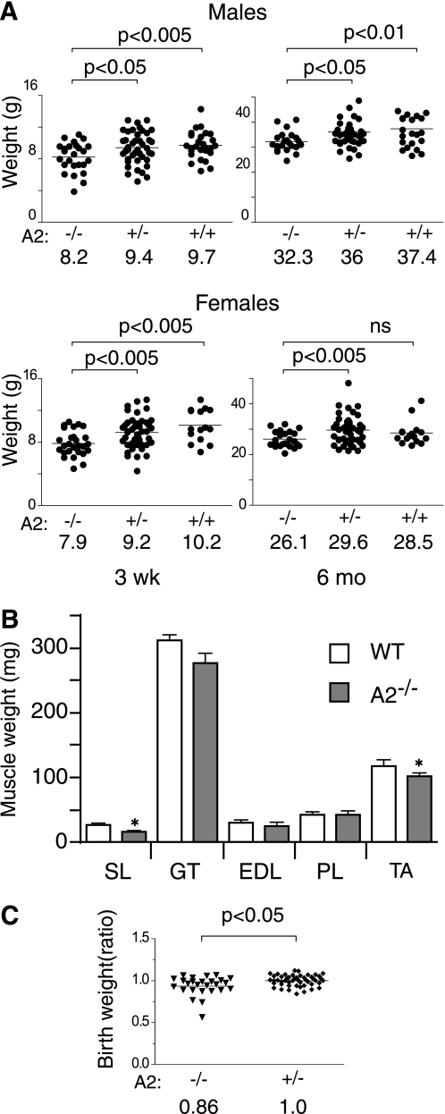
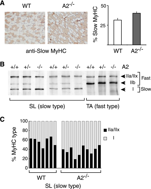
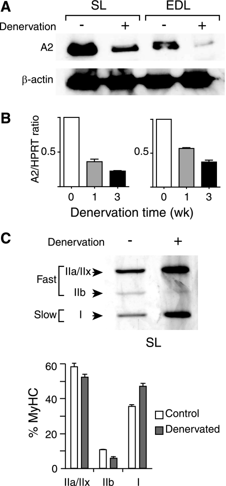
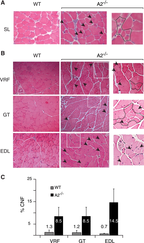
The apoB RNA-editing enzyme, catalytic polypeptide-like (APOBEC) family of proteins includes APOBEC1, APOBEC3, and activation-induced deaminase, all of which are zinc-dependent cytidine deaminases active on polynucleotides and involved in RNA editing or DNA mutation. In contrast, the biochemical and physiological functions of APOBEC2, a muscle-specific member of the family, are unknown, although it has been speculated, like APOBEC1, to be an RNA-editing enzyme. Here, we show that, although expressed widely in striated muscle (with levels peaking late during myoblast differentiation), APOBEC2 is preferentially associated with slow-twitch muscle, with its abundance being considerably greater in soleus compared with gastrocnemius muscle and, within soleus muscle, in slow as opposed to fast muscle fibers. Its abundance also decreases following muscle denervation. We further show that APOBEC2-deficient mice harbor a markedly increased ratio of slow to fast fibers in soleus muscle and exhibit an approximately 15-20% reduction in body mass from birth onwards, with elderly mutant animals revealing clear histological evidence of a mild myopathy. Thus, APOBEC2 is essential for normal muscle development and maintenance of fiber-type ratios; although its molecular function remains to be identified, biochemical analyses do not especially argue for any role in RNA editing.


Figures

FIGURE 1.
FIGURE 1.
APOBEC2 is highly expressed in slow myofibers. A, lateral and longitudinal sections of hind limb muscle from a 3-month-old C57BL/6 mouse (left and middle panels) and parallel staining of longitudinal sections of hind limb muscle from an APOBEC2-deficient (A2−/−) mouse to demonstrate the specificity of staining (right panel). WT, wild-type. B, panel i, staining of serial sections of hind limb muscle from 3-month-old wild-type mice for fast and slow myosin HC (MyHC) isoforms and for APOBEC2; panel ii, regions of the sections depicted in panel i shown in higher magnification but with pseudo-coloring such that fibers staining strongly for both slow myosin HC and APOBEC2 are seen as green in the merged panel. C, comparison of serial sections of soleus muscle from 6-month-old wild-type mice stained for the fast myosin HC isoform and APOBEC2. The two panels stained for APOBEC2 are identical except that, in the one on the left, slow fibers (which lack fast myosin HC) are enclosed by a black line. Scale bar = 200 μm (left panel). D, comparison of serial sections of soleus muscle from 6-month-old male mdx mice stained for the fast myosin HC isoform and APOBEC2. Examples of damaged/regenerating fibers are outlined in black, with region A comprising slow fibers and region B comprising fast fibers. GT, gastrocnemius muscle; SL, soleus muscle.
FIGURE 2.
FIGURE 2.
APOBEC2 expression increases during myoblast differentiation in vitro. Primary mouse myoblasts were cultured under low serum conditions to induce muscle cell differentiation. At days 1–5 after the start of serum starvation, the ratio of multinucleated myotubes to mononucleated myoblasts (fusion index) was determined, and cell extracts were analyzed by Western blotting for expression of APOBEC2 (A2), myosin HC (MyHC), and histone H3.
FIGURE 3.
FIGURE 3.
APOBEC2 is highly expressed in soleus muscle. A, abundance of APOBEC2 in extracts of different muscles from a 6-week (wk)-old male C57BL/6 mouse as determined by Western blot analysis (upper panel), controlling for loading by staining for total protein with Ponceau reagent (lower panel). SL, soleus muscle; GT, gastrocnemius muscle; HT, heart. B, Western blot comparison of APOBEC2 abundance in extracts of soleus and gastrocnemius muscle from 3-week-old or 6-month (mo)-old wild-type (WT) and mdx mice, with APOBEC2-deficient (A2−/−) mice serving as a negative control. C, Northern blot analysis of APOBEC2 RNA in various muscles, with β-actin serving as a control.
FIGURE 4.
FIGURE 4.
APOBEC2-deficient mice weigh less than control siblings. A, the body weights of APOBEC2 (A2)-deficient (−/−), heterozygous (+/−), and wild-type (+/+) mice from heterozygous breedings were determined at the time of weaning (21 days after birth) and at 6 months. The mean weights for the different groups are indicated. Data were analyzed using Student's t test, with the significance for each comparison shown. ns, not significant. B, the weights of individual muscles from groups of five male 15-week-old wild-type (WT) and five male 15-week-old APOBEC2-deficient mice are shown. SL, soleus muscle; GT, gastrocnemius muscle; PT, plantaris muscle; TA, tibialis anterior muscle. Error bars are means ± S.E., with asterisks denoting differences significant at p < 0.05 (Student's t test). C, the body weights of newborn mice born to breedings of an APOBEC2-deficient father and a heterozygous mother are presented, with the individual weights given as a ratio of the absolute weight of an individual animal to that of the mean of APOBEC2+/− heterozygotes within the same litter.
FIGURE 5.
FIGURE 5.
Increased proportion of slow fibers in APOBEC2−/− mouse soleus. A, immunohistochemical staining with an anti-slow myosin HC (MyHC) mAb of transverse sections of soleus muscle from 15-week-old wild-type (WT) and APOBEC2−/− (A2/) mice. Analysis of five pairs of 15-week-old mice revealed that the proportion of soleus fibers staining with the anti-slow myosin HC mAb increased from 31% in control mice to 40% in APOBEC2-deficient mice. B, comparison of the relative abundance of slow and fast myosin HC isoforms in soleus muscle (SL; left and middle panels) and tibialis anterior muscle (TA; right panel) from 3-month-old litter-matched wild-type, APOBEC2+/−, and APOBEC2−/− mice analyzed by silver staining of muscle extracts subjected to SDS-PAGE in low cross-linked gels. C, histogram showing the ratios of fast (MyHCIIa+IIx) to slow (MyHCI) myosins in soleus muscle of seven wild-type and nine APOBEC2-deficient mice as judged by densitometry of silver-stained SDS-polyacrylamide gels.
FIGURE 6.
FIGURE 6.
Reduced APOBEC2 expression and fiber-type switch following denervation. A, Western blot analysis of APOBEC2 expression in soleus (SL) and EDL muscle from 15-week-old male mice that had or had not been subjected to sciatic nerve denervation 2 weeks previously. Protein loading was controlled by reprobing for β-actin. B, reverse transcription-PCR analysis of APOBEC2 (A2) expression in soleus and EDL muscle from 15-week-old male mice that had or had not been subjected to sciatic nerve denervation 1 or 3 weeks (wk) previously. The reverse transcription-PCR signal was calculated relative to that given by hypoxanthine-guanine phosphoribosyltransferase (HPRT). C, analysis of change in myosin HC (MyHC) isoform expression in soleus muscle 3 weeks following denervation in male C57BL/6 mice by high resolution SDS-PAGE. Histograms present the results of the analysis of five mice.
FIGURE 7.
FIGURE 7.
Histological evidence of myopathy in 8–10-month-old APOBEC2−/− mice. A, examples of a region of soleus muscle (SL) in 15-week-old wild-type (WT) or APOBEC2-deficient (A2/) mice. The arrowheads indicate fibers with centrally located nuclei, and the right panel shows a region with immature myotubes. B, comparison of transverse sections of vastus and rectus femoris (VRL), gastrocnemius (GT), and EDL muscle from 9-month-old APOBEC2-deficient and wild-type mice. The boxed areas in the middle panels are shown in higher magnification in the right panels, where outlined regions contain smaller fibers. C, histogram comparing the percentage of centrally nucleated fibers (CNF) in different muscles from 9-month-old APOBEC2-deficient and control mice.
FIGURE 8.
FIGURE 8.
APOBEC2 is a soluble homotetramer showing little evidence of RNA binding. A, APOBEC2 from gastrocnemius muscle extracts was eluted from a calibrated Sephadex gel filtration column and detected by Western blotting following SDS-PAGE. Preincubation of the extract with RNase did not affect the APOBEC2 elution profile (not shown). B, MBP-APOBEC1 (A1) and MBP-APOBEC2 (A2) were compared for binding to AU repeat RNA and apolipoprotein B targets. The MBP fusion proteins produced from E. coli were incubated at two different concentration (differing 10-fold) with 32P-labeled RNA. After UV cross-linking and digestion with RNase, samples were subjected to SDS-PAGE, and proteins were identified by staining (Total), whereas cross-linked RNA (X-linked) was detected by autoradiography.

Similar articles

Cited by

References

    1. Conticello S. G., Thomas C. J., Petersen-Mahrt S. K., Neuberger M. S. (2005) Mol. Biol. Evol. 22, 367–377 - PubMed
    1. Navaratnam N., Sarwar R. (2006) Int. J. Hematol. 83, 195–200 - PubMed
    1. Conticello S. G. (2008) Genome Biol. 9, 229. - PMC - PubMed
    1. Liao W., Hong S. H., Chan B. H., Rudolph F. B., Clark S. C., Chan L. (1999) Biochem. Biophys. Res. Commun. 260, 398–404 - PubMed
    1. Anant S., Mukhopadhyay D., Sankaranand V., Kennedy S., Henderson J. O., Davidson N. O. (2001) Am. J. Physiol. Cell Physiol. 281, C1904–C1916 - PubMed

Publication types

MeSH terms