. 2010 Feb;9(2):278-87.
doi: 10.1128/EC.00307-09. Epub 2009 Dec 18.

Hgc1 mediates dynamic Candida albicans-endothelium adhesion events during circulation

Affiliations

Hgc1 mediates dynamic Candida albicans-endothelium adhesion events during circulation

Duncan Wilson et al. Eukaryot Cell. 2010 Feb.

Abstract

Common iatrogenic procedures can result in translocation of the human pathogenic fungus Candida albicans from mucosal surfaces to the bloodstream. Subsequent disseminated candidiasis and infection of deep-seated organs may occur if the fungus is not eliminated by blood cells. In these cases, fungal cells adhere to the endothelial cells of blood vessels, penetrate through endothelial layers, and invade deeper tissue. In this scenario, endothelial adhesion events must occur during circulation under conditions of physiological blood pressure. To investigate the fungal and host factors which contribute to this essential step of disseminated candidiasis, we have developed an in vitro circulatory C. albicans-endothelium interaction model. We demonstrate that both C. albicans yeast and hyphae can adhere under flow at a pressure similar to capillary blood pressure. Serum factors significantly enhanced the adhesion potential of viable but not killed C. albicans cells to endothelial cells. During circulation, C. albicans cells produced hyphae and the adhesion potential first increased, then decreased with time. We provide evidence that a specific temporal event in the yeast-to-hyphal transition, regulated by the G(1) cyclin Hgc1, is critical for C. albicans-endothelium adhesion during circulation.


Figures
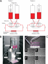
Fig. 1.
Fig. 1.
The circulatory model. (A) Schematic representation of the circulatory model. Air pressure in the reservoirs and opening/closing of the valve set are controlled via an air pump and computer software. Note that in both position 1 and position 2 unidirectional flow through an endothelial channel is maintained. (B) Picture of the circulatory model (ibidi pump system) with reservoirs, valve set, tubing, and μ-Slide labeled. (C) Closeup picture of the μ-Slide. Note that each μ-Slide houses six independent channels, which can be seeded with endothelial cells and connected to the circulatory model via Luer adaptors. (D to G) Micrograph images of the development of endothelial cells within a μ-Slide channel, 1 (D), 2 (E), and 3 (F) days postseeding and following 20 min under flow (G). Note that monolayers are confluent by 3 days postincubation and that 20 min flow does not overtly effect the morphology of the endothelial monolayer.
Fig. 2.
Fig. 2.
Adhesion dynamics of circulating C. albicans. C. albicans cells were circulated for 120 min, and the kinetics of cells adhering to endothelial channels (per mm2 per min) were determined for each 20-min interval. As a control (Static), cells taken from the perfusion system at the indicated time points were applied to a static endothelial monolayer for 20 min. The x axis represents the time of circulation, and each data point is the kinetics of adhesion within 20 min of exposure to a sterile endothelial monolayer. Data points are the means of results of three independent experiments, and error bars indicate the standard errors of the means. Note that adhesion under flow conditions reach maximum kinetics between 40 and 60 min.
Fig. 3.
Fig. 3.
Population dynamics of circulating cells. The concentration of circulating C. albicans cells in the system was determined every 20 min by counting using a hemocytometer. Data represent the means of results of at least two independent experiments, and error bars indicate the standard errors of the means between experiments.
Fig. 4.
Fig. 4.
Adhesion is dose dependent. C. albicans cells were inoculated into the perfusion system at either 2 × 10⁵, 5 × 10⁵, or 1 × 10⁶ cells/ml and circulated for 60 min. Adhesion kinetics were determined for 0 to 20, 20 to 40, and 40 to 60 min of perfusion. Data represent the means of results of at least two independent experiments, and error bars indicate the standard errors of the means between experiments.
Fig. 5.
Fig. 5.
Morphological distribution. The morphology of C. albicans cells in circulation at 60 min and of cells which had adhered to the endothelial monolayer between 40 and 60 min was determined. At least 130 cells, over two independent experiments, for each condition were scored. Data represent the percentage of indicated germ tube length. Note that germ tubes of 3 to 7 μm in length adhered to the endothelium under flow at higher frequency. The variance of germ tube length of endothelium-adherent cells was significantly lower than that for circulating cells (P < 0.0001, by F-test, P = 0.0024 upon exclusion of zero [yeast] values).
Fig. 6.
Fig. 6.
Germ tube orientation. (A) The orientation of germ tubes adhering to the endothelium between 40 and 60 min of circulation with the direction of flow as the comparative vector was determined and displayed as a radar plot. The orientations of germ tubes from the same population incubated on a static endothelial monolayer are included as a control. (B) Micrograph images (40× magnification) of cells which had undergone adhesion to an endothelial monolayer under flow (left panel) or under static conditions (right panel). Note that circulating germ tubes adhere with the mother cells aligned with the direction of flow, suggesting germ tube tip anchor events.
Fig. 7.
Fig. 7.
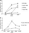
Circulatory adhesion is HGC1 dependent. (A) Wild-type CAI4+CIp10 (Wt) or hgc1Δ homozygous mutant strains were circulated for 60 min, and the adhesion kinetics occurring between 0 and 20, 20 and 40, and 40 and 60 min were determined. At 20 to 40 and 40 to 60 min, the adhesion kinetics of hgc1Δ were significantly lower than those of the Wt (P < 0.0001). The adhesion kinetics of cells taken from the perfusion system at the indicated time points and applied to a static endothelial monolayer are included as a control. Note that between 40 and 60 min, hgc1Δ adheres to the static endothelium at a rate similar to that of the Wt (P = 0.51) but not under conditions of flow. Data points are the means of results of at least two independent experiments, and error bars indicate the standard errors of the means. (B) hgc1Δ and hgc1Δ+HGC1 strains were circulated for 100 min, and adhesion kinetics were determined for 0 to 20, 20 to 40, 40 to 60, 60 to 80, and 80 to 100 min. At all time points except for 0 to 20 min, hgc1Δ adhered significantly less than hgc1Δ+HGC1 (P < 0.0001). Data points are the means of results of at least two independent experiments, and error bars indicate the standard errors of the means.
Fig. 8.
Fig. 8.
Effect of serum on adhesion. C. albicans cells were circulated for 60 min in either DMEM (w/o FBS) or DMEM supplemented with 10% fetal bovine serum (+ FBS), and the adhesion kinetics occurring between 0 and 20, 20 and 40, and 40 and 60 min were determined. As a (Static) control, cells from the perfusion system, taken at the indicated time points, were applied to a static endothelial monolayer for the 20-min intervals. Data points are the means of results of at least two independent experiments, and error bars indicate the standard errors of the means.

Similar articles

Cited by

References

    1. Almeida R. S., Brunke S., Albrecht A., Thewes S., Laue M., Edwards J. E., Filler S. G., Hube B. 2008. The hyphal-associated adhesin and invasin Als3 of Candida albicans mediates iron acquisition from host ferritin. PLoS Pathog. 4:e1000217. - PMC - PubMed
    1. Banerjee M., Thompson D. S., Lazzell A., Carlisle P. L., Pierce C., Monteagudo C., Lopez-Ribot J. L., Kadosh D. 2008. UME6, a novel filament-specific regulator of Candida albicans hyphal extension and virulence. Mol. Biol. Cell 19:1354–1365 - PMC - PubMed
    1. Bendel C. M., Hess D. J., Garni R. M., Henry-Stanley M., Wells C. L. 2003. Comparative virulence of Candida albicans yeast and filamentous forms in orally and intravenously inoculated mice. Crit. Care Med. 31:501–507 - PubMed
    1. Blankenship J. R., Mitchell A. P. 2006. How to build a biofilm: a fungal perspective. Curr. Opin. Microbiol. 9:588–594 - PubMed
    1. Braun B. R., Johnson A. D. 1997. Control of filament formation in Candida albicans by the transcriptional repressor TUP1. Science 277:105–109 - PubMed

Publication types

MeSH terms

LinkOut - more resources