. 2010 Feb;9(2):288-305.
doi: 10.1128/EC.00220-09. Epub 2009 Dec 18.

Distinct subcellular localization of a group of synaptobrevin-like SNAREs in Paramecium tetraurelia and effects of silencing SNARE-specific chaperone NSF

Affiliations

Distinct subcellular localization of a group of synaptobrevin-like SNAREs in Paramecium tetraurelia and effects of silencing SNARE-specific chaperone NSF

Christina Schilde et al. Eukaryot Cell. 2010 Feb.

Abstract

We have identified new synaptobrevin-like SNAREs and localized the corresponding gene products with green fluorescent protein (GFP)-fusion constructs and specific antibodies at the light and electron microscope (EM) levels. These SNAREs, named Paramecium tetraurelia synaptobrevins 8 to 12 (PtSyb8 to PtSyb12), showed mostly very restricted, specific localization, as they were found predominantly on structures involved in endo- or phagocytosis. In summary, we found PtSyb8 and PtSyb9 associated with the nascent food vacuole, PtSyb10 near the cell surface, at the cytostome, and in close association with ciliary basal bodies, and PtSyb11 on early endosomes and on one side of the cytostome, while PtSyb12 was found in the cytosol. PtSyb4 and PtSyb5 (identified previously) were localized on small vesicles, PtSyb5 probably being engaged in trichocyst (dense core secretory vesicle) processing. PtSyb4 and PtSyb5 are related to each other and are the furthest deviating of all SNAREs identified so far. Because they show no similarity with any other R-SNAREs outside ciliates, they may represent a ciliate-specific adaptation. PtSyb10 forms small domains near ciliary bases, and silencing slows down cell rotation during depolarization-induced ciliary reversal. NSF silencing supports a function of cell surface SNAREs by revealing vesicles along the cell membrane at sites normally devoid of vesicles. The distinct distributions of these SNAREs emphasize the considerable differentiation of membrane trafficking, particularly along the endo-/phagocytic pathway, in this protozoan.


Figures

Fig. 1.
Fig. 1.
Sequence analysis of P. tetraurelia synaptobrevins. (A) Domain structure of PtSybs based on homology to known proteins and identified longin (green), synaptobrevin (blue), and transmembrane (yellow) domains. PtSyb6-2 produces a truncated protein because of the creation of a stop codon by intron splicing (*). (B) Alignment of the SNARE domains and predicted transmembrane domains obtained by using ClustalW (slow, accurate) analysis of Paramecium synaptobrevins, a Tetrahymena thermophila synaptobrevin homolog (Tt2415; TIGR gene identifier 2415), Entamoeba histolytica synaptobrevin-like protein 1 (EhSYBL1; GenBank accession number AY256852), Dictyostelium discoideum SybA (Dictybase identifier DDB0214903), Saccharomyces cerevisiae Snc1 (GenBank accession number AAC05002), Arabidopsis thailiana VAMP711 (GenBank accession number O49377), and Homo sapiens Syb1 (GenBank accession number AAA60603). Conserved heptad repeats are shown in black. Note that the orthodox zero-layer arginine (red) is not present in all PtSybs. The 17- to 24-amino-acid-long transmembrane domains are absent from PtSyb families 6 and 7; instead, these proteins possess a potential carboxy-terminal putative prenylation/palmitoylation motif (yellow).
Fig. 2.
Fig. 2.
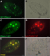
Localization of PtSyb4-1 and PtSyb5-1 as GFP-fusion proteins in vesicles undergoing cyclosis. (A and B) Epifluorescence (A) and bright-field (B) images of a living cell expressing GFP-PtSyb4-1. Arrows indicate GFP-marked small vesicles. (C and D) Epifluorescence of a living cell expressing PtSyb5–1-GFP double stained with LysoTracker red (D). (E) The merged image of panels C and D, PtSyb5-1–GFP with LysoTracker red staining, shows no indication of colocalization of PtSyb5-1–GFP–positive vesicles (e.g., at arrows) with acidic vesicles (pH ≤ 5.2). Note that three of the food vacuoles show strong autofluorescence in both fluorescence channels. (F) Corresponding bright-field image of the same cell. Note that the cell has moved a little between the image capture and has extruded trichocysts which are visible as thin rods surrounding the cell. Bars, 10 μm.
Fig. 3.
Fig. 3.
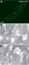
Localization of PtSyb5-1–GFP after fixation on the light microscopic (A) or on the EM (B and C) level. (A) Stack reconstruction of a confocal image series from a fixed PtSyb5-1–GFP cell showing GFP fluorescence on the membrane of numerous small misshapen vesicles. Bar, 10 μm. (B and C) Two examples of EM images of detection of GFP by anti-GFP antibody/protein A-Au5 conjugate in PtSyb5-1–GFP–expressing cells show labeling surrounding undocked trichocysts (t), mainly associated with structures (asterisks) of identical texture as trichocysts. For details, see the text. Bars, 250 nm.
Fig. 4.
Fig. 4.
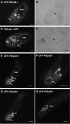
Localization of PtSyb8-1–GFP–fusion constructs. (A and C) GFP–PtSyb8-1 (A) and PtSyb8-1–GFP (C) stain a crescent-shaped structure at the cytopharynx (arrowheads). There was also some staining on the membrane of a single food vacuole (fv; arrows). Note that the bright labeling of the big vacuole in panel A is an artifact due to fluorescence of vacuole contents. (B and D) Corresponding bright-field images of the living cells. (E to H) 3D reconstruction of confocal image stacks (1-μm thickness) from a fixed GFP-PtSyb8–1 cell shows strong staining at the cytopharynx (arrowheads) and of the surface of a previously internalized food vacuole (arrows). (F) Enlargement from panel E. In some images trains of intensely labeled small vesicles leading to the cytopharynx are visible. cs, cytostome; mac, macronucleus. Bars, 10 μm.
Fig. 5.
Fig. 5.
Localization of GFP–PtSyb9-2 in living cells. (A and C) GFP–PtSyb9-2 stains the crescent-shaped site of nascent food vacuole formation at the cytopharynx (arrowheads) as well as the postoral fibers (pf) (C), structures involved in steering the newly formed food vacuole toward the posterior end of the cell. The oral cavity (oc) itself, the macronucleus (mac), and food vacuoles (fv) are not stained. (B and D) Corresponding bright-field images. The anterior end of the cell is oriented toward the top left of the images. Bars, 10 μm.
Fig. 6.
Fig. 6.
Anti-PtSyb8-1 and anti-PtSyb9 antibody staining. (A) A specific antibody against PtSyb8-1 shows strong staining of the peniculi (p) lining the oral cavity (oc). The image shows two cytostomes of a dividing cell. a, the anterior end of the cell. (B) Bright-field image of the same cell. (C) An anti-PtSyb9 antibody stains vesicles along the oral cavity (oc; arrows) as well as the site of nascent food vacuole formation (arrowhead). (C and D) The anti-PtSyb9 antibody also exhibits a rather faint staining of the contractile vacuole system (cv) and some staining at the cell surface. Bars, 10 μm.
Fig. 7.
Fig. 7.
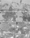
EM localization of PtSyb8-1 and PtSyb9 to two morphologically different nonacidosomal vesicle populations near the cytostome. (A) Labeling of vesicles around the cytostome (cs) with anti-PtSyb8. ci, cilium. (A′) Enlargement from panel A, showing labeling with anti-PtSyb8 antibodies near small vesicles. (B) Labeling in the area of nascent food vacuole formation near the cytopharynx with anti-PtSyb9. (B′) Enlargement of panel B. Bars, 100 nm.
Fig. 8.
Fig. 8.
(A and B) Surface (A) and median (B) focus of epifluorescence of a living cell expressing PtSyb10-1–GFP, showing punctate and partly circular (A, inset) surface staining with clear exclusion of the cytoproct (cp) and strong labeling lining the sides of the oral cavity (oc). The macronucleus (mac) and food vacuoles (fv) are free of label and clearly visible as dark exclusions of staining. a, anterior end of the cell; p, peniculus. (C to E) Confocal image slices (1 μm) of a fixed PtSyb10-1–GFP–expressing cell near the surface (C), a median plane (D), and a 3D reconstruction of the image stack (E), showing a regular pattern of surface ridges. Bars, 10 μm.
Fig. 9.
Fig. 9.
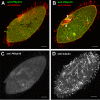
Confocal images of double staining with a specific anti-PtSyb10 and an anti-α-tubulin antibody. (A) Surface slice showing close apposition but no exact overlap of anti-PtSyb10 and anti-α-tubulin staining of basal bodies. (B) Median slice showing strong staining with anti-α-tubulin but less with anti-PtSyb10 antibodies at the cytostome (cs) and cytopharynx, with some overlap along distinct microtubule arrays (arrowheads) and possibly some minor overlap of staining at the contractile vacuole systems (arrows). (C and D) 3D rendering of the anti-PtSyb10-1 label (C) and of the anti-α-tubulin label confocal image stacks (D), clearly showing different patterns. Also note staining of cilia with anti-tubulin but no labeling with anti-PtSyb10 antibodies in panels A, B, and C. Bars, 10 μm.
Fig. 10.
Fig. 10.
Immuno-EM localization of PtSyb10-1–GFP detected with an anti-GFP antibody followed by protein A-Au5. (A) Staining (arrows) of the complex formed by the plasma membrane and peripheral membrane of alveolar sacs (as). ci, cilium. (B and C) Strong labeling on both sides around a cilium at the site of cell membrane/cilium transition. (D and E) Only a small amount of label occurs inside the cytosolic compartment, without any clear correlation with subcellular compartments with the possible exception of the ER, while irrelevant structures such as trichocysts (t) are label free. bb, basal body. Bars, 250 nm.
Fig. 11.
Fig. 11.
Western blot of protein preparations from PtSyb10-silenced cells and wild-type cells. As a loading control, the same blot was probed with antibodies against α-tubulin (α-tub, upper panel) and showed very similar protein amounts (∼ 20 ng) in both lanes. In extracts prepared from PtSyb10-silenced cells, antibodies against PtSyb10 (lower panel) recognized a significantly reduced amount of PtSyb10 antigen compared to wild-type cells.
Fig. 12.
Fig. 12.
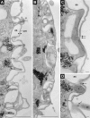
Examples of ultrastructural changes observed in the Paramecium cortex after PtNSF silencing. Note that the normally thin complex made by the cell membrane (cm) and the outer membrane of alveolar sacs (as), as seen at the arrowheads, is frequently interrupted by membrane-bounded structures of different sizes (asterisks) that may occasionally appear as very distinct small vesicles (arrows in panels C and D), e.g., at the base of cilia (ci in panel D). No comparable small vesicles associated with the surface membrane were seen in non-NSF-silenced cells, as discussed in the text. t, trichocyst. Bars, 1 μm (A and B) or 0.1 μm (C and D).
Fig. 13.
Fig. 13.
Localization of GFP–PtSyb11-1 and GFP–PtSyb12-1. (A and B) Surface (A) and median (B) focus of epifluorescence of a living cell expressing GFP–PtSyb11-1. The regular punctate pattern is due to staining of regularly arranged terminal cisternae (early endosomes), situated below ciliary basal bodies, ∼1 μm below the cell surface (arrows). There was little staining of the cell membrane or of a food vacuole (arrowhead) visible in panel B, and strong staining occurred only at one side of the cytostome (cs). (C) Corresponding bright-field image. (D and E) Surface (D) and median (E) focus of epifluorescence of a living cell expressing GFP–PtSyb12-1, which largely appears soluble in the cytosol. Also, some faint staining of “surface ridges,” of the egg case-like cell surface, is observed in panel D. (E) Cytostome (cs), food vacuoles, and the macronucleus (mac) are devoid of label, while some label appears clustered in the cytosolic compartment. (F) Corresponding bright-field image. Bars, 10 μm.
Fig. 14.
Fig. 14.
Scheme of a Paramecium cell superimposed with immunolocalization and GFP localization of the PtSNAREs that we worked with in this study. The trafficking scheme is based on references , , and . The scheme contains elements of the osmoregulatory system (a, ampulla; cv, contractile vacuole; ds, decorated spongiome; ss, smooth spongiome), of the phagosomal apparatus (as, acidosomes mediating acidification of a food vacuole [fv] after pinching off; cp, cytoproct [defecation site]). ci, cilia; dv, discoidal vesicles and other recycling vesicles (rv); ee, early endosomes; er, endoplasmic reticulum; ga, Golgi apparatus; gh, ghosts (from released trichocysts); oc, oral cavity; pm, plasma membrane; ps, parasomal sacs; tr, trichocysts; trpc, trichocyst precursor.

Similar articles

Cited by

References

    1. Adoutte A., Delgado P., Fleury A., Levilliers N., Laine M. C., Marty M. C., Boisvieux-Ulrich E., Sandoz D. 1991. Microtubule diversity in ciliated cells: evidence for its generation by post-translational modification in the axonemes of Paramecium and quail oviduct cells. Biol. Cell 71:227–245 - PubMed
    1. Allen R. D. 1984. Paramecium phagosome membrane: from oral region to cytoproct and back again. J. Protozool. 31:1–6
    1. Allen R. D., Fok A. K. 1993. Endosomal membrane traffic of ciliates, p. 57–83InPlattner H. (ed.), Advances in cell and molecular biology of membranes JAI Press, Greenwich, CT
    1. Allen R. D., Fok A. K. 2000. Membrane trafficking and processing in Paramecium. Int. Rev. Cytol. 198:277–318 - PubMed
    1. Allen R. D., Staehelin L. A. 1981. Digestive system membranes: freeze-fracture evidence for differentiation and flow in Paramecium. J. Cell Biol. 89:9–20 - PMC - PubMed

Publication types

MeSH terms

Substances

LinkOut - more resources