. 2010 Feb;76(4):1251-60.
doi: 10.1128/AEM.01687-09. Epub 2009 Dec 18.

Yeast surface display of trifunctional minicellulosomes for simultaneous saccharification and fermentation of cellulose to ethanol

Affiliations

Yeast surface display of trifunctional minicellulosomes for simultaneous saccharification and fermentation of cellulose to ethanol

Fei Wen et al. Appl Environ Microbiol. 2010 Feb.

Abstract

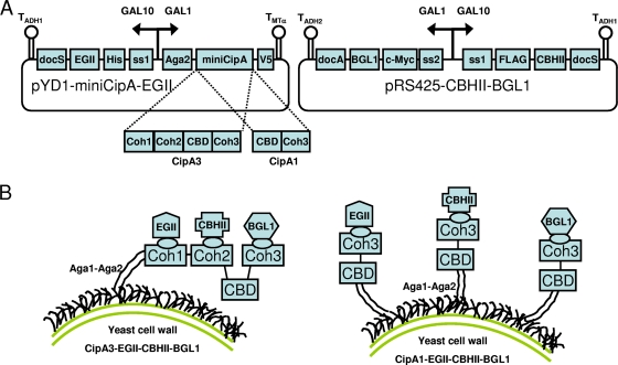
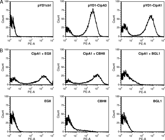
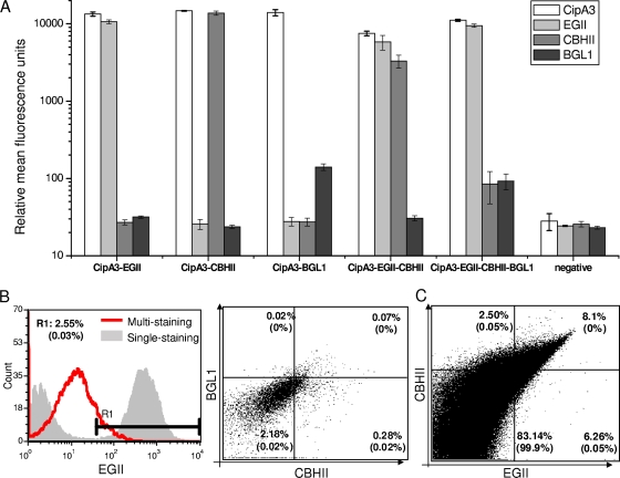
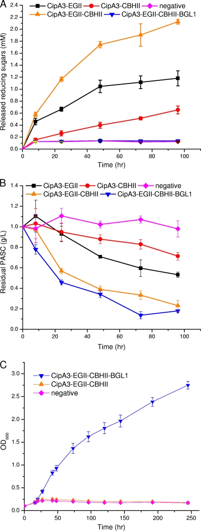
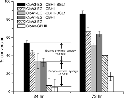
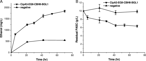
By combining cellulase production, cellulose hydrolysis, and sugar fermentation into a single step, consolidated bioprocessing (CBP) represents a promising technology for biofuel production. Here we report engineering of Saccharomyces cerevisiae strains displaying a series of uni-, bi-, and trifunctional minicellulosomes. These minicellulosomes consist of (i) a miniscaffoldin containing a cellulose-binding domain and three cohesin modules, which was tethered to the cell surface through the yeast a-agglutinin adhesion receptor, and (ii) up to three types of cellulases, an endoglucanase, a cellobiohydrolase, and a beta-glucosidase, each bearing a C-terminal dockerin. Cell surface assembly of the minicellulosomes was dependent on expression of the miniscaffoldin, indicating that formation of the complex was dictated by the high-affinity interactions between cohesins and dockerins. Compared to the unifunctional and bifunctional minicellulosomes, the quaternary trifunctional complexes showed enhanced enzyme-enzyme synergy and enzyme proximity synergy. More importantly, surface display of the trifunctional minicellulosomes gave yeast cells the ability to simultaneously break down and ferment phosphoric acid-swollen cellulose to ethanol with a titer of approximately 1.8 g/liter. To our knowledge, this is the first report of a recombinant yeast strain capable of producing cell-associated trifunctional minicellulosomes. The strain reported here represents a useful engineering platform for developing CBP-enabling microorganisms and elucidating principles of cellulosome construction and mode of action.


Figures

FIG. 1.
FIG. 1.
Design of a yeast surface display system for assembly of minicellulosomes. (A) Plasmids used for constructing strains CipA3-EGII-CBHII-BGL1 and CipA1-EGII-CBHII-BGL1. ss1, synthetic prepro signal peptide (9); ss2, α-factor signal peptide with AG dipeptide spacer (30); T, terminator. V5 (GKPIPNPLLGLDST), His (HHHHHH), FLAG (DYKDDDDK), and c-Myc (EQKLISEEDL) are epitope tags used for detection of minicellulosomal components on the yeast surface. The dockerin modules, docS and docA, were obtained from the two major cellulosomal cellulases of C. thermocellum, CelS (37) and CelA (4), respectively. (B) Two different minicellulosome display schemes using CipA3 (left panel) and CipA1 (right panel). CipA3 enables display of trifunctional minicellulosomes, and CipA1 enables codisplay of three unifunctional minicellulosomes. The cohesin domains are numbered as described elsewhere (29). CBD, cellulose-binding domain; Coh, cohesin.
FIG. 2.
FIG. 2.
Flow cytometric analysis of yeast cells displaying unifunctional minicellulosomes. (A) Both of the miniscaffoldins were successfully displayed on the yeast cell surface, as indicated by V5 epitope detection. Yeast cells transformed with empty plasmids were used as a negative control. (B) Chimeric enzyme display is dependent on the presence of the miniscaffoldin on the cell surface. With CipA1 on the surface (top row), enzymes could be detected. In contrast, without CipA1 on the surface (bottom row), no enzymes were detected on the surface. This CipA1 dependence indicated that there was successful assembly of unifunctional minicellulosomes. The results are representative of three independent experiments using three individual clones. The x axis (PE-A) indicates the expression levels of minicellulosomal proteins as measured by the fluorescence intensity of phycoerythrin.
FIG. 3.
FIG. 3.
Characterization of yeast surface-displayed minicellulosomes. (A) Level of display of all CipA3-based minicellulosomal components on the yeast cell surface. See Table 1 for the phenotype of each yeast strain. (B) Multiantibody staining of strain CipA3-EGII-CBHII-BGL1. The multiantibody staining showed significantly lower efficiency than single-antibody staining (left panel). The right panel was gated on the EGII+ population shown by the R1 region in the left panel. (C) Multiantibody staining of strain CipA3-EGII-CBHII. No gate was set, and the percentage of each population is shown in four quadrants. The corresponding percentages of the negative control are indicated in parentheses. The results were obtained in three independent experiments using three individual clones, and the averages and standard deviations are shown.
FIG. 4.
FIG. 4.
(A and B) Functional analysis of surface-displayed minicellulosomes. Cells displaying different minicellulosomes were tested to determine their abilities to hydrolyze PASC. The concentrations of (A) released reducing sugars and (B) residual PASC were plotted over time. (C) Time courses of the cell growth using cellobiose as the sole carbon source. Samples were taken at the time points indicated, and the optical density at 600 nm was measured using a UV-visible spectrometer. All data were obtained from triplicate experiments, and the averages and standard deviations are shown.
FIG. 5.
FIG. 5.
Enhanced synergy of bifunctional and trifunctional minicellulosomes. The percentages of PASC conversion for six surface-engineered yeast strains were compared after 24 and 73 h. The differences between the CipA1-based minicellulosomes and the unifunctional minicellulosomes reflect the enzyme-enzyme synergy, while the differences between the CipA3- and CipA1-based minicellulosomes reflect the enzyme proximity synergy.
FIG. 6.
FIG. 6.
Simultaneous saccharification and fermentation of PASC to ethanol by yeast strain CipA3-EGII-CBHII-BGL1 displaying trifunctional minicellulosomes. The concentrations of (A) ethanol and (B) residual PASC over time are plotted. Yeast strain HZ1901 was used as a negative control.

Similar articles

Cited by

References

    1. Arai, T., S. Matsuoka, H. Y. Cho, H. Yukawa, M. Inui, S. L. Wong, and R. H. Doi. 2007. Synthesis of Clostridium cellulovorans minicellulosomes by intercellular complementation. Proc. Natl. Acad. Sci. U. S. A. 104:1456-1460. - PMC - PubMed
    1. Bayer, E. A., J. P. Belaich, Y. Shoham, and R. Lamed. 2004. The cellulosomes: multienzyme machines for degradation of plant cell wall polysaccharides. Annu. Rev. Microbiol. 58:521-554. - PubMed
    1. Bayer, E. A., R. Lamed, and M. E. Himmel. 2007. The potential of cellulases and cellulosomes for cellulosic waste management. Curr. Opin. Biotechnol. 18:237-245. - PubMed
    1. Beguin, P., P. Cornet, and J. P. Aubert. 1985. Sequence of a cellulase gene of the thermophilic bacterium Clostridium thermocellum. J. Bacteriol. 162:102-105. - PMC - PubMed
    1. Boder, E. T., J. R. Bill, A. W. Nields, P. C. Marrack, and J. W. Kappler. 2005. Yeast surface display of a noncovalent MHC class II heterodimer complexed with antigenic peptide. Biotechnol. Bioeng. 92:485-491. - PubMed

Publication types

MeSH terms

LinkOut - more resources