. 2010 Feb;298(2):H688-98.
doi: 10.1152/ajpheart.01014.2009. Epub 2009 Dec 18.

Calmodulin kinase II is required for angiotensin II-mediated vascular smooth muscle hypertrophy

Affiliations

Calmodulin kinase II is required for angiotensin II-mediated vascular smooth muscle hypertrophy

Hui Li et al. Am J Physiol Heart Circ Physiol. 2010 Feb.

Abstract

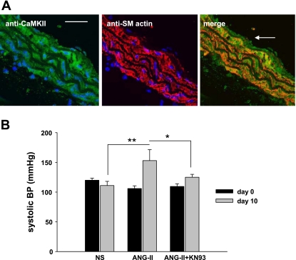
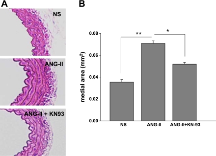
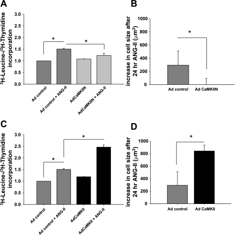
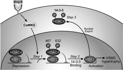
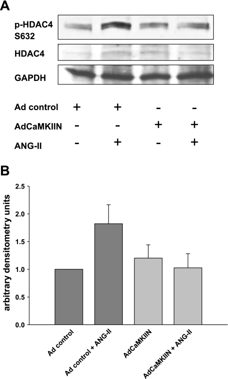
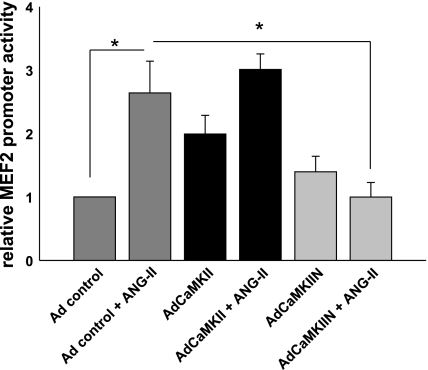
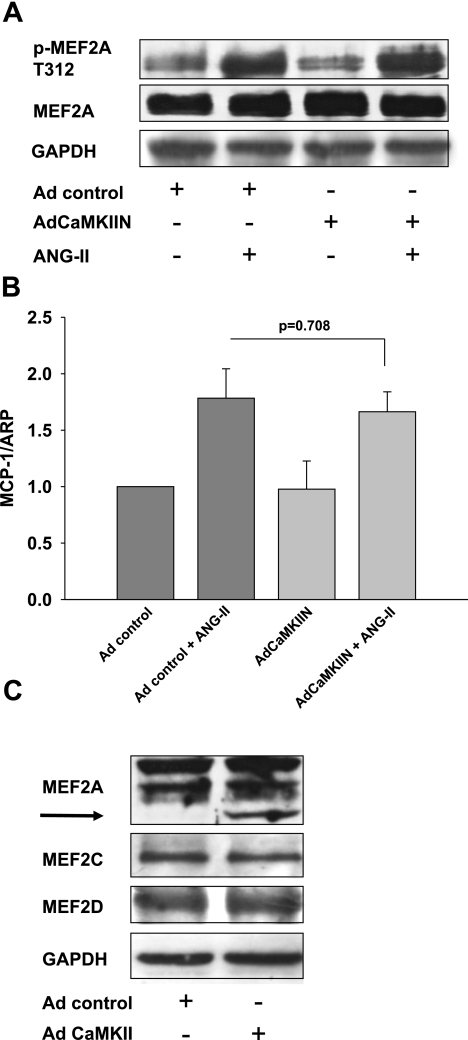
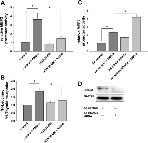
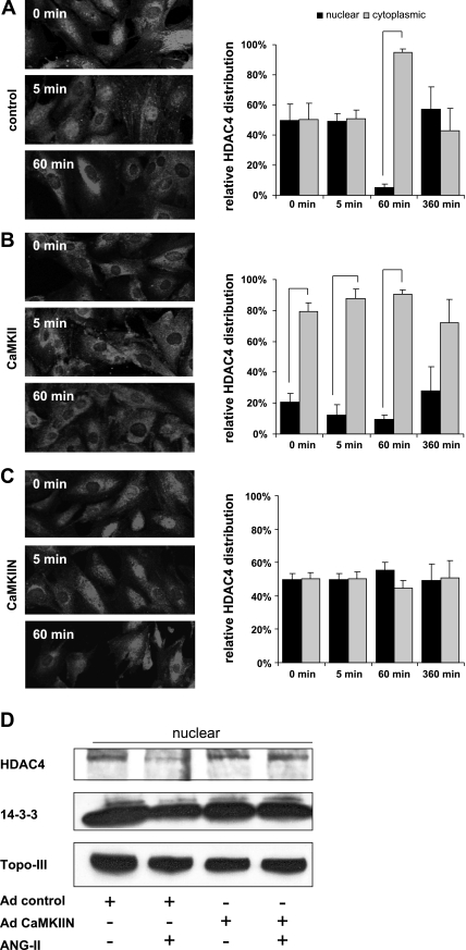
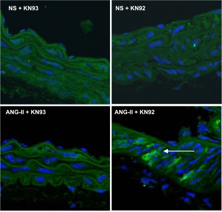
Despite our understanding that medial smooth muscle hypertrophy is a central feature of vascular remodeling, the molecular pathways underlying this pathology are still not well understood. Work over the past decade has illustrated a potential role for the multifunctional calmodulin-dependent kinase CaMKII in smooth muscle cell contraction, growth, and migration. Here we demonstrate that CaMKII is enriched in vascular smooth muscle (VSM) and that CaMKII inhibition blocks ANG II-dependent VSM cell hypertrophy in vitro and in vivo. Specifically, systemic CaMKII inhibition with KN-93 prevented ANG II-mediated hypertension and medial hypertrophy in vivo. Adenoviral transduction with the CaMKII peptide inhibitor CaMKIIN abrogated ANG II-induced VSM hypertrophy in vitro, which was augmented by overexpression of CaMKII-delta2. Finally, we identify the downstream signaling components critical for ANG II- and CaMKII-mediated VSM hypertrophy. Specifically, we demonstrate that CaMKII induces VSM hypertrophy by regulating histone deacetylase 4 (HDAC4) activity, thereby stimulating activity of the hypertrophic transcription factor MEF2. MEF2 transcription is activated by ANG II in vivo and abrogated by the CaMKII inhibitor KN-93. Together, our studies identify a complete pathway for ANG II-triggered arterial VSM hypertrophy and identify new potential therapeutic targets for chronic human hypertension.


Figures

Fig. 1.
Fig. 1.
Calcium/calmodulin-dependent protein kinase II (CaMKII) inhibition prevents angiotensin II (ANG II)-induced hypertension. A: CaMKII expression in mouse aorta. Left: immunofluorescent detection of CaMKII in aortic cross sections from C57BL/6 mice (calibration bar, 20 μm). Center: immunostaining for smooth muscle (SM) actin that demarcates the aortic media. Right: merged image from left and center shows that CaMKII is present in vascular smooth muscle (VSM) (arrow luminal side) (×60). B: increase in systolic blood pressure (BP) by ANG II is blunted by CaMKII inhibition. Mice were infused with 1.25 μg·kg−1·min−1 ANG II or normal saline (NS) for 10 days. ANG II-treated mice were either treated with the CaMKII inhibitory drug KN-93 or with the DMSO vehicle. KN-93 (20 μg/kg) or DMSO was injected intraperitoneally twice daily on days 2–10. Data are presented as means ± SD (n = 4–6/group). *P < 0.05, **P < 0.01.
Fig. 2.
Fig. 2.
CaMKII inhibition prevents ANG II-induced VSM hypertrophy in vivo. A: CaMKII inhibition decreases medial hypertrophy. Aortas were obtained from mice used for studies in Fig. 1B. Five-micrometer cross sections were cut starting distal to the takeoff of the subclavian artery and stained with hematoxylin and eosin. B: summary data for medial descending thoracic aorta thickness from 6 sections for each aorta with n = 6 mice/group. Cross-sectional wall area was determined by tracing the perimeters of the internal and external elastic laminas. The area inside each respective perimeter was determined, and the difference between these areas is reported. All measurements were completed with the use of NIH Image (version 1.62). *P < 0.05, **P < 0.01.
Fig. 3.
Fig. 3.
CaMKII regulates ANG II-induced VSM cell (VSMC) hypertrophy in vitro. CaMKII is necessary for ANG II-induced VSMC hypertrophy. A: rat aortic smooth muscle cells (RASM) were infected with adenovirus (Ad) expressing the CaMKII inhibitor CaMKIIN, grown to 70% confluence, serum starved in serum-free medium for 24 h, and then treated with 100 nM ANG II or vehicle for 24 h in serum-free medium. [3H]leucine incorporation was determined and normalized to [3H]thymidine uptake. Data are expressed as % increase in [3H]leucine incorporation induced by ANG II over the appropriate control. Each bar represents mean of 7 experiments performed in triplicate. *P < 0.05. B: RASM were infected with adenovirus expressing the CaMKII inhibitor CaMKIIN, grown to 70% confluence, serum starved in serum-free medium for 24 h, and then treated with 100 nM ANG II or vehicle for 24 h in serum-free medium. We identified adenovirus-infected cells by green fluorescent protein (GFP) fluorescence and traced 50–100 cells for each condition. No increase in cell size was noted when CaMKII was inhibited by CaMKIIN. *P < 0.05. C: RASM were infected with adenovirus expressing CaMKII and treated as described in A. [3H]leucine incorporation was determined and normalized to [3H]thymidine uptake in cells treated with 100 nM ANG II or vehicle for 24 h. D: RASM were infected with adenovirus expressing CaMKII and treated as described in B. Increase in cell size after ANG II for 24 h of VSMC infected with adenovirus expressing CaMKII is shown. *P < 0.05.
Fig. 4.
Fig. 4.
Model depicting the role of CaMKII in histone deacetylase (HDAC)4/MEF2-dependent VSMC hypertrophy. CaMKII is activated by ANG II and specifically phosphorylates HDAC4 S467 and 632. HDAC4 represses MEF2 transcriptional activity at baseline. Phosphorylation of HDAC4 S467 and 632 by CaMKII generates binding sites for the chaperone 14-3-3. With the subsequent export of HDAC4, MEF2 is derepressed and MEF2-dependent gene transcription activated. MEF2-dependent gene transcription contributes to ANG II-induced VSMC hypertrophy.
Fig. 5.
Fig. 5.
ANG II activates HDAC4 in VSM by CaMKII-mediated phosphorylation. A: immunoblotting for p-HDAC4 S632 phosphorylation in response to ANG II in RASM after adenoviral (Ad) overexpression of CaMKIIN; 20 μg of protein was separated and probed for p-HDAC4 S632, HDAC4, and GAPDH. Representative blot is shown. B: densitometry of 3 independent experiments.
Fig. 6.
Fig. 6.
CaMKII is required for ANG II-induced MEF2 promoter activity. MEF2-luciferase promoter assays after adenoviral (Ad) overexpression of CaMKII or CaMKIIN. VSMC were infected with adenoviruses overnight before transfection with MEF2-luciferase and TK Renilla construct for 8 h. Seventy-two hours after infection, 100 nM ANG II was added for 18 h. MEF2 promoter activity was measured and adjusted for TK Renilla activity (n = 7). *P < 0.05.
Fig. 7.
Fig. 7.
CaMKII does not mediate activation of MEF2 by phosphorylation or change MEF protein levels. A: immunoblotting for p-MEF2A. RASM were infected with control virus or adenovirus expressing the CaMKII inhibitor CAMKIIN. RASM were serum starved for 24 h at 70% confluence. Then 100 nM ANG II was added for 10 min. Twenty micrograms of protein was separated and blotted for pMEF2A T312 and MEF2A (anti-MEF2A antibody; Abcam). Representative blot of 3 independent experiments is shown. B: quantitative real-time PCR for monocyte chemoattractant protein-1 (MCP1) was performed after infection of RASM with adenovirus expressing CaMKIIN or control virus. Total RNA was extracted after RASM were serum starved for 24 h at 70% confluence, followed by treatment with 100 nM ANG II for 12 h. Histograms show the relative amount of MCP1 mRNA (n = 4). ARP, acidic ribosomal phosphoprotein. C: immunoblotting for MEF2A, -C, and -D in RASM with adenoviral overexpression of CaMKII and controls. Fifteen micrograms of protein was separated and blotted for MEF2-A (anti-MEF2A antibody; Santa Cruz), -B, and -C and GAPDH. Representative blot of 4 independent experiments is shown.
Fig. 8.
Fig. 8.
ANG II-induced MEF2 promoter activity and smooth muscle hypertrophy are mediated by CaMKII phosphorylation of HDAC4 S467,632. A: MEF2-luciferase promoter assays were performed after cotransfection of MEF2-luciferase and TK Renilla luciferase cDNA with dominant-negative (DN) HDAC4 mutant S467,632A cDNA lacking the CaMKII phosphorylation sites. Control experiments were transfected with empty vector cDNA. Seventy-two hours after infection, 100 nM ANG II was added for 18 h. MEF2 promoter activity was measured and adjusted for TK Renilla activity (n = 7). *P < 0.05. B: overexpression of CaMKII-resistant HDAC4 S467,632A DN constructs prevented VSM hypertrophy. cDNA for pcDNA3.1 HDAC4-S467,632A and pcDNA3.1(+) control vector was delivered by electroporation into RASM at low density. RASM were grown to 70% confluence, serum starved in serum-free medium for 24 h, and then treated with 100 nM ANG II or vehicle for 24 h in serum-free medium. [3H]leucine incorporation was determined and normalized to [3H]thymidine uptake as described in text (n = 4). *P < 0.05. C: MEF2 promoter activity in RASM infected with adenovirus expressing HDAC4 small interfering RNA (siRNA) or control and transfected with MEF2-luciferase and TK Renilla cDNA constructs. RASM were treated with ANG II for 18 h. MEF2-luciferase was adjusted for TK Renilla activity (n = 5). *P < 0.05. D: immunoblotting for HDAC4 in control and HDAC4 siRNA-infected RASM 72 h after infection. Twenty micrograms of protein was separated and probed for HDAC4 and GAPDH. Representative blot of 3 independent experiments is shown.
Fig. 9.
Fig. 9.
Translocation of HDAC4 from the nucleus to the cytoplasm by ANG II requires CaMKII. A–C, left: growth-arrested 80% confluent RASM were treated with 100 nM ANG II for 0–360 min and fixed with paraformaldehyde. The subcellular localization of HDAC4 was determined in VSMC infected with control adenovirus (A), adenovirus overexpressing CaMKII (B), or CaMKIIN (C) by immunofluorescence with an anti-HDAC4 antibody. Representative frames are shown. A–C, right: a minimum of 100 cells in 5 frames were assessed for HDAC localization and summarized as % of nuclear vs. cytoplasmic localization (all bars represent P < 0.05). D: nuclear protein fractions were prepared from VSMC after infection with adenovirus expressing CaMKIIN or control and treatment with 100 nM ANG II for 1 h. Twenty micrograms of protein was separated and immunoblotted for HDAC-4, 14-3-3, and the nuclear protein marker topoisomerase III (Topo-III).
Fig. 10.
Fig. 10.
Activation of MEF2 by ANG II in vivo is regulated by CaMKII. MEF2 reporter mice that harbor tandem MEF2 consensus DNA-binding sites driving a lacZ reporter gene were infused with 1.25 μg·kg−1·min−1 ANG II or normal saline for 10 days. Reporter mice received either the CaMKII inhibitor KN-93 or the inactive compound KN-92 (20 μg/kg) by intraperitoneal injection twice daily. Immunofluorescence for β-galactosidase and nuclear counterstaining with DAPI is shown (n = 4 mice/group).

Similar articles

Cited by

References

    1. Abraham ST, Benscoter H, Schworer CM, Singer HA. In situ Ca2+ dependence for activation of Ca2+/calmodulin-dependent protein kinase II in vascular smooth muscle cells. J Biol Chem 271: 2506–2513, 1996 - PubMed
    1. Abraham ST, Benscoter HA, Schworer CM, Singer HA. A role for Ca2+/calmodulin-dependent protein kinase II in the mitogen-activated protein kinase signaling cascade of cultured rat aortic vascular smooth muscle cells. Circ Res 81: 575–584, 1997 - PubMed
    1. Anderson RD, Haskell RE, Xia H, Roessler BJ, Davidson BL. A simple method for the rapid generation of recombinant adenovirus vectors. Gene Ther 7: 1034–1038, 2000 - PubMed
    1. Backs J, Backs T, Bezprozvannaya S, McKinsey TA, Olson EN. Histone deacetylase 5 acquires calcium/calmodulin-dependent kinase II responsiveness by oligomerization with histone deacetylase 4. Mol Cell Biol 28: 3437–3445, 2008 - PMC - PubMed
    1. Backs J, Olson EN. Control of cardiac growth by histone acetylation/deacetylation. Circ Res 98: 15–24, 2006 - PubMed

Publication types

MeSH terms

LinkOut - more resources