

Hematopoietic stem cells in Drosophila
- PMID: 20023157
- PMCID: PMC2796932
- DOI: 10.1242/dev.043943
Hematopoietic stem cells in Drosophila
Abstract
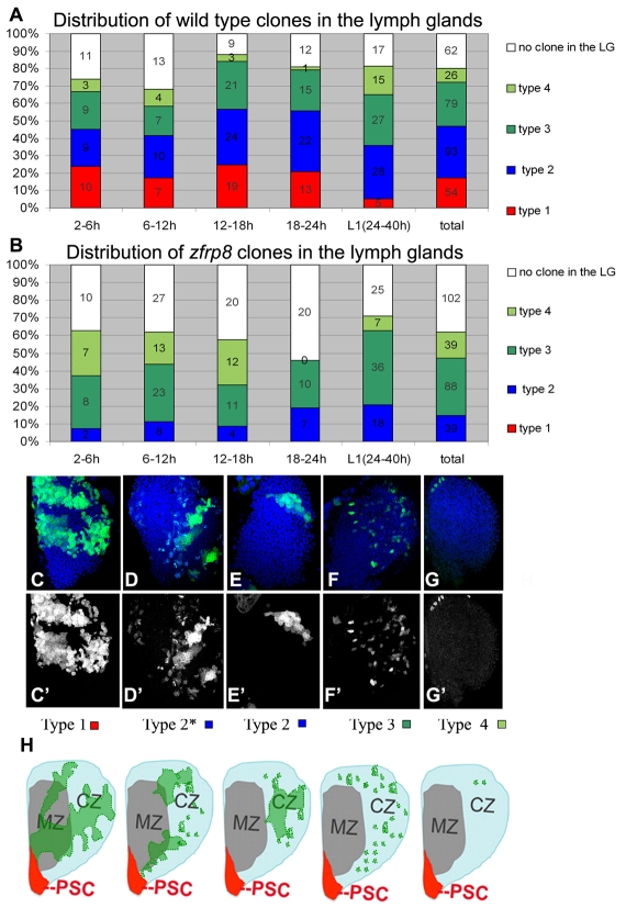
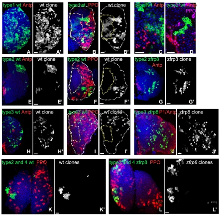
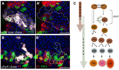
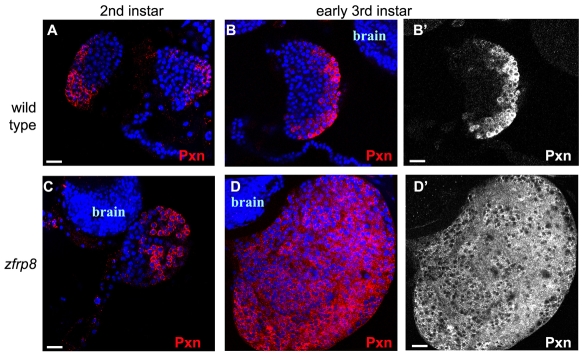
The Drosophila lymph gland, the source of adult hemocytes, is established by mid-embryogenesis. During larval stages, a pool of pluripotent hemocyte precursors differentiate into hemocytes that are released into circulation upon metamorphosis or in response to immune challenge. This process is controlled by the posterior signaling center (PSC), which is reminiscent of the vertebrate hematopoietic stem cell niche. Using lineage analysis, we identified bona fide hematopoietic stem cells (HSCs) in the lymph glands of embryos and young larvae, which give rise to a hematopoietic lineage. These lymph glands also contain pluripotent precursor cells that undergo a limited number of mitotic divisions and differentiate. We further find that the conserved factor Zfrp8/PDCD2 is essential for the maintenance of the HSCs, but dispensable for their daughter cells, the pluripotent precursors. Zfrp8/PDCD2 is likely to have similar functions in hematopoietic stem cell maintenance in vertebrates.
Figures
Similar articles
-
Zfrp8, the Drosophila ortholog of PDCD2, functions in lymph gland development and controls cell proliferation.Development. 2007 Jul;134(13):2387-96. doi: 10.1242/dev.003616. Epub 2007 May 23. Development. 2007. PMID: 17522156
-
Hematopoietic progenitors and hemocyte lineages in the Drosophila lymph gland.Dev Biol. 2010 Oct 15;346(2):310-9. doi: 10.1016/j.ydbio.2010.08.003. Epub 2010 Aug 10. Dev Biol. 2010. PMID: 20707995
-
Gene regulatory networks controlling hematopoietic progenitor niche cell production and differentiation in the Drosophila lymph gland.PLoS One. 2012;7(7):e41604. doi: 10.1371/journal.pone.0041604. Epub 2012 Jul 24. PLoS One. 2012. PMID: 22911822 Free PMC article.
-
Drosophila as a Model to Study Cellular Communication Between the Hematopoietic Niche and Blood Progenitors Under Homeostatic Conditions and in Response to an Immune Stress.Front Immunol. 2021 Aug 16;12:719349. doi: 10.3389/fimmu.2021.719349. eCollection 2021. Front Immunol. 2021. PMID: 34484226 Free PMC article. Review.
-
The hemogenic endothelium: a critical source for the generation of PSC-derived hematopoietic stem and progenitor cells.Cell Mol Life Sci. 2021 May;78(9):4143-4160. doi: 10.1007/s00018-021-03777-y. Epub 2021 Feb 9. Cell Mol Life Sci. 2021. PMID: 33559689 Free PMC article. Review.
Cited by
-
Hedgehog signaling from the Posterior Signaling Center maintains U-shaped expression and a prohemocyte population in Drosophila.Dev Biol. 2018 Sep 1;441(1):132-145. doi: 10.1016/j.ydbio.2018.06.020. Epub 2018 Jul 11. Dev Biol. 2018. PMID: 29966604 Free PMC article.
-
Kinetics of blood cell differentiation during hematopoiesis revealed by quantitative long-term live imaging.Elife. 2023 Mar 31;12:e84085. doi: 10.7554/eLife.84085. Elife. 2023. PMID: 37000163 Free PMC article.
-
The Friend of GATA Transcriptional Co-Regulator, U-Shaped, Is a Downstream Antagonist of Dorsal-Driven Prohemocyte Differentiation in Drosophila.PLoS One. 2016 May 10;11(5):e0155372. doi: 10.1371/journal.pone.0155372. eCollection 2016. PLoS One. 2016. PMID: 27163255 Free PMC article.
-
Signal transduction pathways, intrinsic regulators, and the control of cell fate choice.Biochim Biophys Acta. 2013 Feb;1830(2):2375-84. doi: 10.1016/j.bbagen.2012.06.005. Epub 2012 Jun 15. Biochim Biophys Acta. 2013. PMID: 22705942 Free PMC article. Review.
-
Drosophila melanogaster as a Model to Study the Multiple Phenotypes, Related to Genome Stability of the Fragile-X Syndrome.Front Genet. 2019 Feb 13;10:10. doi: 10.3389/fgene.2019.00010. eCollection 2019. Front Genet. 2019. PMID: 30815010 Free PMC article. Review.
References
-
- de Velasco B., Mandal L., Mkrtchyan M., Hartenstein V. (2006). Subdivision and developmental fate of the head mesoderm in Drosophila melanogaster. Dev. Genes Evol. 216, 39-51 - PubMed
-
- Evans C. J., Banerjee U. (2003). Transcriptional regulation of hematopoiesis in Drosophila. Blood Cells Mol. Dis. 30, 223-228 - PubMed
-
- Fox D., Morris L., Nystul T., Spradling A. C. (2008). Lineage analysis of stem cells. In Stem Book (ed. Girard L.). Initiative in Innovative Computing at Harvard University in collaboration with the Harvard Stem Cell Institute; - PubMed
-
- Harrison D. A., Perrimon N. (1993). Simple and efficient generation of marked clones in Drosophila. Curr. Biol. 3, 424-433 - PubMed
-
- Hartenstein V. (2006). Blood cells and blood cell development in the animal kingdom. Annu. Rev. Cell Dev. Biol. 22, 677-712 - PubMed
Publication types
MeSH terms
Substances
Grants and funding
LinkOut - more resources
Full Text Sources
Medical
Molecular Biology Databases
