. 2010 Jan;137(1):133-40.
doi: 10.1242/dev.043547.

Evidence for a role of vertebrate Disp1 in long-range Shh signaling

Affiliations

Evidence for a role of vertebrate Disp1 in long-range Shh signaling

L Alton Etheridge et al. Development. 2010 Jan.

Abstract

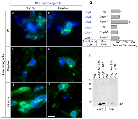
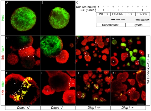
Dispatched 1 (Disp1) encodes a twelve transmembrane domain protein that is required for long-range sonic hedgehog (Shh) signaling. Inhibition of Disp1 function, both by RNAi or dominant-negative constructs, prevents secretion and results in the accumulation of Shh in source cells. Measuring the Shh response in neuralized embryoid bodies (EBs) derived from embryonic stem (ES) cells, with or without Disp1 function, demonstrates an additional role for Disp1 in cells transporting Shh. Co-cultures with Shh-expressing cells revealed a significant reduction in the range of the contact-dependent Shh response in Disp1(-/-) neuralized EBs. These observations support a dual role for Disp1, not only in the secretion of Shh from the source cells, but also in the subsequent transport of Shh through tissue.


Figures

Fig. 1.
Fig. 1.
Disp1 is located basolaterally in polarized epithelial cells and assembles as trimers. (A) Blue-Native gel analysis of Disp1 transfected HEK 293T cells. The 480 kD band corresponds to the predicted size of Disp1 trimers. Disp1 mutants are indicated. (B) Diagram illustrating Disp1 deletion mutants. In addition to deletion of the entire CTD, a series of six ~60-amino-acid-deletion mutants covering the CTD were generated. The box to the right indicates how each mutant localizes relative to the three Disp1 forms shown in C-E, and trimer formation is shown in A. (C-E) Visualization of transiently transfected V5-tagged Disp1 (green) and ZO1 (red) in polarized MDCK cells. ZO1 marks the border between the apical and basolateral aspects of the polarized cells. Wild-type (Wt) Disp1 (C) is located basolaterally, Disp1ΔCTD (D) is located throughout the cell and Disp1 Del2 (E) is primarily located at or near the apical cell surface. x,z-reconstructions are shown on the top and at the left of each panel, yellow As and Bs indicate the apical and basolateral sides, respectively. Scale bar: 20 μm.
Fig. 2.
Fig. 2.
Disp1 is required for the basolateral secretion of Shh. MDCK cells were grown on 12 mm polyester transwell filters (0.4 μm pore size) and transfected with Shh (red) and either control Disp1 miRNA constructs (green, A,E) or Disp1 miRNA constructs (green, B,F,G). (A,B) Shh was visualized after inclusion of 5E1 in the compartment at the basolateral side of the MDCK cells. Secreted Shh is visible in the cells co-transfected with control Disp1 miRNA but is severely reduced in cells co-transfected with Disp1 miRNA1 or 2. (C) Shh staining was quantified by measuring staining intensity in transfected cells (retention) or in a 1.5-cell diameter wide ring around the transfected cells (secretion). Co-transfection of Disp1 miRNA1 or 2 caused significant increase in retention of Shh, whereas the secretion of Shh was significantly reduced. The y-axis is a relative measure of Shh staining. Error bars are s.e.m., P<0.001. (D) Efficacy of the miRNA constructs. Although canine Disp1 can be detected when co-transfected with control miRNA, translation is blocked when co-transfected with Disp1 miRNA1 or 2. (E-G) Shh was visualized after inclusion of 5E1 in the compartment at the apical side. Shh is detected at the surface regardless of the presence of Disp1 miRNA constructs. (E-G′) z-axis reconstructions. Scale bars: 20 μm in A,B; 10 μm in E-G.
Fig. 3.
Fig. 3.
Disp1 mutants act as dominant-negatives. MDCK cells were grown on transwell filters and transfected with Shh and wild-type Disp1 (A), Disp1ΔCTD (B) or Disp1AAA (C). The anti-Shh monoclonal antibody 5E1 was included overnight in the basolateral compartment and visualized. Both Disp1 mutants caused a reduction of Shh secretion and an increase in Shh retention. (D) Quantification of Shh secretion by measuring staining intensity in a 1.5-cell diameter wide ring around the transfected cells, and of Shh retention by measuring staining intensity in transfected cells. The mutant Disp1 co-transfected cells are significantly different from wild-type Disp1-transfected cells both for secretion and retention (P<0.001). The y-axis indicates relative levels of Shh staining. Error bars are s.e.m. Scale bar: 20 μm in A-C.
Fig. 4.
Fig. 4.
Non-acylated Shh (C25S) is secreted in a Disp1-dependent manner. MDCK cells were grown on transwell filters and transfected with wild-type (Wt) Shh (A), Shh lacking cholesterol modification (C*) (B), Shh lacking amino terminal acyl modification (C25S) (C) and Shh with neither modification (C*/C25S) (D). The cells were grown overnight with 5E1 included in the basolateral compartment and visualized. The cholesterol moiety (present in A and C) mediates the retention of Shh outside the cells, whereas the loss of the acyl modification resulted in a shallow Shh gradient (C). Shh C25S was co-transfected either with Wt Disp1 (E) or Disp1AAA (F) into MDCK cells that were grown on transwell filters. Anti-Shh monoclonal 5E1 was included in the basolateral compartment for 16 hours and visualized. Disp1AAA caused significant inhibition of Shh secretion from, and a significant increase of Shh retention in, the transfected cells. (G) Quantification of secretion and retention of Shh. Error bars are s.e.m. Scale bar: 20 μm in A-D; 40 μm in E,F.
Fig. 5.
Fig. 5.
Ptch1 on neighboring cells mediates the transport of Shh secreted in a Disp1-dependent manner. (A-F) Shh-expressing wild-type (Wt) ES cells (blue, A,C,E) and Shh-expressing Disp1−/− cells (blue, B,D,F) were co-cultured with Wt (A,B), Disp1−/− (C,D) and Ptch1−/− cells (E,F). Shh was visualized using directly conjugated 5E1 included in the live cultures for 1 hour, followed by fixation. When Ptch1 was absent from the surrounding cells more Shh could be detected on cells containing Disp1 (E), whereas this had no effect on the Shh present on cells without Disp1 (F). (G) Quantification of the amount of Shh present on cells cultured under the indicated conditions. Error bars indicate s.e.m. Only the amount of Shh present on Wt cells surrounded by Ptch1−/− cells was significantly different from all others. (H) Analysis of the Wt and Disp1−/− Shh-expressing ES cells. Shh was easily detected in lysates, but only a small amount of Shh was detected in the concentrated supernatant from the Wt cells, and not at all in the Disp1−/− cells. Scale bar: 20 μm.
Fig. 6.
Fig. 6.
Long-range Shh signaling is reduced in Disp1−/− EBs. To test whether Disp1 is required outside of Shh-expressing cells for a normal long-range response to cholesterol-modified Shh, wild-type (Wt) EBs expressing Shh were co-cultured for 36 or 48 hours with EBs with or without Disp1 function. Repression of Pax7 was used to measure the range of the Shh response. (A,B) In the absence of a Shh source, Pax7 is expressed throughout both Disp+/− and Disp−/− EBs. (C) The level of Shh present in the lysates and supernatants of Shh AB1 cells. Shh accumulated more efficiently in the medium when suramin (Sur) was present. (D,F) When Shh AB1 EBs were co-cultured with Disp1+/− EBs, Pax7 was significantly reduced throughout the entire EB. (E,G) In Disp1−/− EBs grown in contact with Shh-expressing EBs, Pax7 was only repressed in cells nearest to the Shh source. F and G are low magnification images of the cultures shown in D and E. Induction of HB9 was used to measure the range of Shh response. (H,J) In cases where Shh-expressing EBs contacted Disp1+/− EBs, HB9 was induced close to the Shh source. (I,K) In Disp1−/− EBs grown in contact with Shh-expressing EBs, very few HB9 positive cells were observed. J and K are lower magnification images of the cultures shown in H and I. As Disp1 is not required for the Shh response per se, these results suggest that Disp1 function is important for transmission of the Shh ligand through the responding tissue. Dashed lines, borders between EBs; asterisks, Shh-expressing EBs. Scale bars: 75 μm in A,B,D,E,H,I; 150 μm in F,G,J,K.

Similar articles

Cited by

References

    1. Albright T. D., Jessell T. M., Kandel E. R., Posner M. I. (2000). Neural science: a century of progress and the mysteries that remain. Neuron 25Suppl., S1-S55 - PubMed
    1. Buglino J. A., Resh M. D. (2008). Hhat is a palmitoylacyltransferase with specificity for N-palmitoylation of Sonic Hedgehog. J. Biol. Chem. 283, 22076-22088 - PMC - PubMed
    1. Burke R., Nellen D., Bellotto M., Hafen E., Senti K.-A., Dickson B. J., Basler K. (1999). Dispatched, a novel sterol-sensing domain protein dedicated to the release of cholesterol-modified hedgehog from signaling cells. Cell 99, 803-815 - PubMed
    1. Callejo A., Torroja C., Quijada L., Guerrero I. (2006). Hedgehog lipid modifications are required for Hedgehog stabilization in the extracellular matrix. Development 133, 471-483 - PubMed
    1. Caspary T., Garcia-Garcia M. J., Huangfu D., Eggenschwiler J. T., Wyler M. R., Rakeman A. S., Alcorn H. L., Anderson K. V. (2002). Mouse Dispatched homolog1 is required for long-range, but not juxtacrine, Hh signaling. Curr. Biol. 12, 1628-1632 - PubMed

Publication types