. 2009 Dec;21(12):3850-67.
doi: 10.1105/tpc.109.072363. Epub 2009 Dec 18.

Class I alpha-mannosidases are required for N-glycan processing and root development in Arabidopsis thaliana

Affiliations

Class I alpha-mannosidases are required for N-glycan processing and root development in Arabidopsis thaliana

Eva Liebminger et al. Plant Cell. 2009 Dec.

Abstract

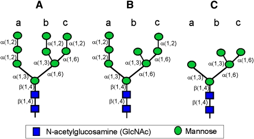
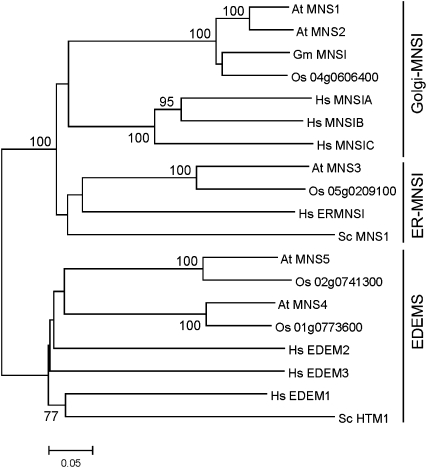
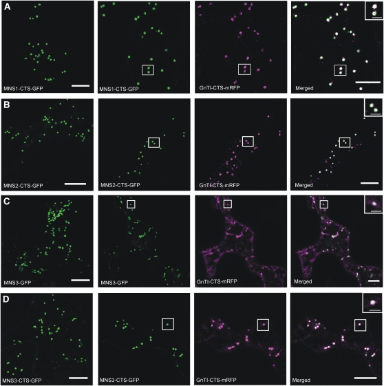
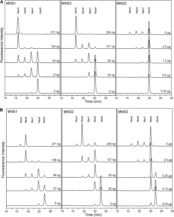
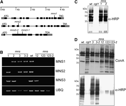
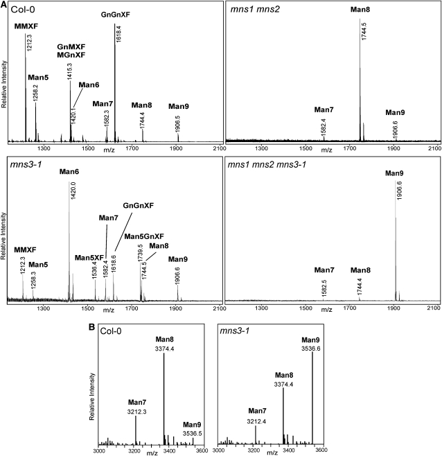
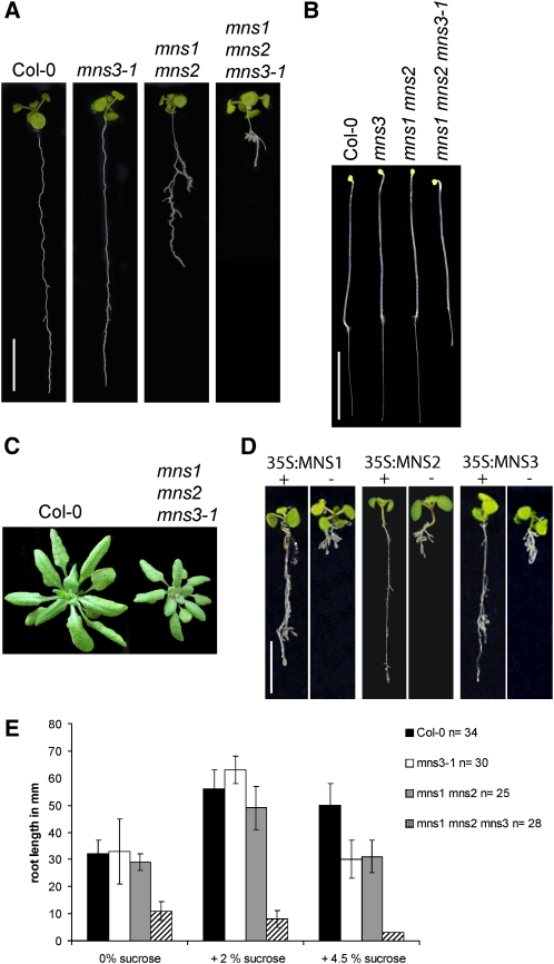
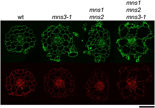
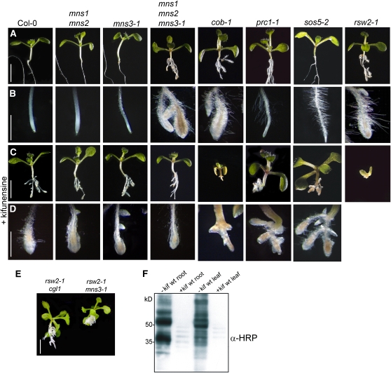
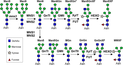
In eukaryotes, class I alpha-mannosidases are involved in early N-glycan processing reactions and in N-glycan-dependent quality control in the endoplasmic reticulum (ER). To investigate the role of these enzymes in plants, we identified the ER-type alpha-mannosidase I (MNS3) and the two Golgi-alpha-mannosidase I proteins (MNS1 and MNS2) from Arabidopsis thaliana. All three MNS proteins were found to localize in punctate mobile structures reminiscent of Golgi bodies. Recombinant forms of the MNS proteins were able to process oligomannosidic N-glycans. While MNS3 efficiently cleaved off one selected alpha1,2-mannose residue from Man(9)GlcNAc(2), MNS1/2 readily removed three alpha1,2-mannose residues from Man(8)GlcNAc(2). Mutation in the MNS genes resulted in the formation of aberrant N-glycans in the mns3 single mutant and Man(8)GlcNAc(2) accumulation in the mns1 mns2 double mutant. N-glycan analysis in the mns triple mutant revealed the almost exclusive presence of Man(9)GlcNAc(2), demonstrating that these three MNS proteins play a key role in N-glycan processing. The mns triple mutants displayed short, radially swollen roots and altered cell walls. Pharmacological inhibition of class I alpha-mannosidases in wild-type seedlings resulted in a similar root phenotype. These findings show that class I alpha-mannosidases are essential for early N-glycan processing and play a role in root development and cell wall biosynthesis in Arabidopsis.


Figures

Figure 1.
Figure 1.
Cartoon of Important Oligosaccharide Structures. (A) Man9GlcNAc2 oligosaccharide (Man9): the substrate for ER-MNSI. (B) Man8GlcNAc2 isomer Man8.1 according to Tomiya et al. (1991): the product of ER-MNSI and substrate for Golgi-MNSI. (C) Man5GlcNAc2 (Man5.1): the product of the mannose trimming reactions. The linkage of the sugar residues is indicated. [See online article for color version of this figure.]
Figure 2.
Figure 2.
Phylogenetic Analysis of Putative Arabidopsis Class I α-Mannosidases. Amino acid sequences of class I α-mannosidases selected from glycoside hydrolase family 47 were aligned using ClustalW. The phylogenetic tree was constructed using the MEGA 4 program. The bar indicates substitutions per residue. Bootstrap values (1000 replicates) above 70% are shown on branches.
Figure 3.
Figure 3.
MNS-GFP Proteins Colocalize with a Golgi Marker. N. benthamiana leaf epidermal cells expressing MNS-GFP fusion proteins either alone (left panel) or in combination with the Golgi marker GnTI-CTS-mRFP. The punctate fluorescence of all MNS-GFP proteins and their colocalization with GnTI-CTS-mRFP indicate Golgi accumulation. Analysis of fluorescent proteins was done by confocal laser scanning microscopy. Boxed region in each row is enlarged in the inset. Bar = 10 μm for all images. Bar of insets = 2 μm. (A) MNS1-CTS-GFP. (B) MNS2-CTS-GFP. (C) MNS3-GFP (full-length MNS3). (D) MNS3-CTS-GFP.
Figure 4.
Figure 4.
The Substrate Specificity of MNS3 Is Different from That of MNS1/MNS2. Activity assays of recombinant MNS proteins were performed with Man8GlcNAc2-PA ([A]; Man8.1) and Man9GlcNAc2-PA ([B]; Man9). The samples were analyzed by NP-HPLC and fluorescence detection. PA-labeled oligosaccharides were incubated with the indicated amounts of purified recombinant MNS protein for 1 h. The elution positions of Man9GlcNAc2-PA (Man9), Man8GlcNAc2-PA (Man8), Man7GlcNAc2-PA (Man7), Man6GlcNAc2-PA (Man6), and Man5GlcNAc2-PA (Man5) are indicated.
Figure 5.
Figure 5.
The Order of Mannose Removal Is Different for MNS3 and MNS1/MNS2. (A) Man6GlcNAc2-PA (Man6) produced by MNS digestions of Man9GlcNAc2-PA was isolated by NP-HPLC and analyzed by RP-HPLC. The elution position of the single peak from the MNS1 and MNS2 digests coeluted with the Man6GlcNAc2 peak isolated from mns3-1 plants (Man6-PA mns3-1). Man6.1-PA and Man5.1-PA were used as standards. (B) A mixture of Man8-PA isomers was obtained by acid hydrolysis of Man9-PA and exhibited elution times consistent with previous work (Tomiya et al., 1991). The other traces show the Man8-PA isomer isolated from digests of Man9-PA with the three Arabidopsis MNS proteins. The Man8GlcNAc2 peak derived from the MNS3 digest coeluted with Man8.1-PA, while the Man8GlcNAc2 peak derived from the MNS1/2 digest coeluted with Man8.4-PA. All samples were analyzed by RP-HPLC. [See online article for color version of this figure.]
Figure 6.
Figure 6.
mns Mutants Display Changes in N-Glycosylation. (A) Schematic overview of mns alleles. Boxes represent exons (black area represents the coding region), and vertical lines indicate the positions of T-DNA insertions. (B) RT-PCR analysis of mns mutants. RT-PCR (two independent repeats) was performed on RNA isolated from rosette leaves of the indicated lines (1, mns1; 2, mns2; 3, mns3-1; 3-2, mns3-2; 12, mns1 mns2; 123, mns1 mns2 mns3-1; and 123-2, mns1 mns2 mns3-2). Col-0 wild type was used as a control. Primers specific for the indicated transcripts were then used for amplification. UBQ5 served as a positive control. (C) and (D) Protein gel blot analysis. Proteins were extracted from leaves and subjected to SDS-PAGE under reducing conditions. Detection was performed using anti-horseradish peroxidase (α-HRP) antibodies, which recognize β1,2-xylose and core α1,3-fucose residues on N-glycans, and the lectin concanavalin A (ConA; top panel), which binds to terminal mannose residues. The cgl1 mutant line, which accumulates Man5GlcNAc2 and does not produce complex N-glycans, was used as a control. Please note that the anti-HRP antibody displays some unspecific staining.
Figure 7.
Figure 7.
mns Mutants Lead to a Block in Mannose Trimming on Glycoproteins. (A) Matrix-assisted laser desorption ionization time of flight MS spectra of total N-glycans extracted from leaves of wild-type (Col-0), mns3-1, mns1 mns2, and mns1 mns2 mns3-1 plants. (B) LC-ESI-MS of the ER-resident GCSI-CTS-GFPglyc glycoreporter protein. Mass spectra of glycopeptide 2 (TKPREEQYNSTYR) derived from the glycoprotein part are shown.
Figure 8.
Figure 8.
Phenotypic Analysis of mns Mutants. (A) Phenotypes of indicated seedlings grown on Murashige and Skoog medium plus 2% sucrose for 17 d. Bar = 1 cm. (B) Phenotypes of indicated hypocotyls of seedlings grown on Murashige and Skoog medium plus 2% sucrose for 6 d at 22°C in the dark. Bar = 1 cm. (C) Col-0 and mns triple mutant plants grown for 3 weeks on soil under long-day conditions. (D) Complementation of mns1 mns2 mns3-1 with 35S:MNS1, 35S:MNS2, and 35S:MNS3. +, complemented mns1 mns2 mns3-1 plant; −, uncomplemented mns1 mns2 mns3-1 control. Bar = 1 cm. (E) Quantification of root length. The seedlings were grown on Murashige and Skoog medium plus 2% sucrose for 5 d and then transferred to Murashige and Skoog medium containing the indicated amounts of sucrose for 6 d. Root length in millimeters is shown on the y axis. Error bars show the sd. Roots of 34 wild-type, 30 mns3-1, 25 mns1 mns2, and 28 mns1 mns2 mns3-1 seedlings were tested. Compared with the wild type, the mns mutants show a decrease in root length when grown in the presence of high amounts of sucrose.
Figure 9.
Figure 9.
Pectin Content Is Reduced in mns Mutants. Transverse sections of Col-0 wild-type, mns3-1 single, mns1 mns2 double, and mns1 mns2 mns3-1 triple mutant plants (from left to right). Calcofluor staining (top row). JIM7 (antipectin antibody) labeling (bottom row) was reduced in cortical and epidermal cells in mutants compared with the wild type, but labeling intensity of the vasculature was similar in all mutants and the wild-type plants. Bar = 50 μm for the wild-type, single, and double mutants and 42 μm for the triple mutant.
Figure 10.
Figure 10.
Effect of Kifunensine on Wild-Type and Mutant Arabidopsis Plants. (A) Untreated seedlings grown on Murashige and Skoog medium plus 2% sucrose. Bar = 5 mm. (B) Root tips of untreated seedlings. Bar = 2 mm. (C) Kifunensine treatment of seedlings. Bar = 5 mm. (D) Root tips of kifunensine-treated seedlings. Bar = 2 mm. (E) Genetic interaction of rsw2-1 and mns3-1. The rsw2-1 cgl1 double mutant is shown for comparison. Bar = 5 mm. (F) Protein gel blot analysis. Proteins were extracted from kifunensine (+kif) treated and untreated (−kif) wild-type seedlings and subjected to SDS-PAGE under reducing conditions. Detection was performed using anti-horseradish peroxidase (α-HRP) antibodies.
Figure 11.
Figure 11.
Proposed N-Glycan Processing Pathway in mns3 Mutant Plants. The following N-glycan processing enzymes are indicated: GnTI, N-acetylglucosaminyltransferase I; GMII, Golgi-α-mannosidase II; XylT, β1,2-xylosyltransferase; FUT, core α1,3-fucosyltransferase; and HEXO, β-hexosaminidase. In the absence of MNS3, MNS1/MNS2 can trim Man9GlcNAc2 (Man9) either to the aberrant Man6GlcNAc2 (Man6) structure (top row) or to Man5GlcNAc2 (Man5, bottom row), which are either processed into Man5XF or MMXF, respectively. [See online article for color version of this figure.]

Comment in

Similar articles

Cited by

References

    1. Avezov, E., Frenkel, Z., Ehrlich, M., Herscovics, A., and Lederkremer, G. (2008). Endoplasmic reticulum (ER) mannosidase I is compartmentalized and required for N-glycan trimming to Man5-6GlcNAc2 in glycoprotein ER-associated degradation. Mol. Biol. Cell 19 216–225. - PMC - PubMed
    1. Bencúr, P., Steinkellner, H., Svoboda, B., Mucha, J., Strasser, R., Kolarich, D., Hann, S., Köllensperger, G., Glössl, J., Altmann, F., and Mach, L. (2005). Arabidopsis thaliana beta1,2-xylosyltransferase: An unusual glycosyltransferase with the potential to act at multiple stages of the plant N-glycosylation pathway. Biochem. J. 388 515–525. - PMC - PubMed
    1. Boisson, M., Gomord, V., Audran, C., Berger, N., Dubreucq, B., Granier, F., Lerouge, P., Faye, L., Caboche, M., and Lepiniec, L. (2001). Arabidopsis glucosidase I mutants reveal a critical role of N-glycan trimming in seed development. EMBO J. 20 1010–1019. - PMC - PubMed
    1. Burn, J., Hurley, U., Birch, R., Arioli, T., Cork, A., and Williamson, R. (2002). The cellulose-deficient Arabidopsis mutant rsw3 is defective in a gene encoding a putative glucosidase II, an enzyme processing N-glycans during ER quality control. Plant J. 32 949–960. - PubMed
    1. Bush, M.S., and McCann, M.C. (1999). Pectic epitopes are differentially distributed in the cell walls of potato (Solanum tuberosum) tubers. Physiol. Plant. 107 201–213.

Publication types

MeSH terms