. 2010 Feb 26;285(9):6003-11.
doi: 10.1074/jbc.M109.071928. Epub 2009 Dec 20.

Activation of the chloroplast monogalactosyldiacylglycerol synthase MGD1 by phosphatidic acid and phosphatidylglycerol

Affiliations

Activation of the chloroplast monogalactosyldiacylglycerol synthase MGD1 by phosphatidic acid and phosphatidylglycerol

Emmanuelle Dubots et al. J Biol Chem. .

Abstract

One of the major characteristics of chloroplast membranes is their enrichment in galactoglycerolipids, monogalactosyldiacylglycerol (MGDG), and digalactosyldiacylglycerol (DGDG), whereas phospholipids are poorly represented, mainly as phosphatidylglycerol (PG). All these lipids are synthesized in the chloroplast envelope, but galactolipid synthesis is also partially dependent on phospholipid synthesis localized in non-plastidial membranes. MGDG synthesis was previously shown essential for chloroplast development. In this report, we analyze the regulation of MGDG synthesis by phosphatidic acid (PA), which is a general precursor in the synthesis of all glycerolipids and is also a signaling molecule in plants. We demonstrate that under physiological conditions, MGDG synthesis is not active when the MGDG synthase enzyme is supplied with its substrates only, i.e. diacylglycerol and UDP-gal. In contrast, PA activates the enzyme when supplied. This is shown in leaf homogenates, in the chloroplast envelope, as well as on the recombinant MGDG synthase, MGD1. PG can also activate the enzyme, but comparison of PA and PG effects on MGD1 activity indicates that PA and PG proceed through different mechanisms, which are further differentiated by enzymatic analysis of point-mutated recombinant MGD1s. Activation of MGD1 by PA and PG is proposed as an important mechanism coupling phospholipid and galactolipid syntheses in plants.


Figures

FIGURE 1.
FIGURE 1.
Activation of MGDG synthase by PA in Arabidopsis leaf homogenates. Activity was measured as described under “Experimental Procedures.” A, effect of PA (1.5 mol%), DAG (7 mol%), and AEBSF (6 mm) on MGDG synthase activity. B, effect of AEBSF (6 mm) on purified atΔ1–137MGD1. 175 ng of purified protein was incubated with 1.5 mol% PA under the same condition as described for leaf homogenates. C, effect of PA (1.5 mol%), DAG (7 mol%), and AEBSF (6 mm) on DGDG synthase activity. D, kinetic analysis of MGDG synthesis: ■, control; ●, in the presence of AEBSF (6 mm), ▴, with addition of AEBSF after 15 min of incubation (arrow), ♦, with 1.5 mol% of PA. E, comparison of MGDG synthase activity in wild-type and in pldζ2. PA (1.5 mol%), DAG (7 mol%) were added as indicated. The inset shows the ratio of the activities measured with addition of DAG in the absence of PA reported to in the presence of PA. F, comparison of DGDG synthase activity in wild-type and in pldζ2. PA (1.5 mol%), DAG (7 mol%) were added as indicated. Results are average values ± S.D. for three independent replicates.
FIGURE 2.
FIGURE 2.
Effect of PA on MGDG synthase measured in isolated spinach chloroplast envelope. Before the experiment, the envelope fraction was prepared from thermolysin-treated chloroplasts and incubated in conditions favorable for PA phosphatase activity as described under “Experimental Procedures.” MGDG synthase was then measured as described under “Experimental Procedures.” A, kinetic analysis of MGDG synthase activity: control, broken line; with PA 1.5 mol%, solid line. Results are average values ± S.D. for three independent measurements. B, analysis of [14C]galactose-labeled lipids on TLC. C, effect of different lipids on MGDG synthase activity. Activity was measured as described under “Experimental Procedures.” The presence of DAG and additional lipid is indicated below the graph, with for additional lipid fatty acid in sn-1 position on the first line and fatty acid in sn-2 position on the second line. D, effect of 16:0/18:1-PA level on the MGDG synthase activity. Activity was measured as described under “Experimental Procedures.” Results are average values ± S.D. for three independent measurements. The curve equation is: V = 3 × [PA]/(0.5 + [PA]).
FIGURE 3.
FIGURE 3.
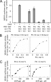
Effect of PA on the MGDG synthase activity of atΔ1–137MGD1. MGDG synthase activity was measured as described under “Experimental Procedures.” A, effect of different lipids on the activity. The presence of DAG and additional lipid is indicated below the graph, with for an additional lipid fatty acid in sn-1 position on the first line and fatty acid in sn-2 position on the second line. B, 16:0/18:1-PA and 18:1/16:0-PA concentration dependence of the activity. The measures were done with 5 ng·μl−1 of protein. The curve equation is V = 7.8 × [PA]1.6/(0.21.6 + [PA]1.6) for 16:0/18:1 − PA and V = 7.1 × [PA]1.9/(0.21.9 + [PA]1.9) for 18:1/16:0 − PA. C, UDP-gal concentration dependence of the activity for two different concentrations of PA, 0.15 mol% and 1.5 mol%. The curve equation is V = 1.8 × [UDP-gal]2.7/(782.7 + [UDP-gal]2.7) for 0.15 mol% of PA and V = 9.2 × [UDP-gal]1.2/(711.2 + [UDP-gal]1.2) for 1.5 mol% of PA. Results are average values ± S.D. for three independent measurements on a single batch of purified protein whereas each part of the figure was done with a separated batch of purified protein.
FIGURE 4.
FIGURE 4.
Lipid binding assay of MGD1. A, lipid-protein overlay assay of three different MGD1 from Arabidopsis, cucumber, and spinach. atΔ1–137MGD1 and csΔ1–104MGD1 harbor a His6 tag at their C terminus, whereas soΔ1–98MGD1 has no His tag. Different lipids were spotted onto nitrocellulose membranes as indicated. The experiment was conducted as described under “Experimental Procedures.” B, different amounts of PA or DAG were spotted onto nitrocellulose membranes. C, liposome binding assay with atΔ1–137MGD1. Liposomes were formed of 100% PC or 5% PA and 95% PC. T, total amount of protein; P, liposome pellet; S supernatant.
FIGURE 5.
FIGURE 5.
Effect of PG on the MGDG synthase activity of atΔ1–137MGD1. Experiments were conducted as described in the legend to Fig. 3. A, PG concentration dependence of the activity. The curve equation is V = 13 × [PG]/(4 + [PG]). B, synergic effect of PG and PA on the activity. The activity was measured with a combination of different mol% concentration of PA and PG as indicated under the graph. Σ indicates the sum of the activities with only PA or PG. C, UDP-gal concentration dependence of the activity for two different concentrations of PG, 0.5 mol% and 2.3 mol%. The curve equation is V = 1.8 × [UDP-gal]/(80 + [UDP-gal]) for 0.5 mol% of PG and V = 4.5 × [UDP-gal]/(80 + [UDP-gal]) for 2.3 mol% of PG. Results are average values ± S.D. for three independent measurements on a single batch of purified protein, whereas each part of the figure was done with a separated batch of purified protein.
FIGURE 6.
FIGURE 6.
Molecular discrimination of PA and PG binding on atΔ1–137MGD1. The activity was measured as described under “Experimental Procedures” without PA or PG (black bars), with 1.5 mol% of PA (gray bars) or PG (white bars). Results are average values ± S.D. for three independent measurements. A, effect of salts on MGDG synthase activity. Concentrations of KH2PO4 and KCl in the incubation medium are indicated below the graph. B, effect of point mutations of the protein on activation by PA and PG. SDS-PAGE analysis of the purified point-mutated atΔ1–137MGD1 proteins used for activity measurement is shown above the graph.

Similar articles

Cited by

References

    1. Block M. A., Douce R., Joyard J., Rolland N. (2007) Photosynth. Res 92, 225–244 - PMC - PubMed
    1. Jarvis P., Dörmann P., Peto C. A., Lutes J., Benning C., Chory J. (2000) Proc. Natl. Acad. Sci. U.S.A. 97, 8175–8179 - PMC - PubMed
    1. Kobayashi K., Kondo M., Fukuda H., Nishimura M., Ohta H. (2007) Proc. Natl. Acad. Sci. U.S.A. 104, 17216–17221 - PMC - PubMed
    1. Maréchal E., Block M. A., Joyard J., Douce R. (1994) J. Biol. Chem. 269, 5788–5798 - PubMed
    1. Miege C., Marechal E., Shimojima M., Awai K., Block M. A., Ohta H., Takamiya K., Douce R., Joyard J. (1999) Eur J Biochem. 265, 990–1001 - PubMed