. 2010 Jan;66(1):144-56; discussion 156-7.
doi: 10.1227/01.NEU.0000363149.58885.2E.

Platelet-derived growth factor BB mediates the tropism of human mesenchymal stem cells for malignant gliomas

Affiliations

Platelet-derived growth factor BB mediates the tropism of human mesenchymal stem cells for malignant gliomas

Nobuhiro Hata et al. Neurosurgery. 2010 Jan.

Abstract

Objective: Bone marrow-derived human mesenchymal stem cells (hMSCs) are capable of localizing to gliomas after systemic delivery and can be used in glioma therapy. However, the mechanism underlying the tropism of hMSCs for gliomas remains unclear. In vitro studies suggest that platelet-derived growth factor BB (PDGF-BB) may mediate this tropism. However, a causal role of PDGF-BB has not been demonstrated in vivo. Therefore, we tested the hypothesis that PDGF-BB mediates the attraction of hMSCs to gliomas in vitro and in vivo.

Methods: U87 or LN229 glioma cells were transfected with plasmids encoding human PDGF-B. Stable transfected clones that secreted large amounts of PDFG-BB and clones that produced low levels of PDGF were chosen. In vitro migration of hMSCs toward PDGF-B or conditioned media from high- and low-secreting PDGF-B tumor cells was assessed using Matrigel invasion assays. For in vivo localization studies, hMSCs were tracked by bioluminescence imaging (BLI) after transduction with an adenovirus containing luciferase cDNA. In other studies, hMSCs were labeled with green fluorescent protein (gfp) and analyzed for intratumoral localization by immunohistochemistry.

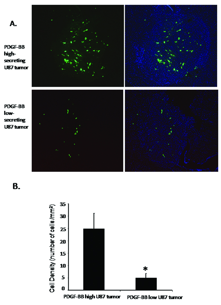
Results: In vitro invasion assays showed that significantly more hMSCs migrated toward glioma cells engineered to secrete high levels of PDGF-BB compared with low-secreting gliomas. Anti-PDGF-BB-neutralizing antibody abrogated this increase in migration. Pretreatment of hMSCs with inhibitory antibodies against PDGF receptor-beta also reduced hMSC migration. To demonstrate that PDGF-BB mediates the localization of hMSCs in vivo, hMSCs-Ad-Luc were injected into the carotid artery of mice harboring orthotopic 7-day-old U87-PDGF-BB-high secreting or U87-PDGF-BB-low secreting xenografts and analyzed by BLI. Statistically significant increases in hMSCs were seen within PDGF-BB-high xenografts compared with PDGF-BB-low xenografts. To control for PDGF-BB-induced differences in tumor size and vascularity, gfp-labeled hMSCs were injected into the carotid arteries of animals harboring 4-day old PDGF-BB-high secreting xenografts or 7-day old PDGF-BB-low secreting xenografts. At these times tumors had similar size and vessel density. Statistically significant more hMSCs localized to PDGF-BB-high secreting xenografts compared with PDGF-BB-low secreting xenografts. Pretreatment of hMSCs with anti-PDGFR-beta-inhibitory antibodies decreased the localization of hMSCs in this intracranial model.

Conclusion: PDGF-BB increases the attraction of hMSCs for gliomas in vitro and in vivo, and this tropism is mediated via PDGF-beta receptors on hMSCs. These findings can be exploited for advancing hMSC treatment.


Figures

Figure 1
Figure 1
Graphs showing the amount of PDGF-BB secreted into the medium of PDGF-B engineered U87 and LN229 clones. A) After transfection with a plasmid containing the cDNA of PDGF-B, selection, and expansion, 106 U87-PDGFB cells (clones #2, #7, #3 and #4) were plated in serum free media, and after 48 hours conditioned media was collected and analyzed for levels of PDGF-BB by ELISA. As a control, serum free media (media) and untransfected U87 cells were assayed. There was significant difference in the levels of PDGF-BB between the high-secretion clones (#2 and #7) and the low secretion clones (#3 and #4). (p<0.01, *). B) After similar transfection, 106 LN229-PDGF-B cells (clones #8, #9 and #12) were plated and after 48 hrs assayed for PDGF-BB expression. A significant difference between high-secretion (#9 and #12) and the low secretion clone (#8) is evident (p < 0.01, *). Bars are mean +/− SD of triplicate experiments.
Figure 2
Figure 2
(A) Graph showing in vitro migration of hMSCs in response to different concentrations of PDGF-BB. hMSCs were plated on matrigel-coated upper wells and exposed to increasing concentrations of PDGF-BB in the lower well. Migration was measured 48 hrs later by counting the number of migrating hMSCs in 10 high-powered fields (HPF). Increased migration of hMSC was seen with 0.1 to 10 ng/ml of PDGF-BB compared with media alone. (B) Graphs showing in vitro invasion of hMSCs in response to conditioned media derived from glioma cell lines engineered to secrete high and low amounts of PDGF-BB. hMSCs were plated on matrigel in the upper wells of transwell plates and exposed to the conditioned media of the indicated U87 cell lines. Invasion was measured 48 hrs later. Increased invasion of hMSC was seen with U87-PDGFB high-secretion clones (#2 and #7) compared with the low-secretion clones (#3 and #4). (C) Graph of hMSCs invasion in response to conditioned media from the indicated LN-229 cell lines. Increased invasion of hMSC was seen with LN229-PDGFB high-secretion clones (#9 and #12) compared with the low-secretion clone (#4). (D) Graph showing effects of PDGF-BB neutralizing antibodies. Conditioned media from U87-PDGF-B-2 cells, along with increasing concentrations of a PDGF-BB neutralizing antibody, was added to the lower well of the transwell experiment described in (B), and after 48 hrs hMSC migration was measured. A dose-dependent inhibition of hMSC migration was observed with the PDGF-BB neutralizing antibody. U87-PDGFB-3 was used as a PDGF-BB-low secreting control. (E) Inhibition of hMSC migration by anti-PDGFR-β antibodies. hMSCs were trypsinized, incubated in 1% serum media with 5% PBS as control (CTR) or with 10µg/ml of anti-PDGFR-β antibodies for 1 hr at 4°C with shaking, twice washed, then placed on matrigel in the upper well. Serum-free media with different concentration of PDGF-BB (5 ng/ml or 10 ng/ml) was placed into the lower wells. hMSC migration was assayed after 16 hrs. hMSC migration was significantly inhibited by pre-treatment of hMSCs with anti-PDGFR- β antibodies. (F) Graph showing inhibition of hMSC migration to conditioned media from U87 PDGF-BB-high secreting clone after pretreatment of hMSC with anti-PDGFR-β antibodies. hMSCs were treated as described in (E) and placed in the upper wells. Conditioned media from U87-PDGFB #2 (high secretion) cells was placed into lower wells and hMSC migration was measured after 16 hrs. hMSC migration was significantly inhibited by treatment of hMSCs with anti-PDGFR-β antibodies. In all graphs bars are mean +/− SD of triplicate experiments (* = p<0.05, ** = p<0.01).
Figure 3
Figure 3
Photomicrograph of mouse brains showing in vivo expression of PDGF-BB in xenografts derived from U87 cells engineered to secrete high and low levels of PDGF-BB. 106 of PDGF-BB high-secreting U87 cells (clone #2) (right panel) or 106 of PDGF-BB low-secreting U87 cells (clone #3) (left panel) were implanted into the right frontal lobes of nude mice and after 14 days, the brains were sectioned and stained with H&E (upper), or by immunohistochemistry (IHC) using an anti-PDGF-BB antibody (lower). High expression level of PDGF-BB was detected in xenografts of PDGF-BB-high secreting U87 xenografts, whereas essentially no staining was observed in the xenografts of PDGF-BB-low secreting U87 xenografts.
Figure 4
Figure 4
Photomicrographs of coronal sections of mice brains bearing high PDGF-BB-secreting and low PDGF-BB-secreting U87 xenografts after treatment with intravascularly-delivered hMSCs transduced with Ad-Luc and analyzed by bioluminescence imaged. A) Mice bearing established U87-PDGF-B-3 (low PDGF-BB-secreting) xenografts (N=9) were treated with 106 hMSCs delivered into the carotid artery. Seven days after delivery animals were treated with Luciferin, the brains were removed and imaged as described in Methods. The scale was fixed between animals and groups to allow comparison. Low levels of signal were observed in these low-secreting xenografts. (B) Mice bearing established U87-PDGF-B-2 (high PDGF-BB-secreting) xenografts (N=9) were treated with hMSCs and analyzed identical to the method in (A). Bright signal was detected in all the xenografts derived from high PDGF-secreting clones. (C) As a control U87-PDGF-B-2 (high PDGF-BB-secreting) xenografts were treated with cell free media. As expected, no signal was detected in these xenografts. (D) Graph showing average signal intensity for animals. The level of signal in the right frontal region was measured by outlining this region of interest, as described in Methods. Values are mean +/− SE (*=p<0.05).
Figure 5
Figure 5
Graphs showing in vitro growth of U87 high and low secreting PDGF-BB clones. 5 ×10³ of U87 parent cells, U87 cells with high (clone #2, 7) or low (clone #3, 4) secretion of PDGF-BB were plated in 6 well plates, then the total cell number of each cell line per well was counted at indicated time points in the triplicate manner, and averaged. The bars are mean +/− SD.
Figure 6
Figure 6
In vivo growth of PDGF-BB-high and PDGF-BB-low secreting xenografts. (A) Photomicrographs of representative H&E coronal sections through the tumors taken at increasing times after cell implantation. 5 ×10⁵ U87-PDGF-BB-high secreting cells (clone#2, lower panel) or –low secreting cells (clone#3, upper panel) were implanted into the right frontal lobe. Mice were sacrificed 4, 7, or 10 days after implantation (N= 3 mice/group/time point). Tumors were manually outlined. (B). Graph showing quantification of tumor size for xenografts derived from U87-PDGF-BB-high secreting cells (clone#2) or –low secreting cells (clone#3). For each mouse the 3 largest cross-sectional areas of tumor were measured under the microscope with a digital camera and software as described in the Methods, and an average tumor area was determined for each mouse. The bars are the mean +/− standard errors of the average areas of 3 mice/group/time point The size of PDGF-BB-high secreting tumors at day 4 was similar to that of PDGF-BB-low secreting tumors at day 7. Likewise, the size of PDGF-BB-high secreting tumors at day 7 also was similar to that of PDGF-BB-low secreting tumors at day 10. (* = p<0.05, ** = p<0.01).
Figure 7
Figure 7
In vivo vascularity of PDGF-BB-high and PDGF-BB-low secreting xenografts. (A) Photomicrographs of representative sections through the tumors taken at increasing times after cell implantation. 5 ×10⁵ U87-PDGF-BB-high secreting cells (clone#2, lower panel) or –low secreting cells (clone#3, upper panel) were implanted into the right frontal lobe. Mice were sacrificed 4, 7, or 10 days after implantation (N= 3 mice/group/time point). Sections were stained for endothelial cells using immunofluorescence with anti-CD31 antibodies and counterstained with DAPI. Photomicrographs were taken using fluorescent microscope at magnification of 400x. (B). Graph showing quantification of vessel density for xenografts derived from U87-PDGF-BB-high secreting cells (clone#2, gray bars) or –low secreting cells (clone#3, black bars). The number of CD31-positive vessels with lumen or branching was counted in 10 high power fields. Bars are mean +/− SE of 3 mice/time point/group. The vascularity of PDGF-BB high-secreting U87 tumors at day 4 was similar to that of PDGF-BB low-secreting U87 tumors at day 7. Likewise the vascularity of PDGF-BB high-secreting U87 tumors at day 7 also was similar to that of PDGF-BB low-secreting U87 tumors at day 10.
Figure 8
Figure 8
(A) Photomicrographs of coronal sections of mice brains bearing PDGF-BB-high secreting and PDGF-BB-low secreting U87 xenografts after treatment with intravascularly-delivered gfp-labeled hMSCs. Seven days after intracranial implantation of U87-PDGF-B-3 cells (PDGF-BB-low) (N=4) and four days after implantation of U87-PDGF-B-2 cells (PDGFBB-high) (N=3), mice were treated with 106 gfp-labeled hMSCs delivered into the carotid artery. Three days after delivery of hMSCs, animals were sacrificed, the brains were removed and analyzed by fluorescent micrsocopy (magnification 50x). The number of hMSCs in the PDGF-BB-high xenografts (upper panel) was qualitatively greater than in the PDGF-BB-low xenografts. (green: gfp cells; blue: nuclei stained with DAPI). Left panels show GFP images, right panels show merged images. (B) Graph showing average hMSC density (number hMSCs per mm2 tumor) within the PDGF-BB-high and PDGF-BB-low xenografts. hMSC density was determined as described in Methods. The number of migrated hMSCs to PDGF-BB high-secreting U87 tumor was significantly increased compared with PDGF-BB low-secreting U87 tumor. Values are mean +/− SE (* = p<0.05).
Figure 9
Figure 9
Decreased hMSC localization in PDGF-BB-high secreting xenografts after treatment of hMSC with anti-PDGFR-β neutralizing antibodies. (A) Mice harboring 4 day old PDGF-BB-high secreting xenografts were injected via the carotid artery with gfp-labeled hMSC that were pre-treated with anti-PDGFR-β antibodies (lower panel, N=3) or with media/PBS as a control (upper panel, N=3). Panel shows representative photographs (magnification 50x) of tumors obtained three days after delivery of hMSCs as viewed under the fluorescent microscope. There is an observable decrease in the number of gfp-labeled hMSCs after inhibition of PDGFR-β. (B) Graph showing quantification of average hMSC density (number hMSCs per mm2 tumor) within the PDGF-BB-high xenografts after injection with hMSCs that were (N=3) or were not (N=3) pretreated with anti-PDGFR-β antibodies. The bars are mean +/− standard errors (* p < 0.05).

References

    1. Birnbaum T, Roider J, Schankin CJ, Padovan CS, Schichor C, Goldbrunner R, Straube A. Malignant gliomas actively recruit bone marrow stromal cells by secreting angiogenic cytokines. J Neurooncol. 2007;83:241–247. - PubMed
    1. Caplan AI, Bruder SP. Mesenchymal stem cells: building blocks for molecular medicine in the 21st century. Trends Mol Med. 2001;7:259–264. - PubMed
    1. Cheng P, Gao ZQ, Liu YH, Xue YX. Platelet-derived growth factor BB promotes the migration of bone marrow-derived mesenchymal stem cells towards C6 glioma and up-regulates the expression of intracellular adhesion molecule-1. Neuroscience letters. 2009;451:52–56. - PubMed
    1. Colter DC, Class R, Di Girolamo CM, Prockop DJ. Rapid expansion of recycling stem cells in cultures of plastic-adherent cells from human bone marrow. Proceedings of the National Academy of Sciences of the United States of America. 2000;97:3213–3218. - PMC - PubMed
    1. Fiedler J, Etzel N, Brenner RE. To go or not to go: Migration of human mesenchymal progenitor cells stimulated by isoforms of PDGF. Journal of cellular biochemistry. 2004;93:990–998. - PubMed

Publication types

MeSH terms

Substances