Wt1 is required for cardiovascular progenitor cell formation through transcriptional control of Snail and E-cadherin
- PMID: 20023660
- PMCID: PMC2799392
- DOI: 10.1038/ng.494
Wt1 is required for cardiovascular progenitor cell formation through transcriptional control of Snail and E-cadherin
Abstract
The epicardial epithelial-mesenchymal transition (EMT) is hypothesized to generate cardiovascular progenitor cells that differentiate into various cell types, including coronary smooth muscle and endothelial cells, perivascular and cardiac interstitial fibroblasts and cardiomyocytes. Here we show that an epicardial-specific knockout of the gene encoding Wilms' tumor-1 (Wt1) leads to a reduction in mesenchymal progenitor cells and their derivatives. We show that Wt1 is essential for repression of the epithelial phenotype in epicardial cells and during embryonic stem cell differentiation through direct transcriptional regulation of the genes encoding Snail (Snai1) and E-cadherin (Cdh1), two of the major mediators of EMT. Some mesodermal lineages do not form in Wt1-null embryoid bodies, but this effect is rescued by the expression of Snai1, underscoring the importance of EMT in generating these differentiated cells. These new insights into the molecular mechanisms regulating cardiovascular progenitor cells and EMT will shed light on the pathogenesis of heart diseases and may help the development of cell-based therapies.
Figures


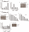
 , □) in the Snai1 and Cdh1 loci,
  (functional binding site), □ (putative but non functional binding site) and ■ (exons). (b) Luciferase activity of reporter construct carrying mouse Snai1 promoter in epicardial cells in the presence of indicated amounts of −KTS Wt1 expression vector. (c) Luciferase activity of wild-type (Control) or mutated Snai1 promoters in the presence of -KTS Wt1 isoform. (d, f) Cell extracts from epicardial cells were chromatin immunoprecipitated (ChIP), using antibodies against Wt1, anti-acetyl-histone H3 (AcH3), anti-H3 tri methyl K4 (K4Me) and anti- H3 tri methyl K27 (K27Me) or an irrelevant antibody (IgG). The input was used as a positive control for PCR of the Snai1 (d) and Cdh1 (f) promoters, intronic regions and 3′UTRs. (g, h) Luciferase activity of constructs carrying DNA fragments identified by the Cdh1 ChIP assay in epicardial cells, together with different concentrations of −KTS Wt1 isoform. (i) Luciferase activity of Control or mutated Cdh1 constructs in the presence of -KTS Wt1 isoform (100ng). (j) ChIP assays of Snail and Wt1 at the endogenous Cdh1 promoter in epicardial cells. The graphs represent the mean values ±s.e.m from three independent experiments.

References
-
- Wessels A, Perez-Pomares JM. The epicardium and epicardially derived cells (EPDCs) as cardiac stem cells. Anat. Rec. A Discov. Mol. Cell Evol. Biol. 2004;276:43–57. - PubMed
-
- Hohenstein P, Hastie ND. The many facets of the Wilms' tumour gene, WT1. Hum. Mol. Genet. 2006;15(Spec No 2):R196–R201. - PubMed
-
- Moore AW, McInnes L, Kreidberg J, Hastie ND, Schedl A. YAC complementation shows a requirement for Wt1 in the development of epicardium, adrenal gland and throughout nephrogenesis. Development. 1999;126:1845–1857. - PubMed
-
- Hosen N, et al. The Wilms' tumor gene WT1-GFP knock-in mouse reveals the dynamic regulation of WT1 expression in normal and leukemic hematopoiesis. Leukemia. 2007;21:1783–1791. - PubMed
Publication types
MeSH terms
Substances
Grants and funding
LinkOut - more resources
Full Text Sources
Other Literature Sources
Medical
Molecular Biology Databases
Research Materials
Miscellaneous

