. 2009 Apr;1(4):301-10.
doi: 10.1039/b900685k. Epub 2009 Feb 26.

Pulling force generated by interacting SNAREs facilitates membrane hemifusion

Affiliations

Pulling force generated by interacting SNAREs facilitates membrane hemifusion

Midhat H Abdulreda et al. Integr Biol (Camb). 2009 Apr.

Abstract

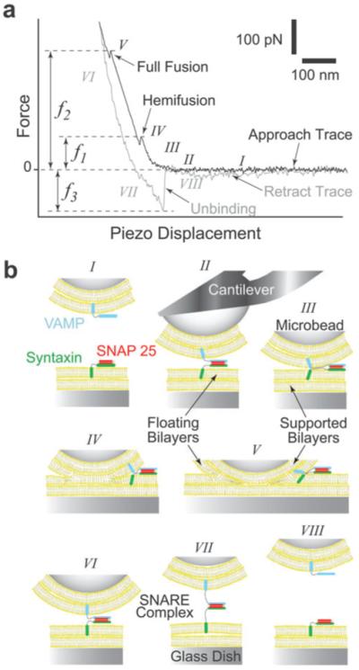
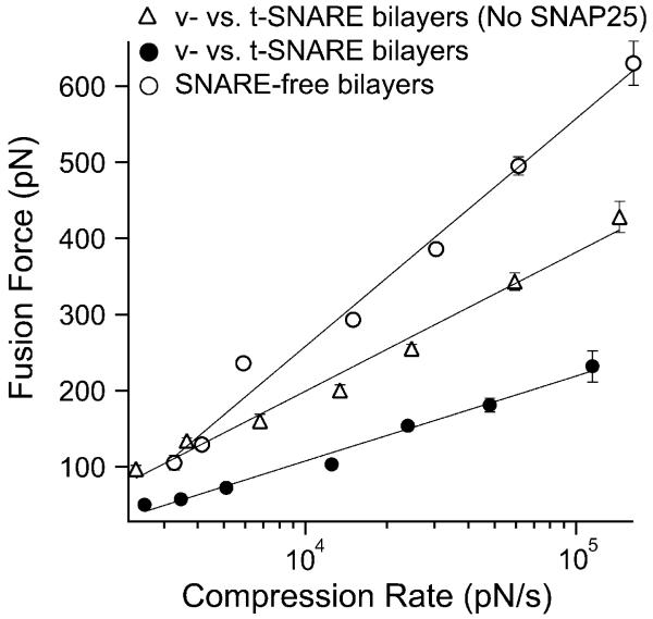
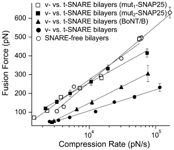
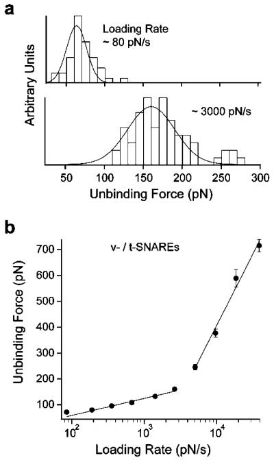
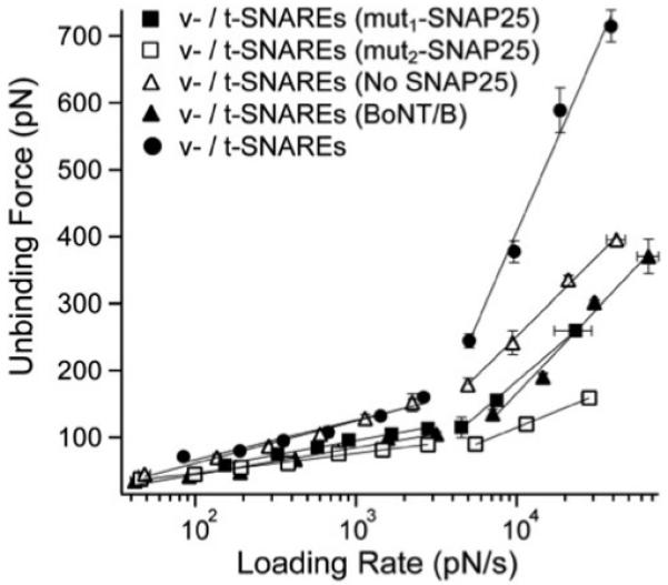
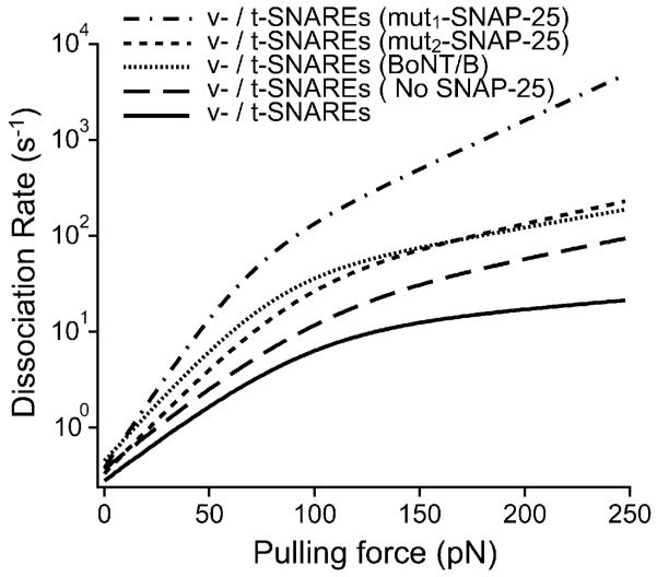
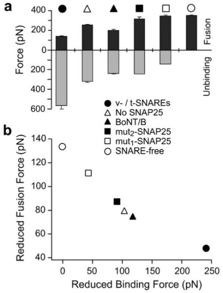
In biological systems, membrane fusion is mediated by specialized proteins. Although soluble N-ethylmaleimide-sensitive factor attachment protein (SNAP) receptors (SNAREs) provide the minimal molecular machinery required to drive membrane fusion, the precise mechanism for SNARE-mediated fusion remains to be established. Here, we used atomic force microscope (AFM) spectroscopy to determine whether the pulling force generated by interacting SNAREs is directly coupled to membrane fusion. The mechanical strength of the SNARE binding interaction was determined by single molecule force measurements. It was revealed that the forced unbinding of the SNARE complex formed between opposing (trans) bilayers involves two activation barriers; where the steep inner barrier governs the transition from the bound to an intermediate state and the outer barrier governs the transition between the intermediate and the unbound state. Moreover, truncation of either SNAP-25 or VAMP 2 reduced the slope of the inner barrier significantly and, consequently, reduced the pulling strength of the SNARE complex; thus, suggesting that the inner barrier determines the binding strength of the SNARE complex. In parallel, AFM compression force measurements revealed that truncated SNAREs were less efficient than native SNAREs in facilitating hemifusion of the apposed bilayers. Together, these findings reveal a mechanism by which a pulling force generated by interacting trans-SNAREs reduces the slope of the hemifusion barrier and, subsequently, facilitates hemifusion and makes the membranes more prone to fusion.


Figures

Fig. 1
Fig. 1
AFM force vs. piezo displacement measurement of interactions between opposing lipid bilayers and SNAREs. (a) A typical AFM force scan measurement showing hemifusion and full fusion of the compressed lipid bilayers and the unbinding of the SNARE complex during approach and retraction of the cantilever, respectively. The compression force required to induce bilayer hemifusion (f1) and fusion (f2) is measured at the onset of each event. Here, we generically refer to f1 as the fusion force. Alternatively, the unbinding force (f3) is measured during the sharp transition in the retraction trace as the SNARE complex dissociates under pulling. (b) Cartoon of our experimental system (not to scale) depicting the different steps (roman numerals) during the force scan measurement shown in (a). Lipid bilayers were formed on the glass dish and glass microbead attached to the cantilever tip. Upon approach of the cantilever toward the substrate, SNAREs embedded in the opposing bilayers form a complex (II) and the bilayers hemifuse (IV) and fully fuse (V) under compression. During the retraction phase, the SNARE complex is extended (VII) before it dissociates (VIII) under pulling.
Fig. 2
Fig. 2
Dynamic force spectrum (DFS) of bilayer hemifusion. With SNAREs in the opposing bilayers, the compression (fusion) force was significantly reduced as compared to SNARE-free bilayers. This revealed facilitation of membrane fusion due to the interaction of VAMP (v-SNARE) with syntaxin/SNAP-25 (v-SNAREs) in the opposite bilayers. Omission of SNAP-25 from the t-SNARE bilayers resulted in partial fusion facilitation due to the direct weaker binary interaction of VAMP with syntaxin. Lines are fits of eqn (1) to the data points. Force values at the different compression rates were derived from distribution histograms of force measurements that were acquired in triplicate experiments on different days (see Materials and methods section). Error bars represent the standard error of the mean (s.e.m.) of all the force values in the distribution histograms of the same compression rates (n; 50 < n < 312).
Fig. 3
Fig. 3
SNARE perturbations suppress the observed facilitation of membrane fusion. An upward shift in the fusion force spectrum (DFS) was observed upon cleavage of VAMP 2 in the v-SNARE bilayers or substitution of SNAP-25 with mutant forms in the t-SNARE bilayers. Such shift in the DFS is due to the increased compression force that is required to induce bilayer hemifusion and is indicative of the increased overall energy requirements for membrane fusion pursuant to the SNARE perturbations in our experimental system. Lines are fits of eqn (1) to the data points. Error bars are the s.e.m. (33 < n < 289).
Fig. 4
Fig. 4
Dynamic force spectrum (DFS) of the forced unbinding of the SNARE complex. (a) Distribution histograms of unbinding forces measured at the specified loading rates. The most probable unbinding force is derived from the Gaussian fit to the histogram. It is evident that the unbinding force increases with increasing loading rate. (b) Two force loading regimes were revealed in the DFS, which indicated the presence of two energy barriers in the dissociation pathway of the SNARE complex. Lines are fits of eqn (1) to the data points. Similar to the compression force experiments, the error bars are the s.e.m. of all the unbinding force values measured at the corresponding loading rates (40 < n < 283). The DFS did not fit well to a single barrier model with either a harmonic or cubic potential.,
Fig. 5
Fig. 5
SNARE perturbations interfere with the mechanical strength of the SNARE complex. The unbinding force of the SNARE complex is significantly reduced upon cleavage of VAMP 2 in the v-SNARE bilayers with BoNT/B or omission of SNAP-25 from the t-SNARE bilayers. A further reduction in the SNARE binding strength was observed in presence of mut1-SNAP-25 or mut2-SNAP-25 in the t-SNARE bilayers as compared to the absence of SNAP-25. This was interpreted as the result of the interference of truncated SNAP-25 mutants with the direct binary interaction of VAMP with syntaxin; hence, a weaker SNARE interaction takes place in presence of mut1-SNAP-25 or mut2-SNAP-25 as compared to the complete absence of SNAP-25. However, the strongest v-/t-SNARE interaction takes place in presence of the full-length SNAP-25 in the a binary syntaxin/SNAP-25 complex in the t-SNARE bilayers. Error bars are the s.e.m. (16 < n < 139).
Fig. 6
Fig. 6
The kinetic profile for the dissociation of the SNARE complex reveals increased dissociation kinetics pursuant to SNARE perturbations. Based on the energy barrier parameters under the different experimental conditions (Table 1), the overall dissociation rate (koffo) of the SNARE complex was derived using eqn (3). The slope of the kinetic profile reflects the dissociation kinetics of the complex; a steeper profile indicates fast dissociation kinetics and a weak interaction. Thus, the native v-/t-SNARE complex has the slowest dissociation kinetics which corresponds to a strong binding interaction. The minimal change in the dissociation rate of the SNARE complex above ~100 pN, where the inner energy barrier dominates the SNARE dissociation process indicates that the inner barrier chiefly determines the mechanical strength of the SNARE binding interaction.
Fig. 7
Fig. 7
Facilitation of membrane fusion is coupled to the pulling strength of interacting SNAREs in the opposite bilayers. (a) Fusion forces (black) were derived from the DFS (see Fig. 2 and 3) at a compression rate of 20 000 pN s−1 in the compression experiments under the specified experimental conditions. The unbinding force measurements (gray) were derived from the DFS (see Fig. 5) at loading rate of 20 000 pN s−1 in the unbinding experiments under the same conditions. It is evident that an inverse relationship between the fusion force and the SNARE unbinding force exists under these conditions. Error bars are the s.e.m. of either the fusion or the unbinding forces measured at the compression/loading rate of 20 000 pN s−1, respectively. (b) Membrane fusion facilitation quantified by the reduced fusion force (fϕ) correlates with the pulling force of interacting SNAREs as characterized by the reduced unbinding force (fβ). This reveals that the pulling force generated by interacting SNAREs facilitates membrane fusion in a force dependent manner.

Similar articles

Cited by

References

    1. Weber T, Zemelman BV, McNew JA, Westermann B, Gmachl M, Parlati F, Sollner TH, Rothman JE. Cell. 1998;92:759–772. - PubMed
    1. Liu T, Tucker WC, Bhalla A, Chapman ER, Weisshaar JC. Biophys. J. 2005;89:2458–2472. - PMC - PubMed
    1. Bhalla A, Chicka MC, Tucker WC, Chapman ER. Nat. Struct. Mol. Biol. 2006;13:323–330. - PubMed
    1. Nickel W, Weber T, McNew JA, Parlati F, Sollner TH, Rothman JE. Proc. Natl. Acad. Sci. U. S. A. 1999;96:12571–12576. - PMC - PubMed
    1. Parlati F, Weber T, McNew JA, Westermann B, Sollner TH, Rothman JE. Proc. Natl. Acad. Sci. U. S. A. 1999;96:12565–12570. - PMC - PubMed

Publication types

MeSH terms

Substances