. 2009 Jun;1(5-6):371-81.
doi: 10.1039/b904878b. Epub 2009 May 11.

Systemic in vivo distribution of activatable cell penetrating peptides is superior to that of cell penetrating peptides

Affiliations

Systemic in vivo distribution of activatable cell penetrating peptides is superior to that of cell penetrating peptides

Todd A Aguilera et al. Integr Biol (Camb). 2009 Jun.

Abstract

Cell penetrating peptides (CPPs) have been developed as vehicles for payload delivery into cells in culture and in animals. However several biologic features limit their usefulness in living animals. Activatable cell penetrating peptides (ACPPs) are polycationic CPPs whose adsorption and cellular uptake are minimized by a covalently attached polyanionic inhibitory domain. Cleavage of the linker connecting the polyanionic and polycationic domains by specific proteases (tumor associated matrix metalloproteases discussed herein) dissociates the polyanion and enables the cleaved ACPP to enter cells. In contrast to their CPP counterpart, ACPPs are relatively nonadherent and distributed uniformly to normal tissues. While nonaarginine (r(9)) CPP administered intravenously into mice initially bind to the local vasculature and redistribute to the liver, where >90% of the injected dose accumulates 30 min after injection. Regardless of the presence of the polyanionic inhibitory domain, confocal imaging of live tissues reveals that the majority of the ACPP and CPP remain in punctate organelles, presumably endosomes. Therefore further improvements in the efficiency of delivery to the cytosol and nucleus are necessary. In addition to improved target specificity, a major advantage of ACPPs over CPPs for potential clinical applications is reduced toxicity. Systemically administered r(9) CPP causes acute toxicity in mice at a dose 4-fold lower than the MMP cleavable ACPP, a complication not observed with an uncleavable ACPP presumably because the polycationic charge remains masked systemically. These data suggest that ACPPs have greater potential than CPPs for systemic delivery of imaging and therapeutic agents.


Figures
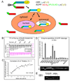
Fig. 1
Fig. 1
ACPPs selectively unmask CPPs upon protease cleavage of a linker, which is selective for MMPs when the linker sequence is PLGLAG. (A) General scheme for ACPPs. While the linker (green) between the polyanion (red) and polycation (blue) sequences remains intact, cell uptake is blocked and the entire molecule can enter the extracellular space of tissues and wash out. Once a protease (symbolized by a scissor) cuts the linker, the polyglutamate dissociates, allowing the polyarginine and its payload (yellow, in current examples Cy5) to immediately adhere to cells and eventually become endocytosed. (B) Isothermal titration calorimetry raw data showing the change in enthalpy as 230 µM Suc-e8 is titrated into a 13 µM solution of r9. (C) These raw data can be corrected for and the change in enthalpy can be plotted as a function of molar ratio of the two peptides yielding determination of the Kd. (D) The percentage by which the polycation is released as a result of cleaving the PLGLAG linker after 30 minute incubation with 50 nM protease. Cleavage of the peptide was detected by tricine SDS-PAGE, a representative fluorescence image of a gel is shown below with first lane being uncleaved peptide. The following lanes line up with the chart above. Arrows point to uncleaved peptide (upper grey arrow) and cleaved peptide (lower black arrow). (E) D-amino acid control Suc-e8-xplglag-r9-c(Cy5) remains uncut by MMP-2 and MMP-9 under conditions where the ACPP Suc-e8-xPLGLAG-r9-c(Cy5) is cleaved.
Fig. 2
Fig. 2
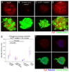
MMP cleavable PLGLAG linker built into the ACPP shows uptake due to endogenous proteases in a 3-D tissue culture model. (A, B) MDA-MB-231 cluster confocal maximum projections of cleavable PLGLAG ACPP versus uncleavable d-amino acid peptide (red) show micro localization of peptide and differential uptake due to the presence of the cleavable linker. (C) Shows decreased cleavable ACPP peptide when co-incubated with 100 µM broad spectrum MMP inhibitor GM6001. (D) r9Cy5 CPP positive control shows uptake of CPP throughout 3-D clusters. Scale bar for A–D is 40 µm. (E) Comparison of the average intensity of multiple 3-D clusters of HT1080 fibrosarcoma (red) or MDA-MB-231 mammary andenocarcinoma (blue) cells treated with PLGLAG cleavable ACPP, ACPP + GM6001 MMP inhibitor, uncleavble d-amino acid ACPP, and r9Cy5 CPP control (t-test p-value for significance labeled accordingly). Cell clusters were incubated with 1.5 µM peptide for 24hrs then washed (3x), counterstained with calcein green AM ester (cell viability, green), and then imaged. (F) Representative confocal slice of a 3-D cluster showing ACPP uptake into subcelluar puncta, cell surface, and extracellular matrix of live cells. ACPP is shown in red, Hoechst 33342 counter stain for cell nuclei (blue), and calcein green AM (green) to show live cells.
Fig. 3
Fig. 3
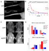
Comparison of pharmacokinetic tissue distribution between CPP and ACPP following intravenous injection into HT-1080 tumor bearing nude mice reveals differences in peptide distribution. (A) Images showing tail veins of animals injected with CPP (top) and ACPP (bottom) at the indicated time points following injection. (B) Cy5 fluorescence in the blood throughout the first 30 minutes after injection as an average of three mice for CPP and ACPP. Tumors are indicated by arrows. (C) Representative HT-1080 mice injected with CPP and ACPP were imaged at 30 minutes and 6 hours (6 hour images brightened 3x). (D) Standardized uptake value (SUV, moles/g in tissue / moles injected/weight of animal) of peptide in tumor, muscle, liver, and kidney, showing changes over time between ACPP and CPP injection. (n=4 for all 6 hour mice, n=5 for all organs of 30 minute mice, n=4 and n=3 for ACPP and CPP tumors of 30 minute mice).
Fig. 4
Fig. 4
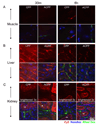
Confocal microscopy of CPP and ACPP in organs revealed peptide localization to endosomes in nude mice. The figure shows confocal slices of Cy5 peptide (red) and then overlay with nuclei (Hoechst 33342- blue) and blood pool (rhodamine dextran -green). Images are of muscle (A), liver (B), and kidney (C) at 30 minutes and 6 hours after IV injection of 10 nmol of peptide as specified. No significant nuclear uptake was observed in any tissue for either the CPP or the ACPP. Peptide signal was scaled equally for each organ to visualize subcellular distribution and a difference in scaling in a particular tissue is labeled accordingly. White arrows point to lumen of renal tubules and green arrows point to basal lateral/blood flow region of renal tubules. Scale bar is 20 µm.
Fig. 5
Fig. 5
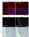
Confocal microscopy reveals no significant nuclear uptake of ACPP or CPP in HT1080 xenografts. Confocal slice of HT1080 tumors from mice injected with CPP and ACPP at 30 minutes (A) and 6 hours (B). Mice were injected with Cy5 peptide (red) at 30 minutes or 6 hours before imaging and Hoechst 33342 nuclear stain (blue) 5 min before imaging to demonstrate that ACPP uptake is in cytoplasmic perinuclear structures but not in nuclei. Upper image is Cy5 peptide alone and lower image shows overlay with Hoechst. The CPP images were scaled brighter by a factor of 4 to visualize intracellular peptide containing punctae. Scale bar is 20µm. (C, D) Show Cy5 fluorescence of frozen sections from mice 6 hours after injection of CPP or ACPP, highlighting greater uptake with ACPP particularly at leading stromal edge of tumors. Scale bar 200 µM.

Similar articles

Cited by

References

    1. Torchilin VP. Tat peptide-mediated intracellular delivery of pharmaceutical nanocarriers. Adv. Drug Deliv. Rev. 2008;60:548–558. - PubMed
    1. Patel LN, Zaro JL, Shen WC. Cell penetrating peptides: intracellular pathways and pharmaceutical perspectives. Pharm. Res. 2007;24:1977–1992. - PubMed
    1. Lundberg M, Johansson M. Is VP22 nuclear homing an artifact? Nat. Biotechnol. 2001;19:713–714. - PubMed
    1. Richard JP, Melikov K, Vives E, Ramos C, Verbeure B, Gait MJ, Chernomordik LV, Lebleu B. Cell-penetrating peptides. A reevaluation of the mechanism of cellular uptake. J. Biol. Chem. 2003;278:585–590. - PubMed
    1. Schwarze SR, Ho A, Vocero-Akbani A, Dowdy SF. In vivo protein transduction: delivery of a biologically active protein into the mouse. Science. 1999;285:1569–1572. - PubMed

Publication types

Substances