

Adhesion and migration of ovarian cancer cells on crosslinked laminin fibers nanofabricated by multiphoton excited photochemistry
- PMID: 20023757
- PMCID: PMC4337955
- DOI: 10.1039/b906310b
Adhesion and migration of ovarian cancer cells on crosslinked laminin fibers nanofabricated by multiphoton excited photochemistry
Abstract
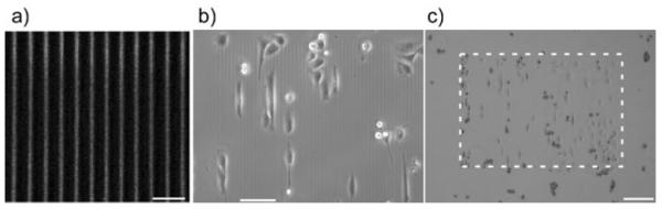
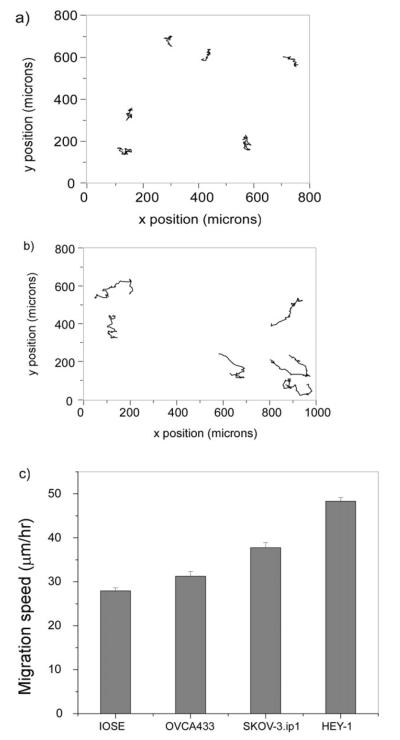
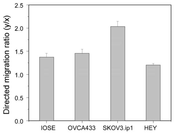
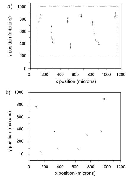
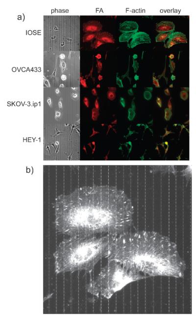
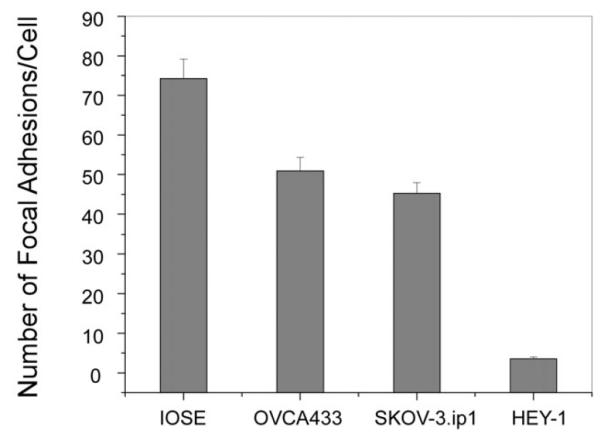
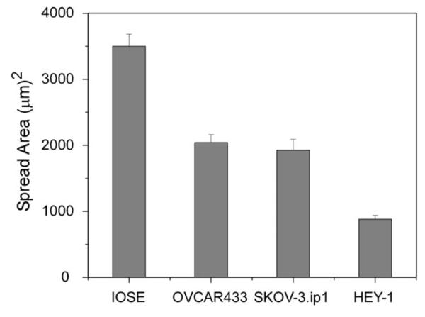
Ovarian cancer is the deadliest gynecological cancer, which may arise in part due to the concurrent invasion and metastasis of high grade tumors. It is thus crucial to gain insight into the adhesion and migration mechanisms in vivo, as this may ultimately lead to new treatment/detection options. To explore this possibility, we have used multiphoton excited photochemistry (MPE) to synthesize models of the ovarian basal lamina consisting of crosslinked laminin nanofibers to quantify the adhesion/migration dynamics. The nanostructured laminin patterns permit the systematic comparison of total migration, directed migration, adhesion, and morphology of "normal" immortalized human ovarian epithelial cells (IOSE) and three lines of varying metastatic potential (OVCA433, SKOV-3.ip1, and HEY-1 cells). We find that the migration of all the cell lines is directed by the crosslinked fibers, and that the contact guidance enhances the total migration rates relative to monolayers. These rates increase with increasing metastatic potential, and the more invasive cells are less rigid and more weakly adhered to the nanofibers. The extent of directed migration also depends on the cell polarity and focal adhesion expression. For the invasive cells, these findings are similar to the integrin-independent ameboid-like migration seen for polar cells in collagen gels. Collectively, the results suggest that contact mediated migration as well as decreased adhesion may be operative in metastasis of ovarian cancer in vivo.
Figures
Similar articles
-
Assessing the roles of collagen fiber morphology and matrix stiffness on ovarian cancer cell migration dynamics using multiphoton fabricated orthogonal image-based models.Acta Biomater. 2022 Nov;153:342-354. doi: 10.1016/j.actbio.2022.09.037. Epub 2022 Sep 21. Acta Biomater. 2022. PMID: 36152908 Free PMC article.
-
Role of integrin receptors for fibronectin, collagen and laminin in the regulation of ovarian carcinoma functions in response to a matrix microenvironment.Clin Exp Metastasis. 2005;22(5):391-402. doi: 10.1007/s10585-005-1262-y. Clin Exp Metastasis. 2005. PMID: 16283482
-
Ovarian Cancer Cell Adhesion/Migration Dynamics on Micro-Structured Laminin Gradients Fabricated by Multiphoton Excited Photochemistry.Bioengineering (Basel). 2015 Jul 16;2(3):139-159. doi: 10.3390/bioengineering2030139. Bioengineering (Basel). 2015. PMID: 28952475 Free PMC article.
-
Invasion of interstitial matrix by a novel cell line from primary peritoneal carcinosarcoma, and by established ovarian carcinoma cell lines: role of cell-matrix adhesion molecules, proteinases, and E-cadherin expression.Gynecol Oncol. 2003 Apr;89(1):60-72. doi: 10.1016/s0090-8258(02)00152-x. Gynecol Oncol. 2003. PMID: 12694655
-
Migration dynamics of ovarian epithelial cells on micro-fabricated image-based models of normal and malignant stroma.Acta Biomater. 2019 Dec;100:92-104. doi: 10.1016/j.actbio.2019.09.037. Epub 2019 Sep 27. Acta Biomater. 2019. PMID: 31568876 Free PMC article.
Cited by
-
The deubiquitinating enzyme USP17 is essential for GTPase subcellular localization and cell motility.Nat Commun. 2011 Mar 29;2:259. doi: 10.1038/ncomms1243. Nat Commun. 2011. PMID: 21448158 Free PMC article.
-
Defining the Role of Metastasis-Initiating Cells in Promoting Carcinogenesis in Ovarian Cancer.Biology (Basel). 2023 Dec 5;12(12):1492. doi: 10.3390/biology12121492. Biology (Basel). 2023. PMID: 38132318 Free PMC article. Review.
-
The traditional Tibetan medicine Yukyung Karne exhibits a potent anti-metastatic activity by inhibiting the epithelial to mesenchymal transition and cell migration.BMC Complement Altern Med. 2015 Jun 13;15:182. doi: 10.1186/s12906-015-0707-3. BMC Complement Altern Med. 2015. Retraction in: BMC Complement Med Ther. 2023 Mar 22;23(1):88. doi: 10.1186/s12906-023-03918-9. PMID: 26070932 Free PMC article. Retracted.
-
Ovarian and Breast Cancer Migration Dynamics on Laminin and Fibronectin Bidirectional Gradient Fibers Fabricated via Multiphoton Excited Photochemistry.Cell Mol Bioeng. 2017 Aug;10(4):295-311. doi: 10.1007/s12195-017-0492-9. Epub 2017 Jul 10. Cell Mol Bioeng. 2017. PMID: 29177019 Free PMC article.
-
Cell Adhesion on Micro-Structured Fibronectin Gradients Fabricated by Multiphoton Excited Photochemistry.Cell Mol Bioeng. 2012 Sep;5(3):307-319. doi: 10.1007/s12195-012-0237-8. Cell Mol Bioeng. 2012. PMID: 23710258 Free PMC article.
References
Publication types
MeSH terms
Substances
Grants and funding
LinkOut - more resources
Full Text Sources
Other Literature Sources
Medical
