Surface topography induces 3D self-orientation of cells and extracellular matrix resulting in improved tissue function
- PMID: 20023803
- PMCID: PMC4487987
- DOI: 10.1039/b820208g
Surface topography induces 3D self-orientation of cells and extracellular matrix resulting in improved tissue function
Abstract
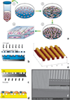
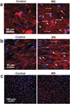
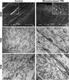
The organization of cells and extracellular matrix (ECM) in native tissues plays a crucial role in their functionality. However, in tissue engineering, cells and ECM are randomly distributed within a scaffold. Thus, the production of engineered-tissue with complex 3D organization remains a challenge. In the present study, we used contact guidance to control the interactions between the material topography, the cells and the ECM for three different tissues, namely vascular media, corneal stroma and dermal tissue. Using a specific surface topography on an elastomeric material, we observed the orientation of a first cell layer along the patterns in the material. Orientation of the first cell layer translates into a physical cue that induces the second cell layer to follow a physiologically consistent orientation mimicking the structure of the native tissue. Furthermore, secreted ECM followed cell orientation in every layer, resulting in an oriented self-assembled tissue sheet. These self-assembled tissue sheets were then used to create 3 different structured engineered-tissue: cornea, vascular media and dermis. We showed that functionality of such structured engineered-tissue was increased when compared to the same non-structured tissue. Dermal tissues were used as a negative control in response to surface topography since native dermal fibroblasts are not preferentially oriented in vivo. Non-structured surfaces were also used to produce randomly oriented tissue sheets to evaluate the impact of tissue orientation on functional output. This novel approach for the production of more complex 3D tissues would be useful for clinical purposes and for in vitro physiological tissue model to better understand long standing questions in biology.
Figures


Similar articles
-
Engineering stem cell cardiac patch with microvascular features representative of native myocardium.Theranostics. 2019 Apr 9;9(8):2143-2157. doi: 10.7150/thno.29552. eCollection 2019. Theranostics. 2019. PMID: 31149034 Free PMC article.
-
Characterization of a Cell-Assembled extracellular Matrix and the effect of the devitalization process.Acta Biomater. 2018 Dec;82:56-67. doi: 10.1016/j.actbio.2018.10.006. Epub 2018 Oct 6. Acta Biomater. 2018. PMID: 30296619
-
Dynamic mechanical stimulations induce anisotropy and improve the tensile properties of engineered tissues produced without exogenous scaffolding.Acta Biomater. 2011 Sep;7(9):3294-301. doi: 10.1016/j.actbio.2011.05.034. Epub 2011 May 30. Acta Biomater. 2011. PMID: 21669302
-
[Porous matrix and primary-cell culture: a shared concept for skin and cornea tissue engineering].Pathol Biol (Paris). 2009 Jun;57(4):290-8. doi: 10.1016/j.patbio.2008.04.014. Epub 2008 Jul 3. Pathol Biol (Paris). 2009. PMID: 18602223 Review. French.
-
The Human Tissue-Engineered Cornea (hTEC): Recent Progress.Int J Mol Sci. 2021 Jan 28;22(3):1291. doi: 10.3390/ijms22031291. Int J Mol Sci. 2021. PMID: 33525484 Free PMC article. Review.
Cited by
-
Construction of vascular graft with circumferentially oriented microchannels for improving artery regeneration.Biomaterials. 2020 Jun;242:119922. doi: 10.1016/j.biomaterials.2020.119922. Epub 2020 Mar 4. Biomaterials. 2020. PMID: 32155476 Free PMC article.
-
Combined technologies for microfabricating elastomeric cardiac tissue engineering scaffolds.Macromol Biosci. 2010 Nov 10;10(11):1330-7. doi: 10.1002/mabi.201000165. Macromol Biosci. 2010. PMID: 20718054 Free PMC article.
-
Corneal stromal stem cells versus corneal fibroblasts in generating structurally appropriate corneal stromal tissue.Exp Eye Res. 2014 Mar;120:71-81. doi: 10.1016/j.exer.2014.01.005. Epub 2014 Jan 15. Exp Eye Res. 2014. PMID: 24440595 Free PMC article.
-
Biofabrication of engineered blood vessels for biomedical applications.Sci Technol Adv Mater. 2024 Mar 21;25(1):2330339. doi: 10.1080/14686996.2024.2330339. eCollection 2024. Sci Technol Adv Mater. 2024. PMID: 38633881 Free PMC article. Review.
-
Modular Fabrication of Intelligent Material-Tissue Interfaces for Bioinspired and Biomimetic Devices.Prog Mater Sci. 2019 Dec;106:100589. doi: 10.1016/j.pmatsci.2019.100589. Epub 2019 Jul 17. Prog Mater Sci. 2019. PMID: 32189815 Free PMC article.
References
-
- Pinsky PM, van der Heide D, Chernyak D. J. Cataract. Refract. Surg. 2005;31:136–145. - PubMed
-
- Ksander GA, Vistnes LM, Rose EH. Plast. Reconstr. Surg. 1977;59:398–406. - PubMed
-
- Bush J, Ferguson MW, Mason T, McGrouther G. J. Plast. Reconstr. Aesthet. Surg. 2007;60:393–399. - PubMed
-
- Langer R, Vacanti JP. Science. 1993;260:920–926. - PubMed
Publication types
MeSH terms
Grants and funding
LinkOut - more resources
Full Text Sources
Other Literature Sources

