

Structural neuroplasticity in the sensorimotor network of professional female ballet dancers
- PMID: 20024944
- PMCID: PMC6870845
- DOI: 10.1002/hbm.20928
Structural neuroplasticity in the sensorimotor network of professional female ballet dancers
Abstract
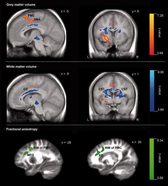
Evidence suggests that motor, sensory, and cognitive training modulates brain structures involved in a specific practice. Functional neuroimaging revealed key brain structures involved in dancing such as the putamen and the premotor cortex. Intensive ballet dance training was expected to modulate the structures of the sensorimotor network, for example, the putamen, premotor cortex, supplementary motor area (SMA), and the corticospinal tracts. We investigated gray (GM) and white matter (WM) volumes, fractional anisotropy (FA), and mean diffusivity (MD) using magnetic resonance-based morphometry and diffusion tensor imaging in 10 professional female ballet dancers compared with 10 nondancers. In dancers compared with nondancers, decreased GM volumes were observed in the left premotor cortex, SMA, putamen, and superior frontal gyrus, and decreased WM volumes in both corticospinal tracts, both internal capsules, corpus callosum, and left anterior cingulum. FA was lower in the WM underlying the dancers' left and right premotor cortex. There were no significant differences in MD between the groups. Age of dance commencement was negatively correlated with GM and WM volume in the right premotor cortex and internal capsule, respectively, and positively correlated with WM volume in the left precentral gyrus and corpus callosum. Results were not influenced by the significantly lower body mass index of the dancers. The present findings complement the results of functional imaging studies in experts that revealed reduced neural activity in skilled compared with nonskilled subjects. Reductions in brain activity are accompanied by local decreases in GM and WM volumes and decreased FA.
2009 Wiley-Liss, Inc.
Figures
References
-
- Alexander MP, Stuss DT, Picton T, Shallice T, Gillingham S ( 2007): Regional frontal injuries cause distinct impairments in cognitive control. Neurology 68: 1515–1523. - PubMed
-
- Ashburner J, Friston KJ ( 2000): Voxel‐based morphometry—The methods. Neuroimage 11( 6 Part 1): 805–821. - PubMed
-
- Ashburner J, Friston KJ ( 2005): Unified segmentation. Neuroimage 26: 839–851. - PubMed
-
- Ashburner J, Csernansk JG, Davatzikos C, Fox NC, Frisoni GB, Thompson PM ( 2003): Computer‐assisted imaging to assess brain structure in healthy and diseased brains. Lancet Neurol 2: 79–88. - PubMed
-
- Assaf Y, Pasternak O ( 2008): Diffusion tensor imaging (DTI)‐based white matter mapping in brain research: A review. J Mol Neurosci 34: 51–61. - PubMed
Publication types
MeSH terms
LinkOut - more resources
Full Text Sources
Medical
Miscellaneous
