

Beyond neurons: Involvement of urothelial and glial cells in bladder function
- PMID: 20025015
- PMCID: PMC2910110
- DOI: 10.1002/nau.20747
Beyond neurons: Involvement of urothelial and glial cells in bladder function
Abstract
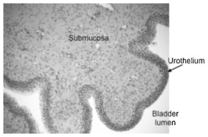
Aim: The urothelium, or epithelial lining of the lower urinary tract (LUT), is likely to play an important role in bladder function by actively communicating with bladder nerves, smooth muscle, and cells of the immune and inflammatory systems. Recent evidence supports the importance of non-neuronal cells that may extend to both the peripheral and central processes of the neurons that transmit normal and nociceptive signals from the urinary bladder. Using cats diagnosed with a naturally occurring syndrome termed feline interstitial cystitis (FIC), we investigated whether changes in physiologic parameters occur within 3 cell types associated with sensory transduction in the urinary bladder: 1) the urothelium, 2) identified bladder dorsal root ganglion (DRG) neurons and 3) grey matter astrocytes in the lumbosacral (S1) spinal cord. As estrogen fluctuations may modulate the severity of many chronic pelvic pain syndromes, we also examined whether 17beta-estradiol (E2) alters cell signaling in rat urothelial cells.
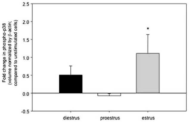
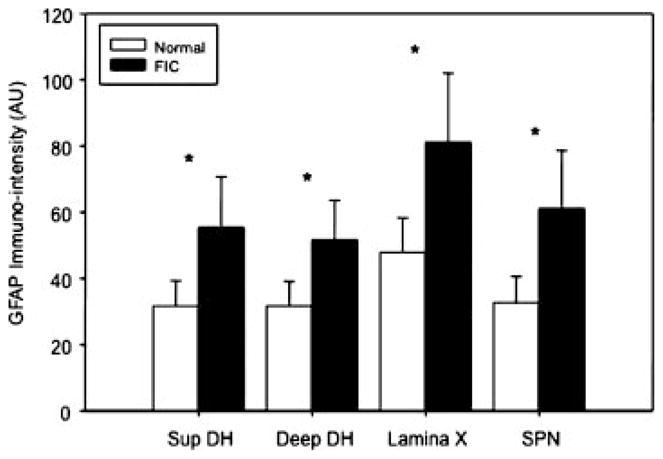
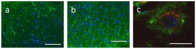
Results: We have identified an increase in nerve growth factor (NGF) and substance P (SP) in urothelium from FIC cats over that seen in urothelium from unaffected (control) bladders. The elevated NGF expression by FIC urothelium is a possible cause for the increased cell body size of DRG neurons from cats with FIC, reported in this study. At the level of the spinal cord, astrocytic GFAP immuno-intensity was significantly elevated and there was evidence for co-expression of the primitive intermediate filament, nestin (both indicative of a reactive state) in regions of the FIC S1 cord (superficial and deep dorsal horn, central canal and laminae V-VIl) that receive input from pelvic afferents. Finally, we find that E2 triggers an estrus-modifiable activation of p38 MAPK in rat urothelial cells. There were cyclic variations with E2-mediated elevation of p38 MAPK at both diestrus and estrus, and inhibition of p38 MAPK in proestrous urothelial cells.
Conclusion: Though urothelial cells are often viewed as bystanders in the processing of visceral sensation, these and other findings support the view that these cells function as primary transducers of some physical and chemical stimuli. In addition, the pronounced activation of spinal cord astrocytes in an animal model for bladder pain syndrome (BPS) may play an important role in the pain syndrome and open up new potential approaches for drug intervention.
Conflict of interest statement
Conflicts of interest: none.
Figures
References
-
- Steers WD, Tuttle JB. Mechanisms of disease: The role of nerve growth factor in the pathophysiology of bladder disorders. Nat Clin Pract Urol. 2006;3:101–10. - PubMed
-
- Okragly AJ, Niles AL, Saban R, et al. Elevated tryptase, nerve growth factor, neurotrophin-3 and glial cell line-derived neurotrophic factor levels in the urine of interstitial cystitis and bladder cancer patients. J Urol. 1999;161:438–41. - PubMed
-
- Lowe EM, Anand P, Terenghi G, et al. Increased nerve growth factor levels in the urinary bladder of women with idiopathic sensory urgency and interstitial cystitis. Br J Urol. 1997;79:572–7. - PubMed
Publication types
MeSH terms
Substances
Grants and funding
LinkOut - more resources
Full Text Sources
Medical
Research Materials
Miscellaneous
