Review
. 2010;29(1):63-76.
doi: 10.1002/nau.20761.

Changes in afferent activity after spinal cord injury

Affiliations
Review

Changes in afferent activity after spinal cord injury

William C de Groat et al. Neurourol Urodyn. 2010.

Abstract

Aims: To summarize the changes that occur in the properties of bladder afferent neurons following spinal cord injury.

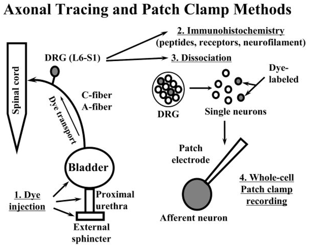
Methods: Literature review of anatomical, immunohistochemical, and pharmacologic studies of normal and dysfunctional bladder afferent pathways.

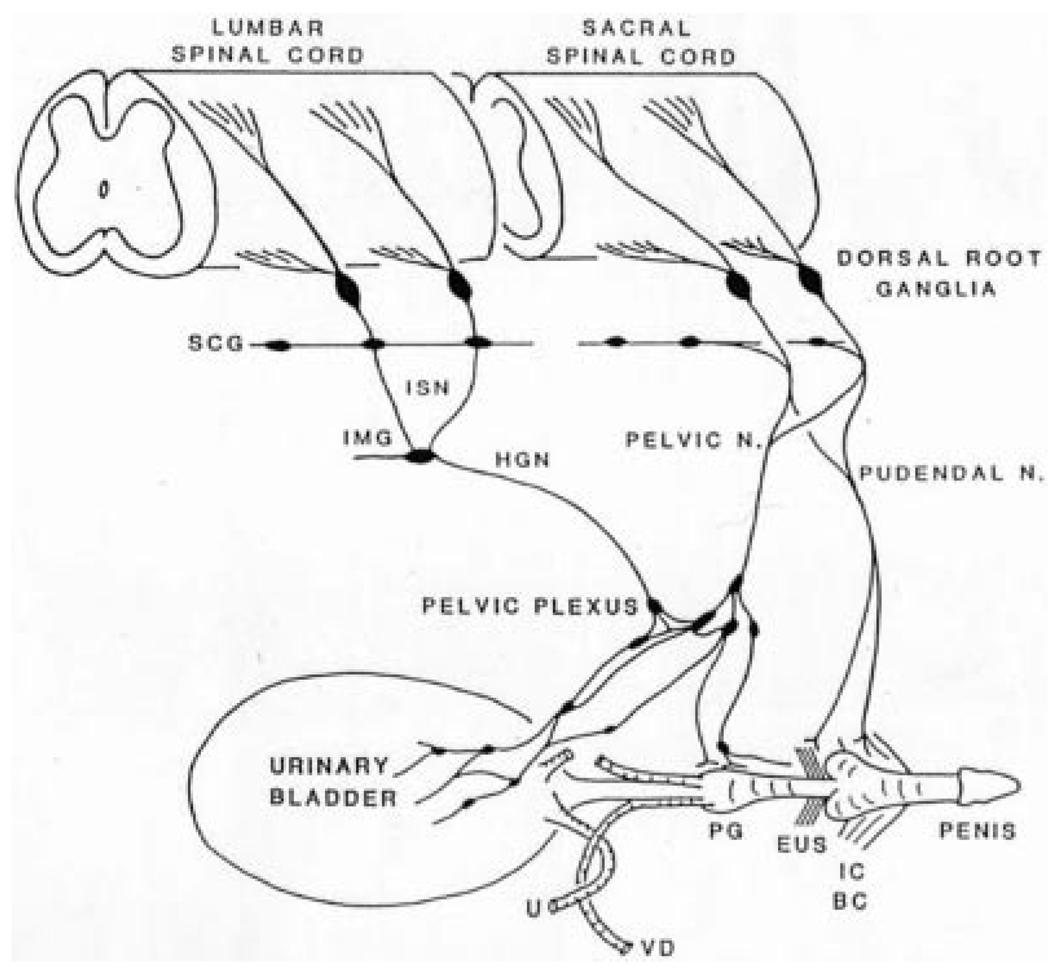
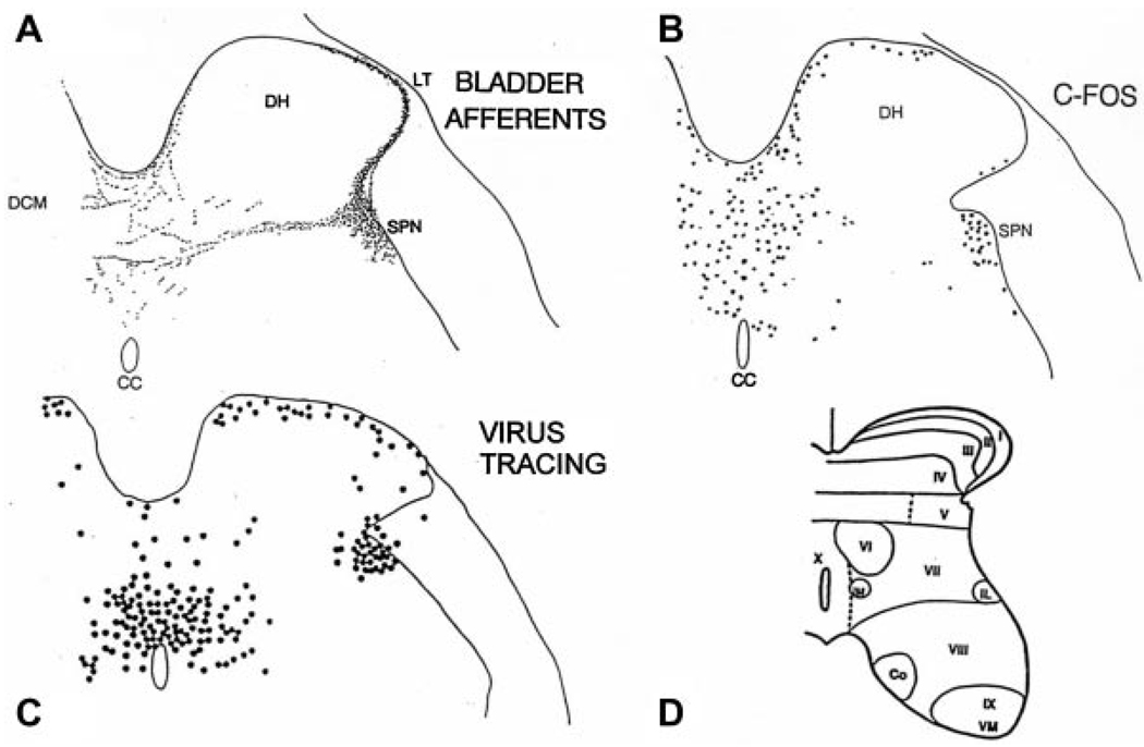
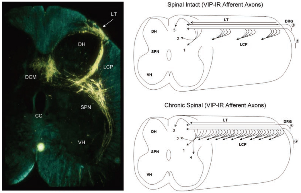
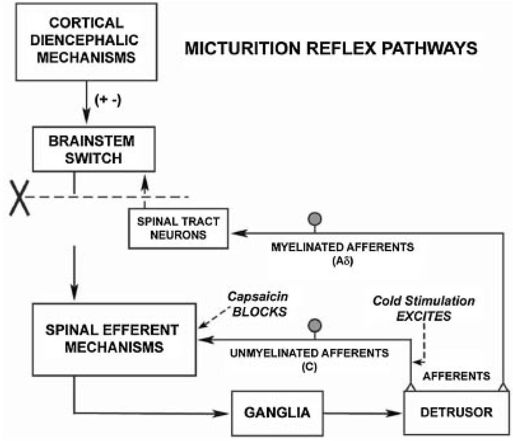
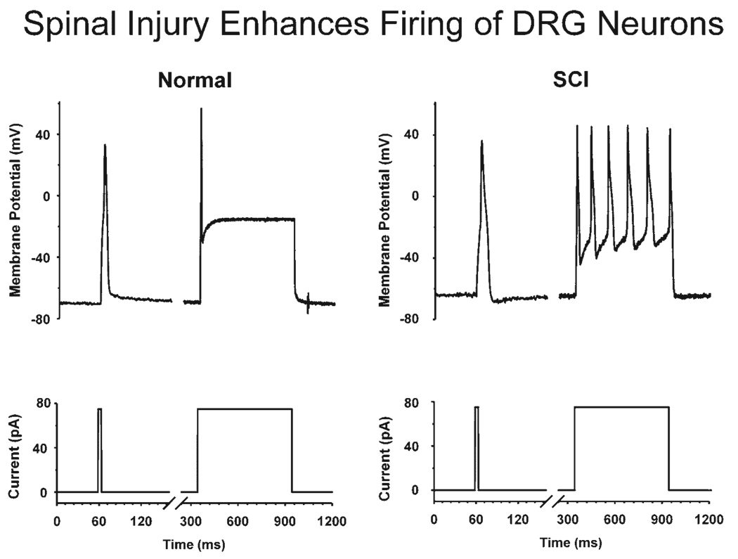
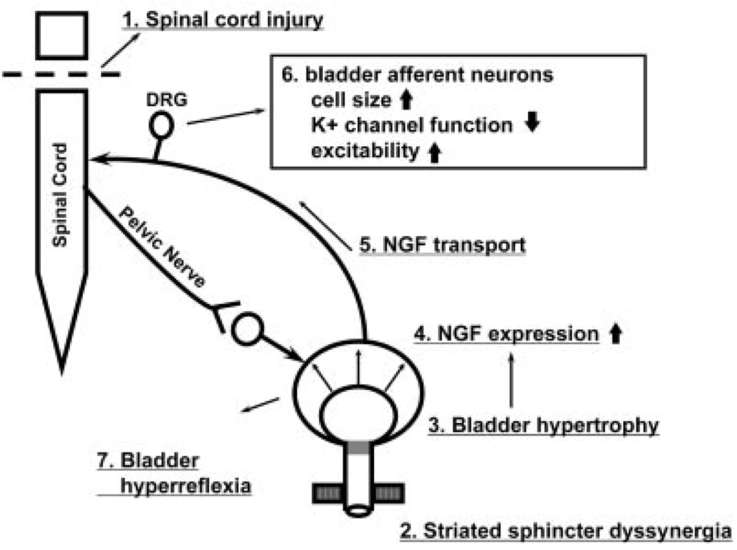
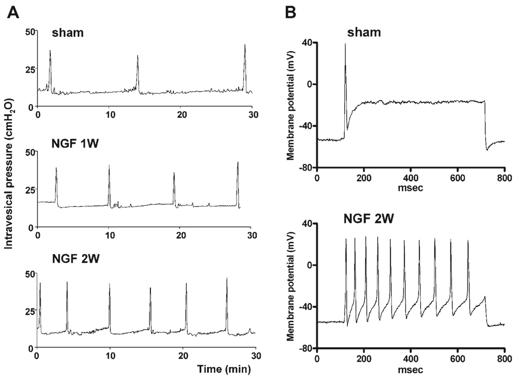
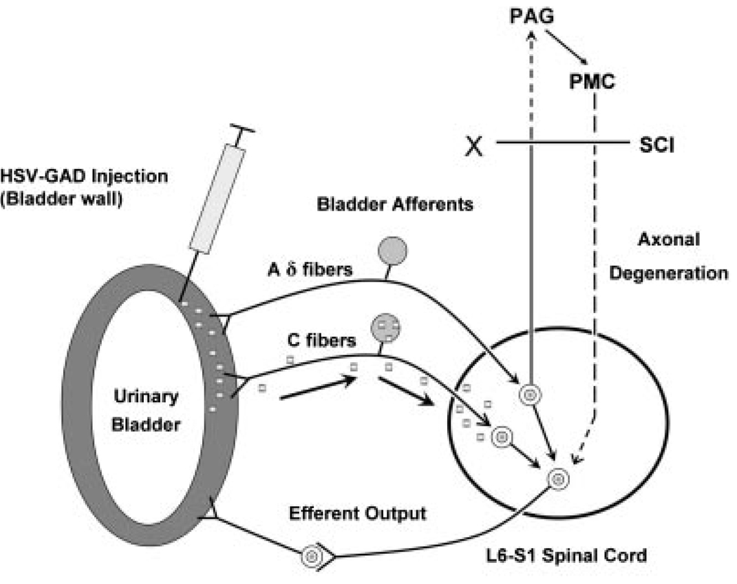
Results: Studies in animals indicate that the micturition reflex is mediated by a spinobulbospinal pathway passing through coordination centers (periaqueductal gray and pontine micturition center) located in the rostral brain stem. This reflex pathway, which is activated by small myelinated (Adelta) bladder afferent nerves, is in turn modulated by higher centers in the cerebral cortex involved in the voluntary control of micturition. Spinal cord injury at cervical or thoracic levels disrupts voluntary voiding, as well as the normal reflex pathways that coordinate bladder and sphincter function. Following spinal cord injury, the bladder is initially areflexic but then becomes hyperreflexic due to the emergence of a spinal micturition reflex pathway. The recovery of bladder function after spinal cord injury is dependent in part on the plasticity of bladder afferent pathways and the unmasking of reflexes triggered by unmyelinated, capsaicin-sensitive, C-fiber bladder afferent neurons. Plasticity is associated with morphologic, chemical, and electrical changes in bladder afferent neurons and appears to be mediated in part by neurotrophic factors released in the spinal cord and the peripheral target organs.

Conclusions: Spinal cord injury at sites remote from the lumbosacral spinal cord can indirectly influence properties of bladder afferent neurons by altering the function and chemical environment in the bladder or the spinal cord.


Conflict of interest statement

Conflicts of interest: none.

Figures

Fig. 1
Fig. 1
Diagram showing the sympathetic, parasympathetic, and somatic innervation of the urogenital tract of the male cat. Sympathetic preganglionic pathways emerge from the lumbar spinal cord and pass to the sympathetic chain ganglia (SCG) and then via the inferior splanchnic nerves (ISN) to the inferior mesenteric ganglia (IMG). Preganglionic and postganglionic sympathetic axons then travel in the hypogastric nerve (HGN) to the pelvic plexus and the urogenital organs. Parasympathetic preganglionic axons, which originate in the sacral spinal cord, pass in the pelvic nerve to ganglion cells in the pelvic plexus and to distal ganglia in the organs. Sacral somatic pathways are contained in the pudendal nerve, which provides an innervation to the penis, the ischiocavernosus (IC), bulbocavernosus (BC), and external urethral sphincter (EUS) muscles. The pudendal and pelvic nerves also receive postganglionic axons from the caudal SCG. These three sets of nerves contain afferent axons from the lumbosacral dorsal root ganglia. U, ureter; PG, prostate gland; VD, vas deferens.
Fig. 2
Fig. 2
Diagram illustrating the method for studying identified lower urinary tract afferent neurons in the L6-S1 dorsal root ganglia (DRG) using immunohistochemistry or patch-clamp techniques. (1) DRG neurons are labeled by axonal transport of fluorescent dyes injected into the bladder, urethra, or external sphincter several days prior to the experiment. (2) DRGs are removed for histologic experiments or (3) enzymatically dissociated to liberate single neurons. (4) Individual dye-labeled neurons are studied with whole cell patch-clamps methods.
Fig. 3
Fig. 3
Comparison of the distribution of bladder afferent projections to the L6 spinal cord of the rat (A) with the distribution of c-fos positive cells in the L6 spinal segment following chemical irritation of the lower urinary tract of the rat (B), and the distribution of interneurons in the L6 spinal cord labeled by transneuronal transport of pseudorabies virus injected into the urinary bladder (C). Afferents labeled by WGA-HRP injected into the urinary bladder. c-fos immunoreactivity is present in the nuclei of cells. DH, dorsal horn; SPN, sacral parasympathetic nucleus; CC, central canal. D: Drawing shows the laminar organization of the cat spinal cord.
Fig. 4
Fig. 4
Transverse section of the S2 spinal cord of the cat showing primary afferent axons and preganglionic neurons after application of horseradish peroxidase (HRP) to the pelvic nerve. Afferents enter Lissauer’s tract (LT) and then send collaterals through lamina 1 laterally around the dorsal horn (DH) in a large bundle (the lateral collateral pathway, LCP) into the area of the sacral parasympathetic nucleus (SPN). A smaller group of afferents extend medially into the dorsal gray commissure (DCM). Axons of preganglionic neurons in the SPN project into the ventral horn (VH). Diagram on the top right side shows that the distribution of VIP-immunoreactive (VIP-IR) afferent axons in the sacral spinal cord of the cat is similar to the distribution of pelvic visceral afferent axons in the LCP, lamina V (1) and in the DCM (2, 3). In addition, the VIP-IR axons and pelvic afferent axons in the LCP, which arise as collaterals from longitudinal axons in LT, occur in bundles distributed at regular intervals along the rostrocaudal axis of the cord. After chronic spinal cord injury (lower left diagram) the VIP-IR afferent pathways expand and reorganize, leading to a continuous band of axons in the LCP and more extensive projections into region of the SPN (4).
Fig. 5
Fig. 5
Comparison of the electrophysiologic characteristics of C-fiber (top traces) and Aδ-fiber (lower traces) bladder afferent neurons isolated from the lumbar dorsal root ganglion (DRG) of the rat. The left panels are voltage responses and action potentials evoked by 20 msec depolarizing current pulses. Asterisks with dashed lines indicate thresholds for spike activation (20 and 29 mV in the top and bottom traces, respectively). C-fiber neurons have higher thresholds and longer duration action potentials. The second panels show that C-fiber and Aδ-fiber afferent neurons have tetrodotoxin (TTX)-resistant and -sensitive action potentials, respectively. The third panel shows the firing patterns during long duration (600 msec) depolarizing currents pulses. C-fiber neurons fire phasically (1–2 spikes), whereas Aδ-fiber neurons fire tonically (12–15 spikes). The fourth panel shows that C-fiber neurons are capsaicin sensitive, whereas Aδ-fiber neurons are capsaicin insensitive.
Fig. 6
Fig. 6
Diagram showing the organization of the parasympathetic excitatory reflex pathway to the detrusor muscle. Scheme is based on electrophysiologic studies in cats. In animals with an intact spinal cord, micturition is initiated by a supraspinal reflex pathway passing through a center in the brain stem. The pathway is triggered by myelinated afferents (Aδ-fibers), which are connected to the tension receptors in the bladder wall. Injury to the spinal cord above the sacral segments (X) interrupts the connections between the brain and spinal autonomic centers and initially blocks micturition. However, over a period of several weeks following spinal cord injury, a spinal reflex mechanism emerges, which is triggered by unmyelinated vesical afferents (C-fibers); the A-fiber afferent inputs are ineffective. The C-fiber reflex pathway is usually weak or undetectable in animals with an intact nervous system. Stimulation of the C-fiber bladder afferents by instillation of ice water into the bladder (cold stimulation) activates voiding responses in patients with spinal cord injury. Capsaicin (20–30 mg, subcutaneously) blocks the C-fiber reflex in chronic spinal cord injured cats, but does not block micturition reflexes in intact cats. Intravesical capsaicin also suppresses detrusor hyperreflexia and cold-evoked reflexes in patients with neurogenic bladder dysfunction.
Fig. 7
Fig. 7
Effect of chronic spinal cord injury on the firing of a bladder sacral dorsal root ganglion neuron. Records on the left side, which are from an afferent neuron from a cat with an intact spinal cord, show action potentials (top trace) elicited by short (10 msec) and long (600 msec) duration depolarizing current pulses (bottom trace). This neuron is typical of small diameter bladder neurons which exhibit phasic firing. Records on the right side show tonic firing during a long duration depolarizing current pulse in a small diameter bladder neuron from a spinal cord injured (SCI) cat.
Fig. 8
Fig. 8
Diagram showing hypothetical mechanisms inducing lower urinary tract dysfunction following spinal cord injury (SCI). Injury to the spinal cord (1) causes detrusor-sphincter-dyssynergia (DSD) (2), which leads to functional urethral obstruction, reduced voiding efficiency, urinary retention, and bladder hypertrophy (3), resulting in increased levels of NGF in the bladder wall (4). NGF is taken up by afferent nerves and transported to the dorsal root ganglion cells (5) where it changes gene expression, which leads to increased cell size; regulation of ion channels, including a decrease in K+ channel function; and increased neuronal excitability (6). The levels of NGF also increase in the spinal cord after SCI. Hyperexcitability of bladder afferent pathways causes or enhances neurogenic detrusor overactivity and DSD. Intrathecal application of NGF antibodies reduces NGF levels in DRGs and suppresses neurogenic detrusor overactivity and DSD.
Fig. 9
Fig. 9
The effect of nerve growth factor (NGF) treatment on voiding function (A) and bladder afferent neuron firing pattern (B). NGF (2.5 µg/µl) was continuously infused into the intrathecal space at the L6-S1 level of the spinal cord for 1–2 weeks using osmotic minipumps (0.5 µl/hr). A: Cystometrograms showing continuous infusion cystometry in awake vehicle-treated (sham) and NGF-treated animals one week (1 w) and two weeks (2 w) after the start of treatment. Note the increased frequency of voiding after infusion of NGF. B: Firing patterns in response to 600 msec duration depolarizing current pulses in bladder afferent neurons, with TTX-resistant spikes from sham- and NGF-treated (2 w) rats. The current intensity was set to the threshold value for inducing single spikes with 10 msec current pulses. Note NGF-induced tonic firing, whereas the sham animal exhibited phasic firing as noted for C-fiber neurons from normal untreated animals (see Fig. 5).
Fig. 10
Fig. 10
Experimental gene therapy using a herpes simplex viral vector to increase the expression of glutamic acid decarboxylase (GAD) in the afferent pathways to the bladder. GAD is an enzyme involved in the synthesis of gamma aminobutyric acid (GABA), an inhibitory neurotransmitter in the spinal cord. Following injection of HSV-GAD into the bladder wall in SCI rats, the HSV-GAD is transported to the L6-S1 bladder afferents in the dorsal root ganglia and increases GABA synthesis and release from afferent terminals in the spinal cord. Three weeks after the injection, cystometry in awake rats revealed decreased number and amplitude of non-voiding contractions compared to the measurements in SCI rats receiving control vector (HSV-LacZ). HSV-GAD treatment did not alter the amplitude of voiding contractions. Since the effect of HSV-GAD is similar to the effect of capsaicin pretreatment, which targets C-fiber afferent pathways, it is possible that HSV-GAD selectively suppresses reflex responses induced by capsaicin-sensitive C-fiber afferents and does not affect Aδ-fiber-evoked reflexes which elicit voiding reflexes in spinal cord intact and chronic SCI rats.

Similar articles

Cited by

References

    1. Barrington FJF. The effect of lesion of the hind-and mid brain on micturition in the cat. Q J Exp Physiol. 1925;15:81–102.
    1. Chancellor MB, Yoshimura N. Physiology and pharmacology of the bladder and urethra. In: Wein AJ, editor. Campbell-walsh urology. 9th edition. Philadelphia, PA, USA: B. Saunders Elserion; 2006. pp. 1922–1972. Chapter 56.
    1. de Groat WC, Booth AM, Yoshimura N. Neurophysiology of micturition and its modification in animal models of human disease. In: Maggi CA, editor. Nervous control of the urogenital system. London, UK: Harwood Academic Publishers; 1993. pp. 227–290.
    1. Everaerts W, Gevaert T, Nilius B, et al. On the origin of bladder sensing: Tr(i)ps in urology. Neurourol Urodyn. 2008;27:264–273. - PubMed
    1. Fowler CJ, Griffiths D, de Groat WC. The neural control of micturition. Nat Rev Neurosci. 2008;9:453–466. - PMC - PubMed

Publication types

MeSH terms

Substances