

Cells on pores: a simulation-driven analysis of transcellular small molecule transport
- PMID: 20025248
- PMCID: PMC2920490
- DOI: 10.1021/mp9001969
Cells on pores: a simulation-driven analysis of transcellular small molecule transport
Abstract
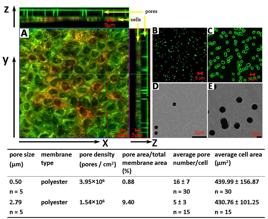
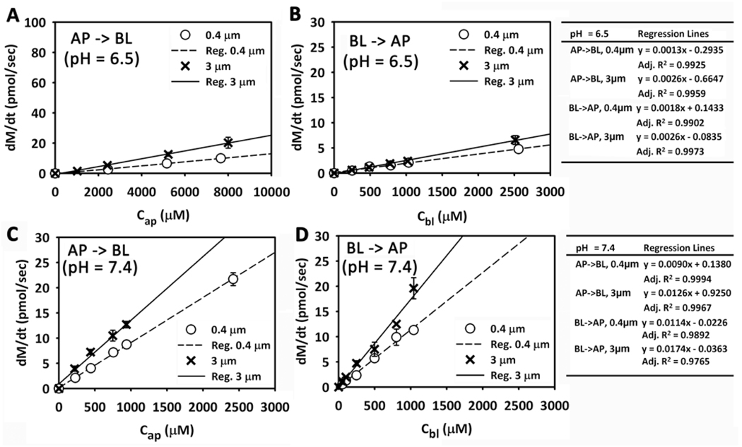
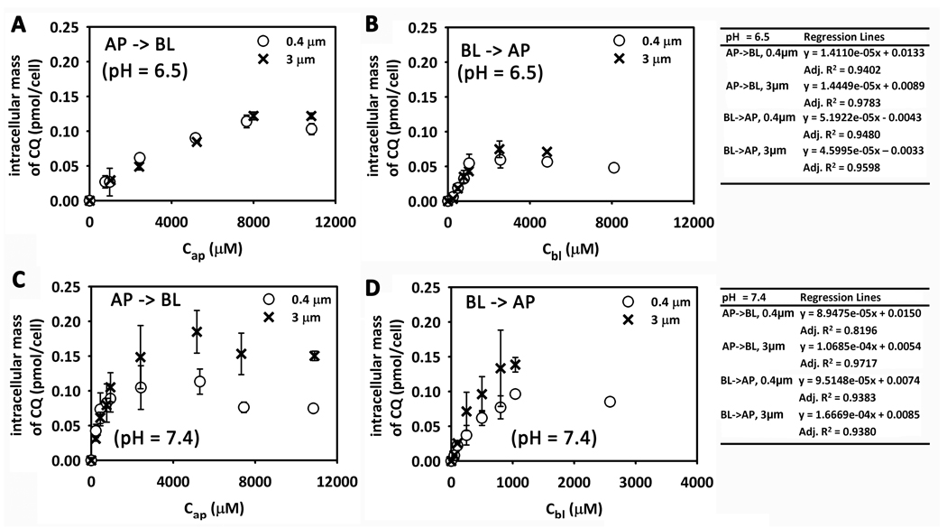
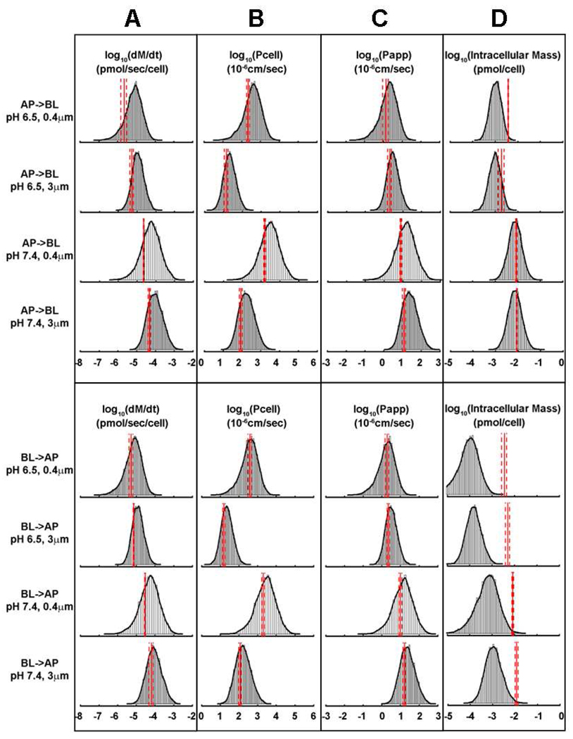
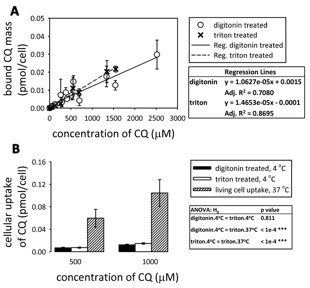
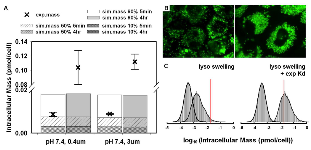
A biophysical, computational model of cell pharmacokinetics (1CellPK) is being developed to enable prediction of the intracellular accumulation and transcellular transport properties of small molecules using their calculated physicochemical properties as input. To test if 1CellPK can generate accurate, quantitative hypotheses and guide experimental analysis of the transcellular transport kinetics of small molecules, epithelial cells were grown on impermeable polyester membranes with cylindrical pores and chloroquine (CQ) was used as a transport probe. The effect of the number of pores and their diameter on transcellular transport of CQ was measured in apical-to-basolateral or basolateral-to-apical directions, at pH 7.4 and 6.5 in the donor compartment. Experimental and simulation results were consistent with a phospholipid bilayer-limited, passive diffusion transport mechanism. In experiments and 1CellPK simulations, intracellular CQ mass and the net rate of mass transport varied <2-fold although total pore area per cell varied >10-fold, so by normalizing the net rate of mass transport by the pore area available for transport, cell permeability on 3 mum pore diameter membranes was more than an order of magnitude less than on 0.4 mum pore diameter membranes. The results of simulations of transcellular transport were accurate for the first four hours of drug exposure, but those of CQ mass accumulation were accurate only for the first five minutes. Upon prolonged incubation, changes in cellular parameters such as lysosome pH rise, lysosome volume expansion, and nuclear shrinkage were associated with excess CQ accumulation. Based on the simulations, lysosome volume expansion alone can partly account for the measured, total intracellular CQ mass increase, while adding the intracellular binding of the protonated, ionized forms of CQ (as reflected in the measured partition coefficient of CQ in detergent-permeabilized cells at physiological pH) can further improve the intracellular CQ mass accumulation prediction.
Figures
Similar articles
-
Effect of phospholipidosis on the cellular pharmacokinetics of chloroquine.J Pharmacol Exp Ther. 2011 Mar;336(3):661-71. doi: 10.1124/jpet.110.175679. Epub 2010 Dec 14. J Pharmacol Exp Ther. 2011. PMID: 21156819 Free PMC article.
-
A cell-based molecular transport simulator for pharmacokinetic prediction and cheminformatic exploration.Mol Pharm. 2006 Nov-Dec;3(6):704-16. doi: 10.1021/mp060046k. Mol Pharm. 2006. PMID: 17140258 Free PMC article.
-
Simulation of kinetic data on the influx and efflux of chloroquine by erythrocytes infected with Plasmodium falciparum. Evidence for a drug-importer in chloroquine-sensitive strains.Biochem Pharmacol. 1991 Dec 11;42 Suppl:S167-79. doi: 10.1016/0006-2952(91)90407-v. Biochem Pharmacol. 1991. PMID: 1768274
-
Transport of cadmium across the apical membrane of epithelial cell lines.Comp Biochem Physiol C Toxicol Pharmacol. 2002 Mar;131(3):223-9. doi: 10.1016/s1532-0456(02)00009-1. Comp Biochem Physiol C Toxicol Pharmacol. 2002. PMID: 11912047 Review.
-
Computer simulations of transport through membranes: passive diffusion, pores, channels and transporters.Clin Exp Pharmacol Physiol. 2006 Oct;33(10):893-903. doi: 10.1111/j.1440-1681.2006.04461.x. Clin Exp Pharmacol Physiol. 2006. PMID: 17002665 Review.
Cited by
-
Effect of phospholipidosis on the cellular pharmacokinetics of chloroquine.J Pharmacol Exp Ther. 2011 Mar;336(3):661-71. doi: 10.1124/jpet.110.175679. Epub 2010 Dec 14. J Pharmacol Exp Ther. 2011. PMID: 21156819 Free PMC article.
-
A cell-based computational modeling approach for developing site-directed molecular probes.PLoS Comput Biol. 2012;8(2):e1002378. doi: 10.1371/journal.pcbi.1002378. Epub 2012 Feb 23. PLoS Comput Biol. 2012. PMID: 22383866 Free PMC article.
-
Toxicological assessment of inhaled nanoparticles: role of in vivo, ex vivo, in vitro, and in silico studies.Int J Mol Sci. 2014 Mar 18;15(3):4795-822. doi: 10.3390/ijms15034795. Int J Mol Sci. 2014. PMID: 24646916 Free PMC article. Review.
-
Quantitative Raman chemical imaging of intracellular drug-membrane aggregates and small molecule drug precipitates in cytoplasmic organelles.Adv Drug Deliv Rev. 2023 Nov;202:115107. doi: 10.1016/j.addr.2023.115107. Epub 2023 Sep 26. Adv Drug Deliv Rev. 2023. PMID: 37769851 Free PMC article. Review.
-
Visualizing chemical structure-subcellular localization relationships using fluorescent small molecules as probes of cellular transport.J Cheminform. 2013 Oct 5;5(1):44. doi: 10.1186/1758-2946-5-44. J Cheminform. 2013. PMID: 24093553 Free PMC article.
References
Publication types
MeSH terms
Substances
Grants and funding
LinkOut - more resources
Full Text Sources
