

HDAC inhibitor, valproic acid, induces p53-dependent radiosensitization of colon cancer cells
- PMID: 20025549
- PMCID: PMC2920759
- DOI: 10.1089/cbr.2009.0629
HDAC inhibitor, valproic acid, induces p53-dependent radiosensitization of colon cancer cells
Abstract
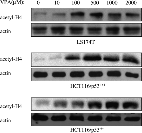
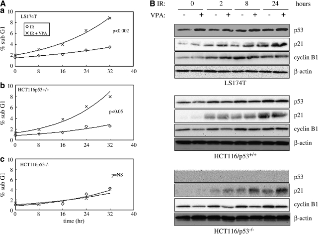
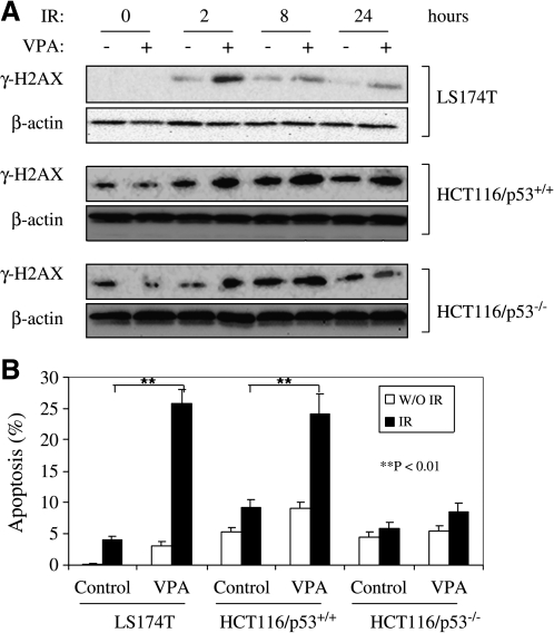
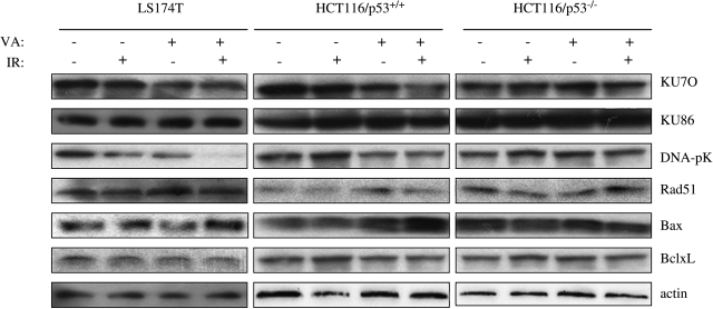
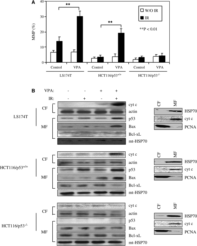
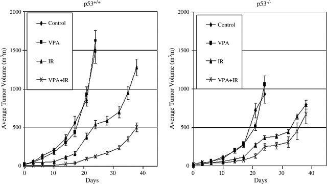
Agents that inhibit histone deacetylases (HDAC inhibitors) have been shown to enhance radiation response. The aim of this study was to evaluate the effects of low, minimally cytotoxic concentrations of the HDAC inhibitor, valproic acid (VPA), on radiation response of colorectal cancer cells. Cell lines LS174T and an isogenic pair of HCT116, which differed only for the presence of wild-type p53, were exposed to ionizing radiation (IR) alone, VPA alone, or the combination. Clonogenic survival, gamma-H2AX induction, apoptosis, changes in mitochondrial membrane potential, and mitochondrial levels of p53 and Bcl-2 family proteins were assessed. In vivo studies monitored tumor growth suppression after therapy in mice bearing HCT116/p53(+/+) and HCT116/p53(-/-) tumor xenografts. VPA led to radiosensitization, which was dependent on p53 status. A decrease in clonogenic survival, an increase in apoptosis, and an increase in levels of gamma-H2AX were observed after VPA+IR, compared to IR alone, in wild-type p53 cells (LS174T and HCT116/p53(+/+)), as opposed to p53 null cells (HCT116/p53(-/-)). Exposure to VPA resulted in enhancement of IR-induced mitochondrial localizations of Bax and Bcl-xL, mitochondrial membrane potential, and cytochrome c release only in wild-type p53 cell lines. VPA also enhanced tumor growth suppression after IR only in wild-type p53 xenografts. These data suggest that VPA may have an important role in enhancing radiotherapy response in colorectal cancer, particularly in tumors with the wild-type p53 genotype.
Figures

Similar articles
-
Synergistic antitumor interaction between valproic acid, capecitabine and radiotherapy in colorectal cancer: critical role of p53.J Exp Clin Cancer Res. 2017 Dec 6;36(1):177. doi: 10.1186/s13046-017-0647-5. J Exp Clin Cancer Res. 2017. PMID: 29212503 Free PMC article.
-
Low-dose valproic acid enhances radiosensitivity of prostate cancer through acetylated p53-dependent modulation of mitochondrial membrane potential and apoptosis.Mol Cancer Res. 2011 Apr;9(4):448-61. doi: 10.1158/1541-7786.MCR-10-0471. Epub 2011 Feb 8. Mol Cancer Res. 2011. PMID: 21303901 Free PMC article.
-
Histone deacetylase inhibitors valproic acid and depsipeptide sensitize retinoblastoma cells to radiotherapy by increasing H2AX phosphorylation and p53 acetylation-phosphorylation.Int J Oncol. 2010 Oct;37(4):787-95. doi: 10.3892/ijo_00000728. Int J Oncol. 2010. PMID: 20811699
-
The small molecule AU14022 promotes colorectal cancer cell death via p53-mediated G2/M-phase arrest and mitochondria-mediated apoptosis.J Cell Physiol. 2018 Jun;233(6):4666-4676. doi: 10.1002/jcp.26234. Epub 2018 Jan 15. J Cell Physiol. 2018. PMID: 29030986
-
Enhanced radiosensitization of p53 mutant cells by oleamide.Int J Radiat Oncol Biol Phys. 2006 Apr 1;64(5):1466-74. doi: 10.1016/j.ijrobp.2005.11.033. Int J Radiat Oncol Biol Phys. 2006. PMID: 16580498
Cited by
-
HD-MB03 is a novel Group 3 medulloblastoma model demonstrating sensitivity to histone deacetylase inhibitor treatment.J Neurooncol. 2012 Dec;110(3):335-48. doi: 10.1007/s11060-012-0978-1. Epub 2012 Oct 6. J Neurooncol. 2012. PMID: 23054560
-
Synergistic antitumor interaction between valproic acid, capecitabine and radiotherapy in colorectal cancer: critical role of p53.J Exp Clin Cancer Res. 2017 Dec 6;36(1):177. doi: 10.1186/s13046-017-0647-5. J Exp Clin Cancer Res. 2017. PMID: 29212503 Free PMC article.
-
The Effect of VPA on Increasing Radiosensitivity in Osteosarcoma Cells and Primary-Culture Cells from Chemical Carcinogen-Induced Breast Cancer in Rats.Int J Mol Sci. 2017 May 10;18(5):1027. doi: 10.3390/ijms18051027. Int J Mol Sci. 2017. PMID: 28489060 Free PMC article.
-
Role of HDAC3 on p53 expression and apoptosis in T cells of patients with multiple sclerosis.PLoS One. 2011 Feb 8;6(2):e16795. doi: 10.1371/journal.pone.0016795. PLoS One. 2011. PMID: 21346816 Free PMC article.
-
Histone deacetylase inhibitor potentiates chemotherapy-induced apoptosis through Bim upregulation in Burkitt's lymphoma cells.J Cancer Res Clin Oncol. 2012 Feb;138(2):317-25. doi: 10.1007/s00432-011-1093-y. Epub 2011 Dec 1. J Cancer Res Clin Oncol. 2012. PMID: 22131152 Free PMC article.
References
-
- Howell SB. Resistance to apoptosis in prostate cancer cells. Mol Urol. 2000;4:225. - PubMed
-
- Gordon AT. McMillan TJ. A role for molecular radiobiology in radiotherapy? Clin Oncol (R Coll Radiol) 1997;9:70. - PubMed
-
- Ward JF. The complexity of DNA damage: Relevance to biological consequences. Int J Radiat Biol. 1994;66:427. - PubMed
-
- Kastan MB. Bartek J. Cell-cycle checkpoints and cancer. Nature. 2004;432:316. - PubMed
-
- Rogakou EP. Pilch DR. Orr AH, et al. DNA double-stranded breaks induce histone H2AX phosphorylation on serine 139. J Biol Chem. 1998;273:5858. - PubMed
Publication types
MeSH terms
Substances
Grants and funding
LinkOut - more resources
Full Text Sources
Medical
Research Materials
Miscellaneous
