. 2009 Dec 21:9:70.
doi: 10.1186/1471-213X-9-70.

Isolation and characterization of a novel plasma membrane protein, osteoblast induction factor (obif), associated with osteoblast differentiation

Affiliations

Isolation and characterization of a novel plasma membrane protein, osteoblast induction factor (obif), associated with osteoblast differentiation

Takashi Kanamoto et al. BMC Dev Biol. .

Abstract

Background: While several cell types are known to contribute to bone formation, the major player is a common bone matrix-secreting cell type, the osteoblast. Chondrocytes, which plays critical roles at several stages of endochondral ossification, and osteoblasts are derived from common precursors, and both intrinsic cues and signals from extrinsic cues play critical roles in the lineage decision of these cell types. Several studies have shown that cell fate commitment within the osteoblast lineage requires sequential, stage-specific signaling to promote osteoblastic differentiation programs. In osteoblastic differentiation, the functional mechanisms of transcriptional regulators have been well elucidated, however the exact roles of extrinsic molecules in osteoblastic differentiation are less clear.

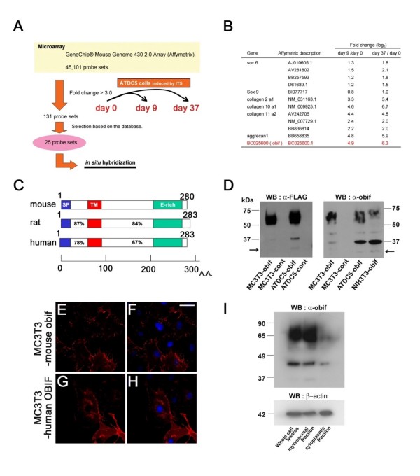
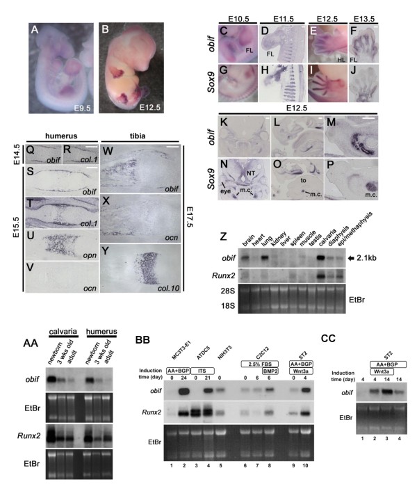
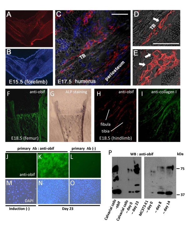
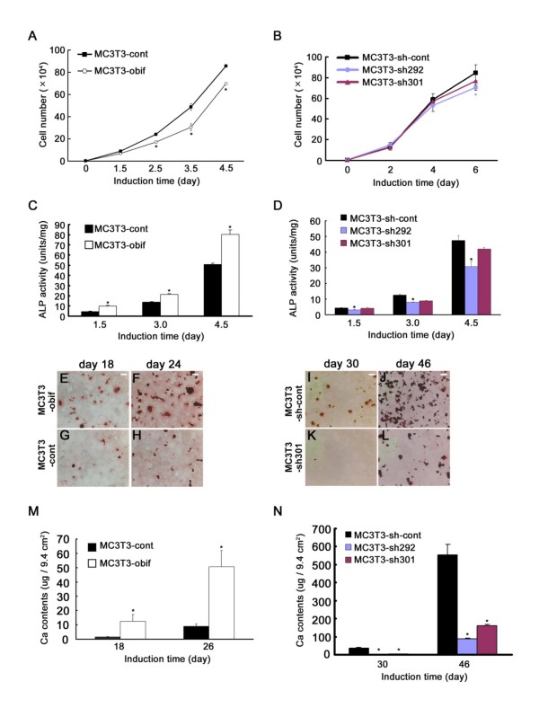
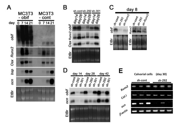
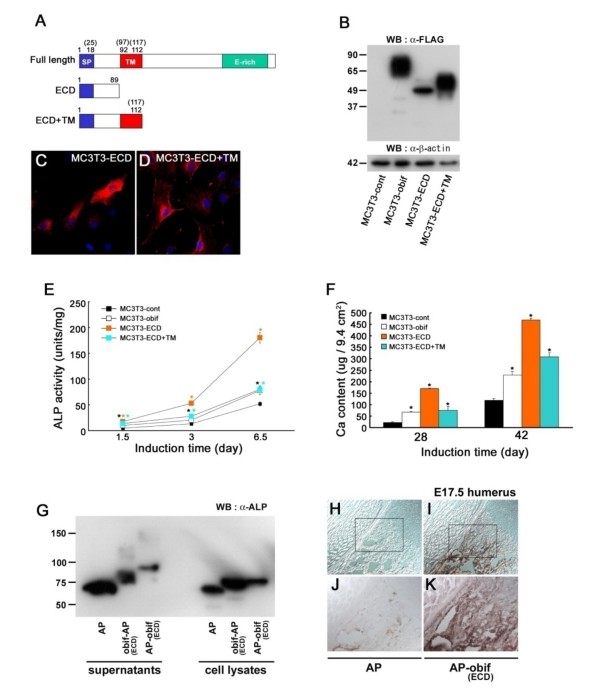
Results: We identify a novel gene, obif (osteoblast induction factor), encoding a transmembrane protein that is predominantly expressed in osteoblasts. During mouse development, obif is initially observed in the limb bud in a complementary pattern to Sox9 expression. Later in development, obif is highly expressed in osteoblasts at the stage of endochondral ossification. In cell line models, obif is up-regulated during osteoblastic differentiation. Exogenous obif expression stimulates osteoblastic differentiation and obif knockdown inhibits osteoblastic differentiation in preosteblastic MC3T3-E1 cells. In addition, the extracellular domain of obif protein exhibits functions similar to the full-length obif protein in induction of MC3T3-E1 differentiation.

Conclusions: Our results suggest that obif plays a role in osteoblastic differentiation by acting as a ligand.


Figures

Figure 1
Figure 1
Identification of a novel plasma membrane protein, obif, and its subcellular localization. (A) The strategy of microarray-based screening to identify up-regulated genes in chondrocyte differentiations of ATDC5 cell line. (B) Fold changes of representative chondrocyte markers (black) and obif (red) transcripts which are up-regulated in the microarray analysis. (C) Schematic diagram of obif protein structure. The percent sequence identities of each region are shown: SP, signal peptide; TM, transmembrane domain; E-rich, glutamic acid-rich domain. Numbers represent amino acid residues. (D) Detection of FLAG-tagged obif protein. Whole cell lysates of MC3T3-obif, MC3T3-cont, ATDC5-obif, ATDC5-cont, and NIH3T3-obif were electrophoresed by SDS-PAGE. Immunoblots were probed with anti-FLAG M2 antibody or anti-obif antibody. Arrows represent the molecular size of obif protein predicted from its amino acid sequences (29.4 kDa). (E-H) Confocal analysis of overexpressed mouse and human obif in MC3T3-E1 cells. Cells expressing FLAG-tagged mouse obif were stained with anti-obif antibody (red) (E, F). Cells expressing FLAG-tagged human OBIF were stained with anti-FLAG M2 antibody (red) (G, H). Nuclei were stained with DAPI (blue). Scale bar = 50 μm. (I) Subcellular localization of obif protein. Subcellular fractions of ST2 cells expressing exogenous mouse obif were analyzed by Western blotting. Immunoblots were probed with an anti-obif antibody to localize obif (top) and with an antibody against β-actin, an abundant cytoplasmic protein (bottom).
Figure 2
Figure 2
Predominant expression of obif transcript in osteoblast-lineage cells. (A, B) Expressions of obif in mouse embryos examined by whole mount in situ hybridization. (C-P) In situ hybridization analysis of mouse obif and Sox9 in the developing mouse limb, rib, and mandible. Obif expression was complementary to that of Sox9 in the developing limb bud (C-J), rib (D, H) and mandible (K-P). FL, forelimb; HL, hindlimb; NT, neural tube; to, tongue; m.c., Meckel's cartilage. Scale bars = 200 μm. (Q-Y) Section in situ hybridization of mouse obif in endochondral ossification. At E14.5 obif transcripts were detected in the perichondrium but were absent from more centrally located chondrocytic cells (Q, R). In E15.5 humerus and E17.5 tibia, obif expression was found in cells associated with bone trabeculae and in cells associated with the formation of bone collars (S-Y). Scale bars = 200 μm. (Z) Northern blot analysis of obif and Runx2 expression in 4 week-old mouse tissues. The arrow corresponds to a 2.1 kb obif transcript. (AA) Northern blot analysis of obif and Runx2 expression in the calvaria and humeri harvested from newborn, 3 week-old, and adult mice. (BB, CC) Northern blot analysis of obif and Runx2 transcripts in cell differentiation models of osteoblasts and chondrocytes. Total RNAs, extracted from MC3T3-E1 cells (BB: lane 1, undifferentiated cells; lane 2, cells cultured in differentiation medium for 24 days), ATDC5 cells (BB: lane 3, undifferentiated cells; lane 4, cells cultured in differentiation medium for 21 days), NIH3T3 cells (BB, lane 5), C2C12 cells (BB: lane 6, undifferentiated cells; lane 7, cells cultured in myogenic differentiation medium for 6 days; lane 8, cells cultured in osteoblastic differentiation medium for 6 days), and ST2 cells (BB: lane 9, cells cultured in growth medium; lane 10, cells cultured in medium supplemented with Wnt3a and ascorbic acid for 4 days; and CC: lane 1, cells cultured in growth medium for 4 days; lane 2, cells cultured in medium with Wnt3a and ascorbic acid for 4 days; lane 3, cells cultured in medium with Wnt3a and ascorbic acid for 14 days; lane 4, cells cultured in medium with ascorbic acid for 14 days) were used for Northern blot analysis.
Figure 3
Figure 3
Predominant expression of obif protein in osteoblast-lineage cells. (A-E) Immunostaining of developing limbs using anti-obif antibody. Sections of limbs were stained with anti-obif antibody (red) and nuclei were stained with DAPI (blue). E15.5 forelimb sections (A, B). Scale bars = 200 μm. E17.5 humerus sections (C). Obif was expressed in spindle-shaped osteoblasts lining trabecular bone surfaces and in cuboidal-shaped osteoblasts. Scale bars = 50 μm. Higher magnification of E17.5 humerus sections shows that obif is localized to the plasma membrane (D, E). White arrows indicate typical cells that show the transmembrane pattern. (F, G) E 18.5 femur section was first stained with anti-obif antibody (green) and then stained for ALP. (H, I) E 18.5 hindlimb section was double-stained with anti-obif antibody (red) and anti-collagen I antibody (green). (J-O) Primary calvarial cells cultured in 12-well plates were immunostained with anti-obif antibody (J-L) and DAPI (M-O). Sparsely cultured cells (J, M). Densely cultured cells were incubated in presence of ascorbic acid for 23 days and used for analysis (K-O). L & O are negative controls. (P) Detection of endogenous obif protein. Whole cell lysates of calvarial cells-obif (stably infected), calvarial cells-day 0 (not stimulated), calvarial cells-day 23 (stimulated for 23 days), MC3T3-E1 cells-day 0 (not stimulated), MC3T3-E1 cells-day 8 (stimulated for 8 days), and MC3T3-E1 cells-day 14 (stimulated for 14 days) were electrophoresed by SDS-PAGE. Immunoblots were probed with the anti-obif antibody. Calvarial cells-obif is a positive control.
Figure 4
Figure 4
The role of obif in the osteoblastic differentiation of MC3T3-E1 cells. (A, B) Effect of obif expression on cell growth in the MC3T3-E1 cells. The proliferation rate decreased somewhat in MC3T3-obif compared with MC3T3-cont (A). Cells infected with retroviruses expressing shRNA against obif (MC3T3-sh292, MC3T3-sh301) showed almost the same level of cell growth with MC3T3-sh-cont (B). (C) Effect of obif expression on ALP activity in MC3T3-E1 cells cultured in the presence of ascorbic acid and β-glycerophosphate. (D) Effect of expression of shRNAs against obif in MC3T3-E1 cells. The reduction of obif expression caused a significant decrease of ALP activity at all time points. The decrease was proportionate to the strength of the shRNA's knockdown effects. (E-N) Mineral deposition visualized by Alizarin Red staining (E-L), and calcium contents measured by a colorimetric assay (M, N). The mineral deposition observed in MC3T3-obif cells and MC3T3-cont cells at days 18 and 24 (E-H). The mineral deposition observed in MC3T3-sh301 cells compared to MC3T3-sh-cont cells at days 30 and 46 (I-L). Scale bars = 2 mm. Calcium contents were significantly higher in MC3T3-obif cells than in MC3T3-cont cells both at day 18 and day 26 (M). Calcium deposition in MC3T3-sh292/301 and MC3T3-sh-cont cells at days 30 and 46 (N).
Figure 5
Figure 5
The effect of obif expression on osteoblastic markers. (A-D) Northern blot analyses of osteoblastic differentiation markers in MC3T3-E1 cells. Comparison between MC3T3-obif cells and MC3T3-control cells (A). The overexpression of obif was confirmed in MC3T3-obif cells. Since the coding sequence of obif is connected with an IRES sequence in the retrovirus construct, transcripts produced are longer than endogenous obif transcripts. The mRNA levels of osteoblastic differentiation markers, in particular bsp and ocn increased in MC3T3-obif cells. The comparison among control cells (MC3T3-sh147) and obif knocked down cells (MC3T3-sh292, -sh301) (B-D). At day 14, Osx transcripts decreased in obif knocked down cells whereas Runx2 transcripts were not influenced by obif level (B). At day 28, Runx2 was down-regulated in MC3T3-sh292 cells where we could detect only faint levels of obif mRNA. In MC3T3-shRunx2 cells, obif was down-regulated whereas Runx2 was not affected in MC3T3-sh292 cells at day 8 (C). At day 14 and 28, ocn were down-regulated in both MC3T3-sh292 and -sh301 cells (D). At day 42, ocn mRNA level in MC3T3-sh301 cells is almost the same as that in the control cells, but significantly lower in MC3T3-sh292 cells. (E) Semi-quantitative RT-PCR of osteoblastic differentiation markers in primary calvarial cells. The data presented are derived from three independent experiments.
Figure 6
Figure 6
Extracellular domain of obif can promote osteoblast differentiation. (A) Schematic representation of partial obif proteins overexpressed in MC3T3-E1 cells. Upper: the full-length obif protein, middle: the partial obif fragment containing the N-terminal extracellular domain (ECD), lower: the partial obif fragment composed of the ECD and the transmembrane domain (TM). Numbers represent amino acid residues of mouse (human) obif proteins. (B-F) MC3T3-E1 cells infected with retroviruses were cultured in the differentiation medium and used for assays. Both of bands of partial obif poteins detected by Western blot were larger than predicted sizes (B). Immunocytochemistry analyses using anti-FLAG antibody (C-D). MC3T3-ECD+TM protein localizes to the plasma membranes whereas a large amount of obif-ECD protein localizes to the cytoplasm. Scale bars = 50 μm. MC3T3-E1 cells infected with retroviruses expressing a full-length obif or its partial proteins showed elevated ALP activities at all time points examined (E). Infection with retroviruses expressing full-length and partial proteins significantly promoted mineral deposition both at day 28 and 42 (F). (G) AP fusion proteins (obif-ECD-AP, AP-obif-ECD) and AP protein were detected by Western blotting analysis using anti-AP antibody. Fusion proteins in supernatants are larger than those in lysates from transfected cells. (H-I) AP-obif (ECD) bound to bone tissues whereas control AP protein did not bind at detectable levels. (J, K) Higher magnifications of the squares in H &I, respectively. Scale bars = 100 μm.

Similar articles

Cited by

  • FAD104, a regulatory factor of adipogenesis, acts as a novel regulator of calvarial bone formation.
    Kishimoto K, Nishizuka M, Katoh D, Kato A, Osada S, Imagawa M. Kishimoto K, et al. J Biol Chem. 2013 Nov 1;288(44):31772-83. doi: 10.1074/jbc.M113.452961. Epub 2013 Sep 19. J Biol Chem. 2013. PMID: 24052261 Free PMC article.
  • Parathyroid hormone-responsive Smad3-related factor, Tmem119, promotes osteoblast differentiation and interacts with the bone morphogenetic protein-Runx2 pathway.
    Hisa I, Inoue Y, Hendy GN, Canaff L, Kitazawa R, Kitazawa S, Komori T, Sugimoto T, Seino S, Kaji H. Hisa I, et al. J Biol Chem. 2011 Mar 18;286(11):9787-96. doi: 10.1074/jbc.M110.179127. Epub 2011 Jan 14. J Biol Chem. 2011. PMID: 21239498 Free PMC article.
  • A systematic characterization of microglia-like cell occurrence during retinal organoid differentiation.
    Bartalska K, Hübschmann V, Korkut-Demirbaş M, Cubero RJA, Venturino A, Rössler K, Czech T, Siegert S. Bartalska K, et al. iScience. 2022 Jun 11;25(7):104580. doi: 10.1016/j.isci.2022.104580. eCollection 2022 Jul 15. iScience. 2022. PMID: 35789843 Free PMC article.
  • The role of TMEM119 in gastric adenocarcinoma and its specific effects on immunity.
    Liu Y, Yan X, Qu C, Tang F, Wang Q, Li Y. Liu Y, et al. J Int Med Res. 2025 Apr;53(4):3000605241306668. doi: 10.1177/03000605241306668. Epub 2025 Apr 12. J Int Med Res. 2025. PMID: 40219804 Free PMC article.
  • Microglia states and nomenclature: A field at its crossroads.
    Paolicelli RC, Sierra A, Stevens B, Tremblay ME, Aguzzi A, Ajami B, Amit I, Audinat E, Bechmann I, Bennett M, Bennett F, Bessis A, Biber K, Bilbo S, Blurton-Jones M, Boddeke E, Brites D, Brône B, Brown GC, Butovsky O, Carson MJ, Castellano B, Colonna M, Cowley SA, Cunningham C, Davalos D, De Jager PL, de Strooper B, Denes A, Eggen BJL, Eyo U, Galea E, Garel S, Ginhoux F, Glass CK, Gokce O, Gomez-Nicola D, González B, Gordon S, Graeber MB, Greenhalgh AD, Gressens P, Greter M, Gutmann DH, Haass C, Heneka MT, Heppner FL, Hong S, Hume DA, Jung S, Kettenmann H, Kipnis J, Koyama R, Lemke G, Lynch M, Majewska A, Malcangio M, Malm T, Mancuso R, Masuda T, Matteoli M, McColl BW, Miron VE, Molofsky AV, Monje M, Mracsko E, Nadjar A, Neher JJ, Neniskyte U, Neumann H, Noda M, Peng B, Peri F, Perry VH, Popovich PG, Pridans C, Priller J, Prinz M, Ragozzino D, Ransohoff RM, Salter MW, Schaefer A, Schafer DP, Schwartz M, Simons M, Smith CJ, Streit WJ, Tay TL, Tsai LH, Verkhratsky A, von Bernhardi R, Wake H, Wittamer V, Wolf SA, Wu LJ, Wyss-Coray T. Paolicelli RC, et al. Neuron. 2022 Nov 2;110(21):3458-3483. doi: 10.1016/j.neuron.2022.10.020. Neuron. 2022. PMID: 36327895 Free PMC article. Review.

References

    1. Karsenty G, Wagner EF. Reaching a genetic and molecular understanding of skeletal development. Dev Cell. 2002;2:389–406. doi: 10.1016/S1534-5807(02)00157-0. - DOI - PubMed
    1. Zelzer E, Olsen BR. The genetic basis for skeletal diseases. Nature. 2003;423:343–8. doi: 10.1038/nature01659. - DOI - PubMed
    1. Hartmann C. A Wnt canon orchestrating osteoblastogenesis. Trends Cell Biol. 2006;16:151–8. doi: 10.1016/j.tcb.2006.01.001. - DOI - PubMed
    1. Otto F, Thornell AP, Crompton T, Denzel A, Gilmour KC, Rosewell IR, Stamp GW, Beddington RS, Mundlos S, Olsen BR, Selby PB, Owen MJ. Cbfa1, a candidate gene for cleidocranial dysplasia syndrome, is essential for osteoblast differentiation and bone development. Cell. 1997;89:765–71. doi: 10.1016/S0092-8674(00)80259-7. - DOI - PubMed
    1. Ducy P, Zhang R, Geoffroy V, Ridall AL, Karsenty G. Osf2/Cbfa1: a transcriptional activator of osteoblast differentiation. Cell. 1997;89:747–54. doi: 10.1016/S0092-8674(00)80257-3. - DOI - PubMed

Publication types