. 2010 Feb 15;338(2):270-9.
doi: 10.1016/j.ydbio.2009.12.008. Epub 2009 Dec 16.

Disruption of Paneth and goblet cell homeostasis and increased endoplasmic reticulum stress in Agr2-/- mice

Affiliations

Disruption of Paneth and goblet cell homeostasis and increased endoplasmic reticulum stress in Agr2-/- mice

Fang Zhao et al. Dev Biol. .

Abstract

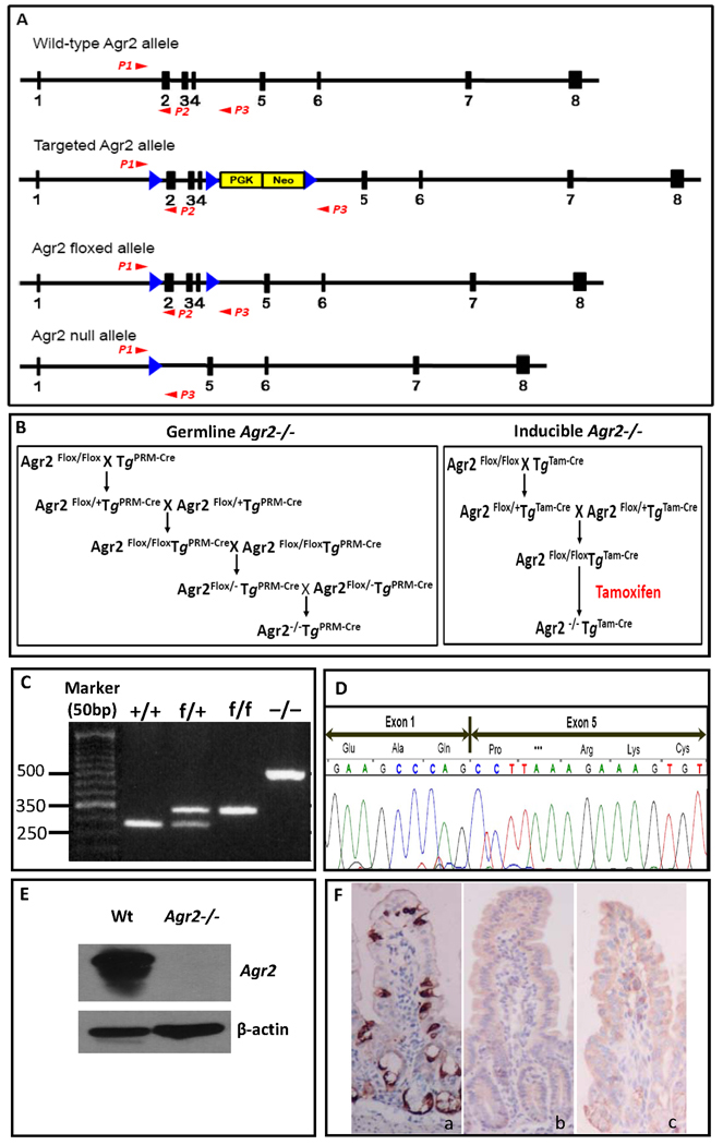
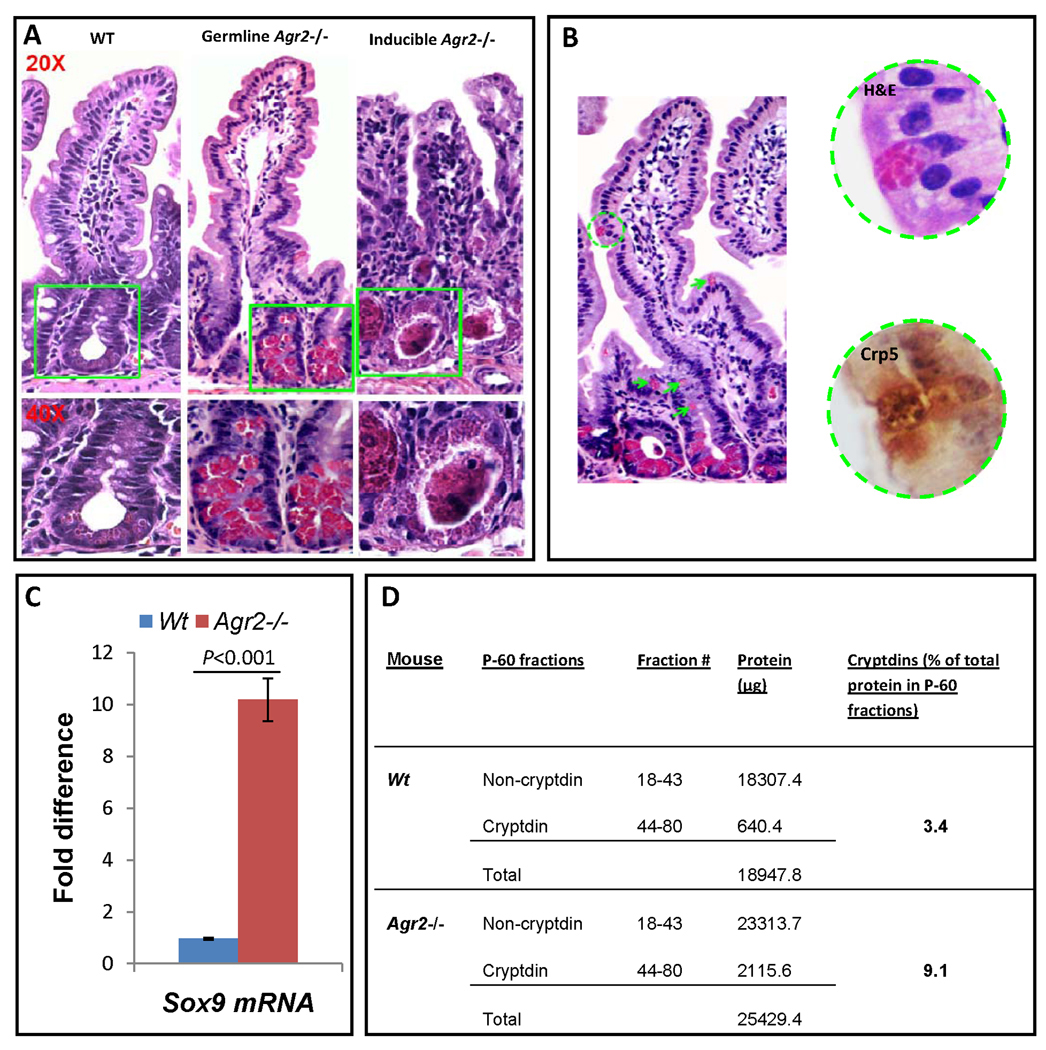
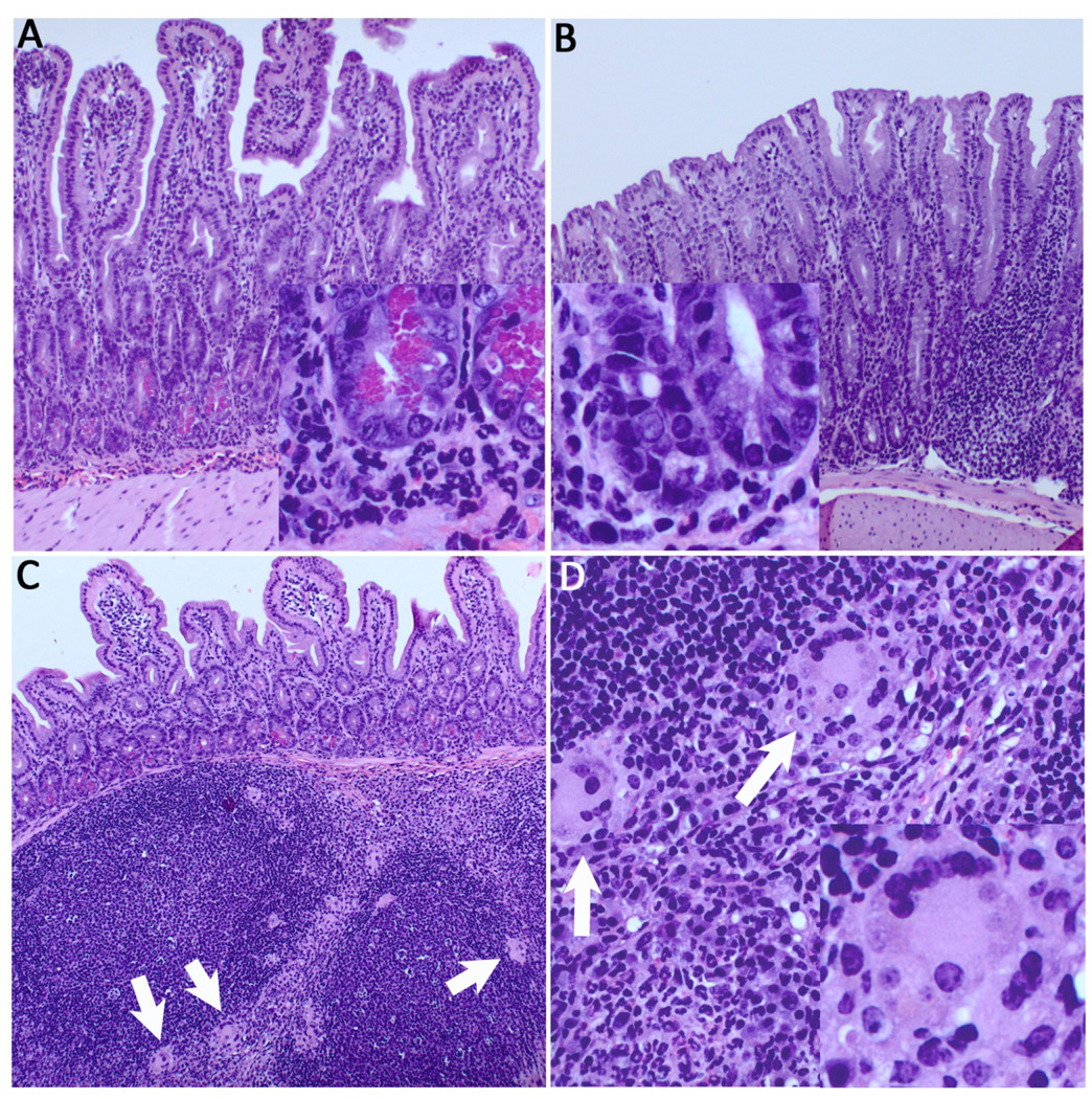
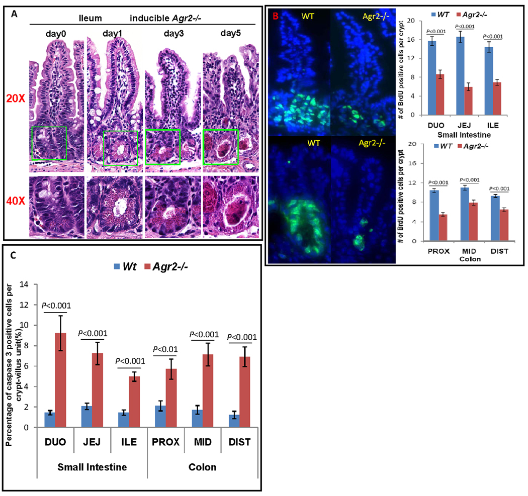
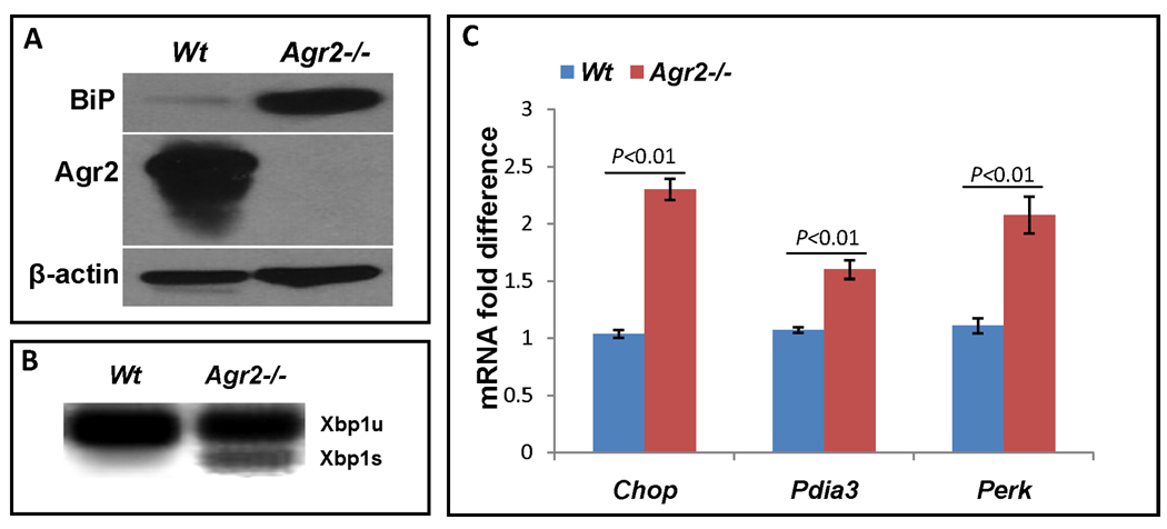
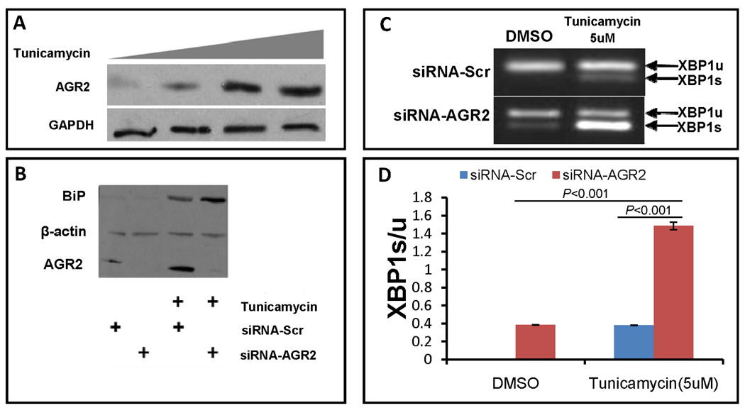
Anterior Gradient 2 (AGR2) is a protein disulfide isomerase that plays important roles in diverse processes in multiple cell lineages as a developmental regulator, survival factor and susceptibility gene for inflammatory bowel disease. Here, we show using germline and inducible Agr2-/- mice that Agr2 plays important roles in intestinal homeostasis. Agr2-/- intestine has decreased goblet cell Mucin 2, dramatic expansion of the Paneth cell compartment, abnormal Paneth cell localization, elevated endoplasmic reticulum (ER) stress, severe terminal ileitis and colitis. Cell culture experiments show that Agr2 expression is induced by ER stress, and that siRNA knockdown of Agr2 increases ER stress response. These studies implicate Agr2 in intestinal homeostasis and ER stress and suggest a role in the etiology of inflammatory bowel disease.


Conflict of interest statement

There are no conflicts of interest to disclose for any author.

Figures

Figure 1
Figure 1
Generation of Agr2−/− mice. (A) Gene-targeting strategy. The four alleles used in the gene targeting of Agr2 are shown. Black boxes with numbers denote Agr2 exons. PGK-Neo, neomycin resistance cassette, is shown as a yellow box. Blue arrows show LoxP sites. Red arrows P1–P3 depict PCR primer locations. Deletion of exon 2, 3 and 4 produces a frame shift in exon 5. (B) Breeding strategies for germline and inducible Agr2 knockout mice. Flox, flanking LoxP site. PRM-Cre, protamine promoter driven Cre recombinase. Tam-Cre, tamoxifen inducible Cre recombinase. (C) PCR products from different Agr2 genotypes. Mouse tail DNA was PCR amplified using three primers whose locations are shown in Panel A. The wild-type allele produced a band of 280bp, the floxed allele a band of 335bp and the null allele a band of 519bp. (D) DNA sequencing of cDNA showing the junction of exons 1 and 5 in the Agr2−/− intestine epithelium. (E) Western analysis shows a 19.9-kDa band specific for Agr2 absent in Agr2−/− mice. β-actin is used as a protein loading control. (F) Immunohistochemistry for Agr2 in small intestine. Agr2 protein is stained dark brown. (a) wild type; (b) germline Agr2−/−; (c) inducible Agr2−/−.
Figure 2
Figure 2
Paneth cell abnormalities in germline and inducible Agr2 −/− mice. (A) H+E stained sections of small intestine from wild type, germline and inducible Agr2−/− mice (day5). Paneth cells containing brightly eosinophilic granules are dramatically expanded in both germline and inducible Agr2−/− mice. Intralumenal Paneth cell granule casts are easily visible in the inducible Agr2−/− small intestine. Representative crypt bases are boxed in green and shown at 20× and 40× magnifications. A marked increase in lamina propria mononuclear infiltrates can also be seen in the inducible Agr2−/− small intestine. (B) Abnormally positioned Paneth cells in the upper crypt and villus epithelium of germline Agr2−/− mice. Multiple Paneth cells are ectopically located in the upper crypt (green arrows). A Paneth cell located on the villus is indicated by a green circle. A 40× closeup image reveals characteristic Paneth cell granules that are cryptdin 5-positive by immunohistochemistry. (C) Increased Paneth cell marker Sox9 mRNA in inducible Agr2−/− small intestine (day6). TaqMan analysis of Sox9 mRNA in small intestine of inducible Agr2−/− and wild type mice. Wild-type Sox9 mRNA expression is normalized to 1.0. Mean bars of standard error are shown. The indicated P-values were determined by Student t-test. (D) Increased relative amount of cryptdins in the total protein extracted from distal small intestine of inducible Agr2−/− mice (day4). Bradford assays were performed to quantitate the amount of total protein in individual P-60 fractions from separations of inducible Agr2−/− and Wt P-60 fractions. Cryptdin P-60 fractions were those that appeared to contain mainly cryptdins by AU-PAGE. The total amount of protein was determined by adding the protein in non-cryptdin containing fractions to the protein in cryptdin containing fractions. Relative differences in the protein quantities between inducible Agr2−/− and Wt mice are listed.
Figure 3
Figure 3
Severe ileitis and colitis in germline Agr2−/− mice. (A) H&E staining reveals severe acute ileitis, with a dense, primarily neutrophilic infiltrate in the lamina propria and submucosa. Inset reveals neutrophils surrounding hypertrophied Paneth cells. (B) Acute colitis in the proximal colon, with loss of goblet cells, crypt elongation, and infiltrating neutrophils (inset). (C) Florid lymphoid hyperplasia of ileal Peyer’s patches, with multinucleated giant cells (white arrows). (D) Multinucleated giant cells suggestive of granulomatous inflammation in interfollicular zones are seen at higher power (inset, lower right).
Figure 4
Figure 4
Sequential abnormalities in morphology in the intestine of inducible Agr2−/− mice. (A) H&E staining shows sequential abnormalities of Paneth cells, goblet cells and enterocytes in inducible Agr2−/− small intestine. Representative crypt bases are boxed in green and shown at 20× and 40× magnifications. Agr2 Tam-Cre flox/flox mice were injected with tamoxifen and mice analyzed over 1, 3 and 5 days. Day 0 (same day as tamoxifen injection), no visible abnormality. Day 1, crypt Paneth cell compartment expansion is seen, while goblet cells and enterocytes are unremarkable. Day 3, decrease in morphologically normal goblet cells, enterocyte dysplasia, further Paneth cell expansion and lamina propria inflammation is seen. Day 5, complete loss of morphologically normal goblet cells, severe loss of enterocytes, appearance of Paneth cell casts and increased lamina propria inflammation. (B) BrdU staining shows decreased proliferation in inducible Agr2−/− small intestine and colon (day6). Mice were injected with BrdU and sacrificed 2 hours after injection. Small intestine (top) and colon (bottom) were visualized with fluorescence microscopy. (Left) Immunoflorescence of BrdU (green) labeled crypts from Wt and induced Agr2−/− mice. (Right) Number of BrdU positive cells per crypt in small intestine (top) and colon (bottom) of Wt mice and inducible Agr2−/− mice. (C) Caspase 3 assay shows increased apoptosis in inducible Agr2−/− small intestine and colon (day6). Mean bars of standard error are shown. The indicated P-values were determined by Student t-test.
Figure 5
Figure 5
Agr2 knockout increases ER stress in the intestine. (A) Germline Agr2−/− small intestine has increased levels of the ER stress response marker BiP. Agr2 protein and actin loading control are shown below. (B) Inducible Agr2−/− small intestine (day6) has increased ER stress response marker Xbp1s. Agr2 deletion was induced with tamoxifen and small intestine was assayed for Xbp1s mRNA on day6. Xbp1u, unspliced, full length form of Xbp1. Xbp1s, spliced form that is induced by ER stress. (C) Increased ER stress marker mRNA expression in induced Agr2−/− mice (day 6). qPCR of ER stress biomarker expression levels of Chop, Pdia3 and Perk. mRNA expressions of wild type are normalized to 1.0. Mean bars of standard error are shown. The indicated P-values were determined by Student t-test.
Figure 6
Figure 6
AGR2 siRNA knock down increases ER stress. (A) AGR2 is an ER stress response gene. Western blot analysis of SU86.86 cells in culture treated with increasing amounts of the ER stress inducer tunicamycin (0, 2.5, 5 and 7.5uM) and probed with anti-AGR2 (Top) and anti-GAPDH as a loading control (bottom). (B) AGR2 siRNA knockdown increases the ER stress biomarker BiP in tunicamycin treated cells. Western blot analysis of SU86.86 cells treated with 2.5uM tunicamycin and transfected with anti-AGR2 siRNA. BiP, ER stress biomarker BiP (Top). Beta-actin is included as a loading control (Middle). AGR2, Anterior gradient 2 (bottom). BiP, beta-actin and AGR2 all have different electrophoretic mobilities and were probed on the same transferred gel. SiRNA-Scr, negative control scrambled siRNA. SiRNA-AGR2, AGR2 specific siRNA (Open biosystems). (C,D) RT-PCR of XBP1 unspliced and spliced isoforms in SU 86.86 cells. AGR2 siRNA knockdown induces XBP1s levels in SU 86.86 cells. Cells were treated with tunicamycin at 5uM, siRNA -AGR2 increases XBP1s levels to much higher levels than siRNA-Scr, P<0.001. Standard error of the mean is shown. The indicated P-values were determined by Student t-test.

References

    1. Aberger F, Weidinger G, Grunz H, Richter K. Anterior specification of embryonic ectoderm: the role of the Xenopus cement gland-specific gene XAG-2. Mech Dev. 1998;72:115–130. - PubMed
    1. Abzhanov A, Tzahor E, Lassar AB, Tabin CJ. Dissimilar regulation of cell differentiation in mesencephalic (cranial) and sacral (trunk) neural crest cells in vitro. Development (Cambridge, England) 2003;130:4567–4579. - PubMed
    1. Andreu P, Peignon G, Slomianny C, Taketo MM, Colnot S, Robine S, Lamarque D, Laurent-Puig P, Perret C, Romagnolo B. A genetic study of the role of the Wnt/beta-catenin signalling in Paneth cell differentiation. Developmental biology. 2008;324:288–296. - PubMed
    1. Badea TC, Wang Y, Nathans J. A noninvasive genetic/pharmacologic strategy for visualizing cell morphology and clonal relationships in the mouse. J Neurosci. 2003;23:2314–2322. - PMC - PubMed
    1. Bamias G, Okazawa A, Rivera-Nieves J, Arseneau KO, De La Rue SA, Pizarro TT, Cominelli F. Commensal bacteria exacerbate intestinal inflammation but are not essential for the development of murine ileitis. J Immunol. 2007;178:1809–1818. - PubMed

Publication types

LinkOut - more resources