Characterization of corneal keratocyte morphology and mechanical activity within 3-D collagen matrices
- PMID: 20025872
- PMCID: PMC2822042
- DOI: 10.1016/j.exer.2009.11.016
Characterization of corneal keratocyte morphology and mechanical activity within 3-D collagen matrices
Abstract
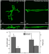
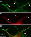
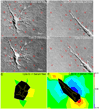
The purpose of this study was to assess quantitatively the differences in morphology, cytoskeletal organization and mechanical behavior between quiescent corneal keratocytes and activated fibroblasts in a 3-D culture model. Primary cultures of rabbit corneal keratocytes and fibroblasts were plated inside type I collagen matrices in serum-free media or 10% FBS, and allowed to spread for 1-5 days. Following F-actin labeling using phalloidin, and immunolabeling of tubulin, alpha-smooth muscle actin or connexin 43, fluorescent and reflected light (for collagen fibrils) 3-D optical section images were acquired using laser confocal microscopy. In other experiments, dynamic imaging was performed using differential interference contrast microscopy, and finite element modeling was used to map ECM deformations. Corneal keratocytes developed a stellate morphology with numerous cell processes that ran a tortuous path between and along collagen fibrils without any apparent impact on their alignment. Fibroblasts on the other hand, had a more bipolar morphology with pseudopodial processes (P </= 0.001). Time-lapse imaging of keratocytes revealed occasional extension and retraction of dendritic processes with only transient displacements of collagen fibrils, whereas fibroblasts exerted stronger myosin II-dependent contractile forces (P < 0.01), causing increased compaction and alignment of collagen at the ends of the pseudopodia (P < 0.001). At high cell density, both keratocytes and fibroblasts appeared to form a 3-D network connected via gap junctions. Overall, this experimental model provides a unique platform for quantitative investigation of the morphological, cytoskeletal and contractile behavior of corneal keratocytes (i.e. their mechanical phenotype) in a 3-D microenvironment.
Copyright 2009 Elsevier Ltd. All rights reserved.
Figures


Similar articles
-
Effect of HDAC Inhibitors on Corneal Keratocyte Mechanical Phenotypes in 3-D Collagen Matrices.Mol Vis. 2015 Apr 29;21:502-14. eCollection 2015. Mol Vis. 2015. PMID: 25999677 Free PMC article.
-
Mechanical interactions and crosstalk between corneal keratocytes and the extracellular matrix.Exp Eye Res. 2015 Apr;133:49-57. doi: 10.1016/j.exer.2014.09.003. Exp Eye Res. 2015. PMID: 25819454 Free PMC article. Review.
-
Growth factor regulation of corneal keratocyte differentiation and migration in compressed collagen matrices.Invest Ophthalmol Vis Sci. 2010 Feb;51(2):864-75. doi: 10.1167/iovs.09-4200. Epub 2009 Oct 8. Invest Ophthalmol Vis Sci. 2010. PMID: 19815729 Free PMC article.
-
MMP regulation of corneal keratocyte motility and mechanics in 3-D collagen matrices.Exp Eye Res. 2014 Apr;121:147-60. doi: 10.1016/j.exer.2014.02.002. Epub 2014 Feb 14. Exp Eye Res. 2014. PMID: 24530619 Free PMC article.
-
Regulation of corneal stroma extracellular matrix assembly.Exp Eye Res. 2015 Apr;133:69-80. doi: 10.1016/j.exer.2014.08.001. Exp Eye Res. 2015. PMID: 25819456 Free PMC article. Review.
Cited by
-
Generation of Functional Immortalized Human Corneal Stromal Stem Cells.Int J Mol Sci. 2022 Nov 2;23(21):13399. doi: 10.3390/ijms232113399. Int J Mol Sci. 2022. PMID: 36362184 Free PMC article.
-
Effect of HDAC Inhibitors on Corneal Keratocyte Mechanical Phenotypes in 3-D Collagen Matrices.Mol Vis. 2015 Apr 29;21:502-14. eCollection 2015. Mol Vis. 2015. PMID: 25999677 Free PMC article.
-
Methylcellulose/agarose hydrogel loaded with short electrospun PLLA/laminin fibers as an injectable scaffold for tissue engineering/3D cell culture model for tumour therapies.RSC Adv. 2023 Apr 17;13(18):11889-11902. doi: 10.1039/d3ra00851g. eCollection 2023 Apr 17. RSC Adv. 2023. PMID: 37077262 Free PMC article.
-
Sphere formation from corneal keratocytes and phenotype specific markers.Exp Eye Res. 2011 Dec;93(6):898-905. doi: 10.1016/j.exer.2011.10.004. Epub 2011 Oct 21. Exp Eye Res. 2011. PMID: 22032988 Free PMC article.
-
Mechanical interactions and crosstalk between corneal keratocytes and the extracellular matrix.Exp Eye Res. 2015 Apr;133:49-57. doi: 10.1016/j.exer.2014.09.003. Exp Eye Res. 2015. PMID: 25819454 Free PMC article. Review.
References
-
- Abbott A. Biology's new dimension. Nature. 2003;424:870–872. - PubMed
-
- Allingham JS, Smith R, Rayment I. The structural basis of blebbistatin inhibition and specificity for myosin II. Nat Struct Mol Biol. 2005;12:378–379. - PubMed
-
- Beales MP, Funderburgh JL, Jester JV, Hassell JR. Proteoglycan synthesis by bovine keratocytes and corneal fibroblasts: maintenance of the keratocyte phenotype in culture. Invest Ophthalmol Vis Sci. 1999:1658–1663. - PubMed
-
- Burridge K, Chrzanowska-Wodnicka C. Focal adhesions, contractility, and signaling. Ann. Rev. Cell Dev. Biol. 1996;12:463–519. - PubMed
Publication types
MeSH terms
Substances
Grants and funding
LinkOut - more resources
Full Text Sources
Miscellaneous

