

Noradrenergic innervation of the rat spinal cord caudal to a complete spinal cord transection: effects of olfactory ensheathing glia
- PMID: 20025875
- PMCID: PMC2838922
- DOI: 10.1016/j.expneurol.2009.12.008
Noradrenergic innervation of the rat spinal cord caudal to a complete spinal cord transection: effects of olfactory ensheathing glia
Abstract
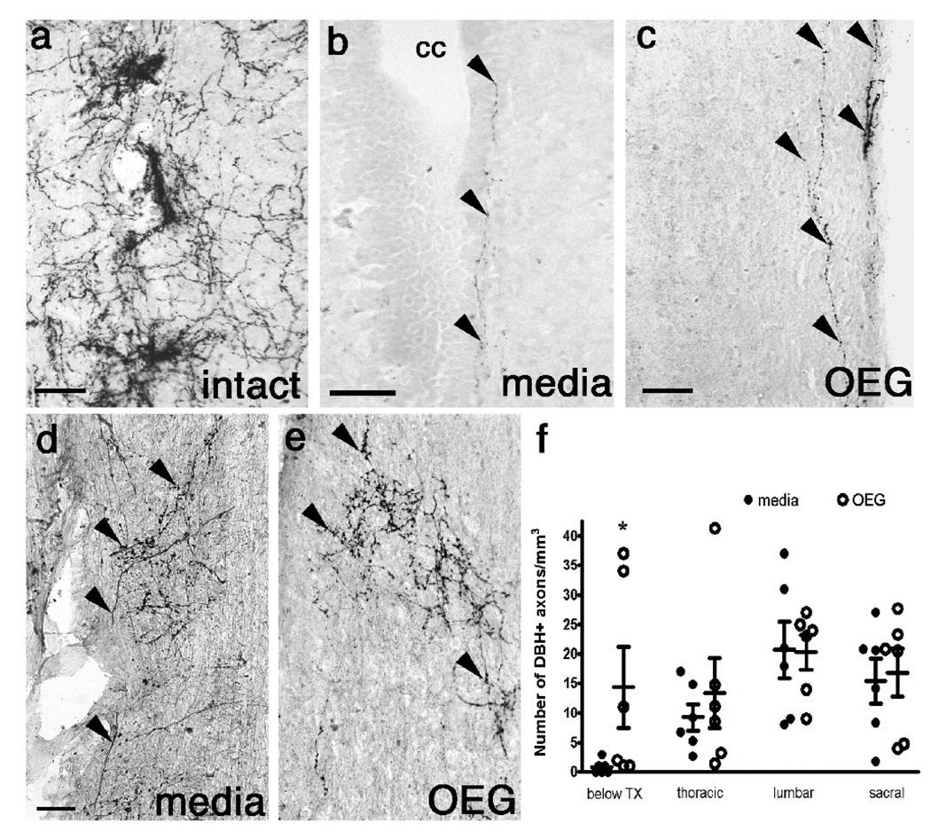
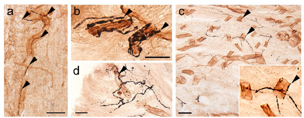
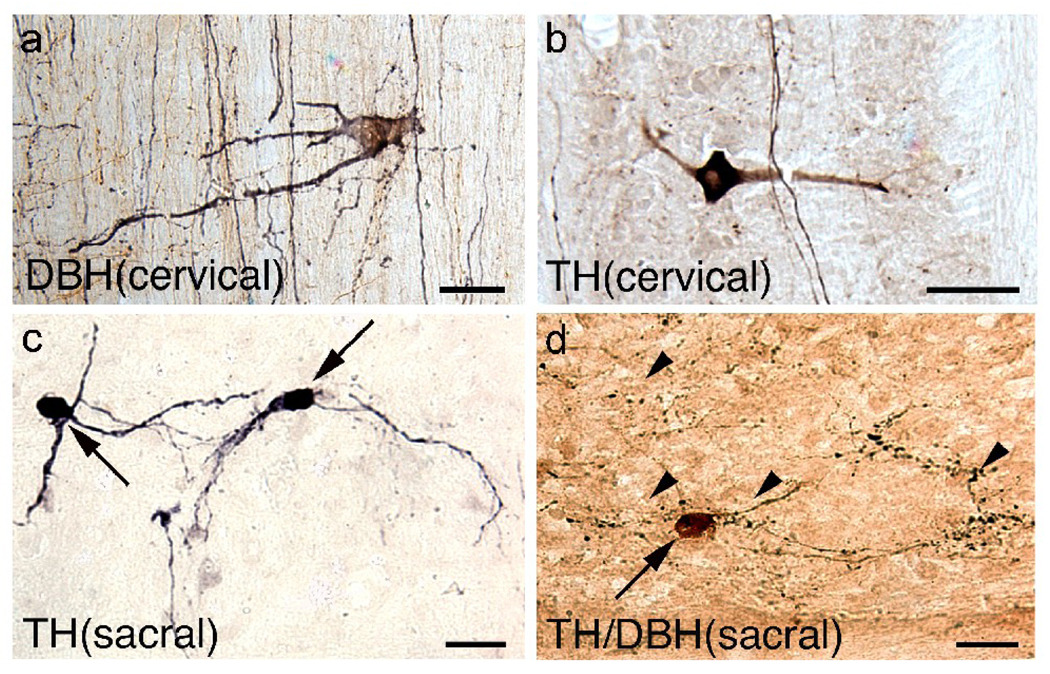
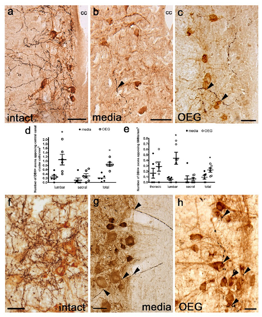
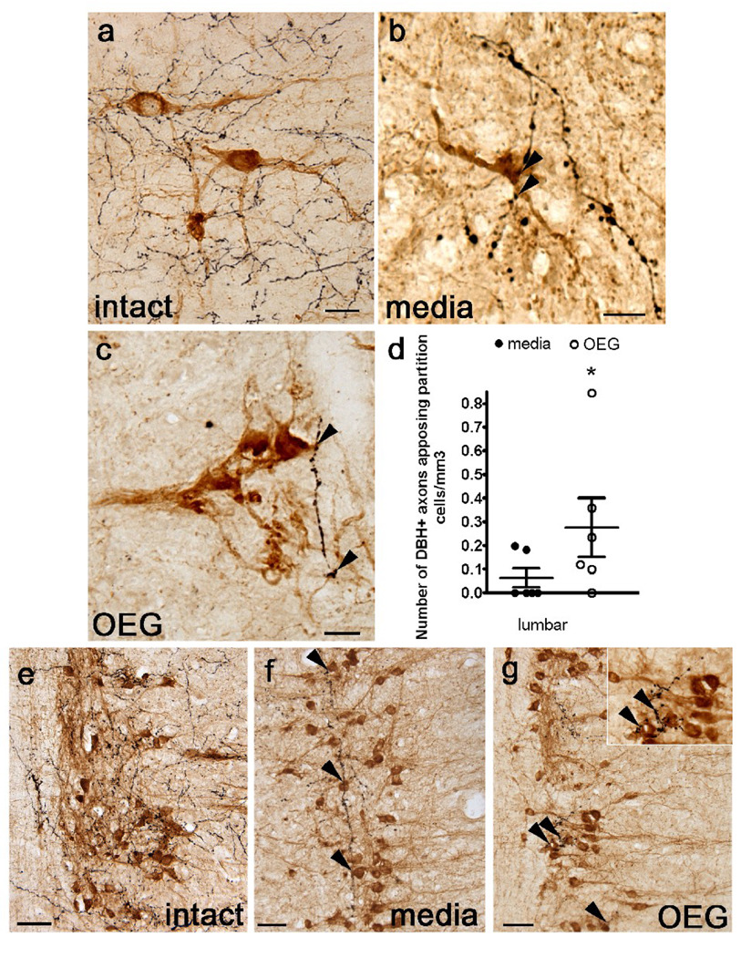
Transplantation of olfactory bulb-derived olfactory ensheathing glia (OEG) combined with step training improves hindlimb locomotion in adult rats with a complete spinal cord transection. Spinal cord injury studies use the presence of noradrenergic (NA) axons caudal to the injury site as evidence of axonal regeneration and we previously found more NA axons just caudal to the transection in OEG- than media-injected spinal rats. We therefore hypothesized that OEG transplantation promotes descending coeruleospinal regeneration that contributes to the recovery of hindlimb locomotion. Now we report that NA axons are present throughout the caudal stump of both media- and OEG-injected spinal rats and they enter the spinal cord from the periphery via dorsal and ventral roots and along large penetrating blood vessels. These results indicate that the presence of NA fibers in the caudal spinal cord is not a reliable indicator of coeruleospinal regeneration. We then asked if NA axons appose cholinergic neurons associated with motor functions, i.e., central canal cluster and partition cells (active during fictive locomotion) and somatic motor neurons (SMNs). We found more NA varicosities adjacent to central canal cluster cells, partition cells, and SMNs in the lumbar enlargement of OEG- than media-injected rats. As non-synaptic release of NA is common in the spinal cord, more associations between NA varicosities and motor-associated cholinergic neurons in the lumbar spinal cord may contribute to the improved treadmill stepping observed in OEG-injected spinal rats. This effect could be mediated through direct association with SMNs and/or indirectly via cholinergic interneurons.
Copyright 2009 Elsevier Inc. All rights reserved.
Figures

Similar articles
-
Further evidence of olfactory ensheathing glia facilitating axonal regeneration after a complete spinal cord transection.Exp Neurol. 2011 May;229(1):109-19. doi: 10.1016/j.expneurol.2011.01.007. Epub 2011 Jan 25. Exp Neurol. 2011. PMID: 21272578 Free PMC article.
-
Serotonergic innervation of the caudal spinal stump in rats after complete spinal transection: effect of olfactory ensheathing glia.J Comp Neurol. 2009 Aug 20;515(6):664-76. doi: 10.1002/cne.22080. J Comp Neurol. 2009. PMID: 19496067 Free PMC article.
-
OEG implantation and step training enhance hindlimb-stepping ability in adult spinal transected rats.Brain. 2008 Jan;131(Pt 1):264-76. doi: 10.1093/brain/awm267. Epub 2007 Dec 3. Brain. 2008. PMID: 18056162 Free PMC article.
-
Olfactory ensheathing glia: their contribution to primary olfactory nervous system regeneration and their regenerative potential following transplantation into the injured spinal cord.Brain Res Rev. 2007 Nov;56(1):236-58. doi: 10.1016/j.brainresrev.2007.07.013. Epub 2007 Aug 14. Brain Res Rev. 2007. PMID: 17884174 Review.
-
Transplants and neurotrophic factors increase regeneration and recovery of function after spinal cord injury.Prog Brain Res. 2002;137:257-73. doi: 10.1016/s0079-6123(02)37020-1. Prog Brain Res. 2002. PMID: 12440372 Review.
Cited by
-
Reliable cell purification and determination of cell purity: crucial aspects of olfactory ensheathing cell transplantation for spinal cord repair.Neural Regen Res. 2020 Nov;15(11):2016-2026. doi: 10.4103/1673-5374.282218. Neural Regen Res. 2020. PMID: 32394949 Free PMC article. Review.
-
Cell transplantation to repair the injured spinal cord.Int Rev Neurobiol. 2022;166:79-158. doi: 10.1016/bs.irn.2022.09.008. Epub 2022 Nov 9. Int Rev Neurobiol. 2022. PMID: 36424097 Free PMC article. No abstract available.
-
Survival and Integration of Transplanted Olfactory Ensheathing Cells are Crucial for Spinal Cord Injury Repair: Insights from the Last 10 Years of Animal Model Studies.Cell Transplant. 2019 Dec;28(1_suppl):132S-159S. doi: 10.1177/0963689719883823. Epub 2019 Nov 15. Cell Transplant. 2019. PMID: 31726863 Free PMC article. Review.
-
Further evidence of olfactory ensheathing glia facilitating axonal regeneration after a complete spinal cord transection.Exp Neurol. 2011 May;229(1):109-19. doi: 10.1016/j.expneurol.2011.01.007. Epub 2011 Jan 25. Exp Neurol. 2011. PMID: 21272578 Free PMC article.
-
Promoting Axon Regeneration in Adult CNS by Targeting Liver Kinase B1.Mol Ther. 2019 Jan 2;27(1):102-117. doi: 10.1016/j.ymthe.2018.10.019. Epub 2018 Nov 1. Mol Ther. 2019. PMID: 30509565 Free PMC article.
References
-
- Amenta F, Bronzetti E, Cavallotti C, Felici L. Quantitative image analysis of the density and pattern of adrenergic innervation of blood vessels of rat spinal cord. J Auton Nerv Syst. 1987;18:261–264. - PubMed
-
- Andén N-E, Engel J, Rubenson A. Central decarboxylation and uptake of L-DOPA. Naunyn Schmiedebergs Arch Pharmacol. 1972;273:11–26. - PubMed
-
- Aramant RB, Giron LT, Jr, Ziegler MG. Postnatal development of dopamine-beta-hydroxylase-immunoreactive fibers of the spinal cord of the rat. Brain Res. 1986;390:161–171. - PubMed
-
- Barbeau H, Chau C, Rossignol S. Noradrenergic agonists and locomotor training affect locomotor recovery after cord transection in adult cats. Brain Res Bull. 1993;30:387–393. - PubMed
-
- Barbeau H, Norman KE. The effect of noradrenergic drugs on the recovery of walking after spinal cord injury. Spinal Cord. 2003;41:137–143. - PubMed
Publication types
MeSH terms
Substances
Grants and funding
LinkOut - more resources
Full Text Sources
Medical
Research Materials
