

Evaluating functional localizers: the case of the FFA
- PMID: 20025980
- PMCID: PMC2825676
- DOI: 10.1016/j.neuroimage.2009.12.024
Evaluating functional localizers: the case of the FFA
Abstract
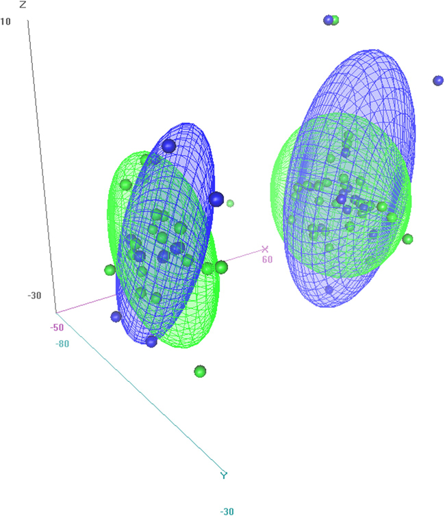
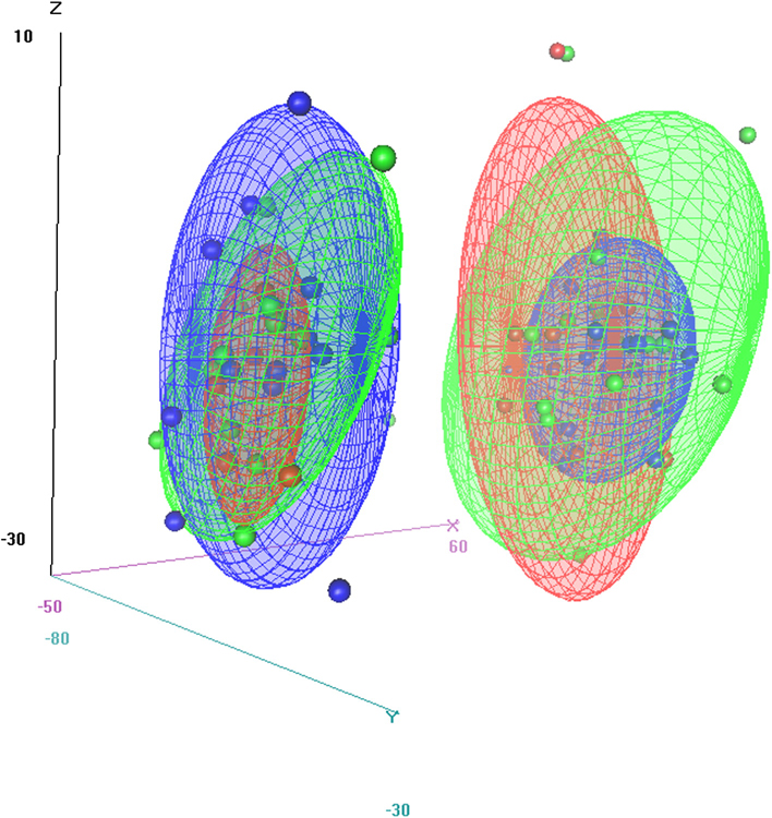
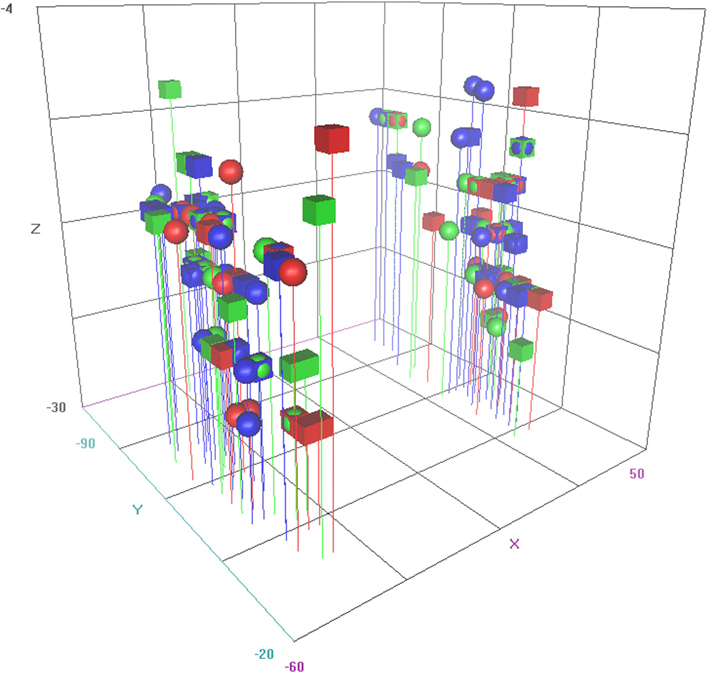
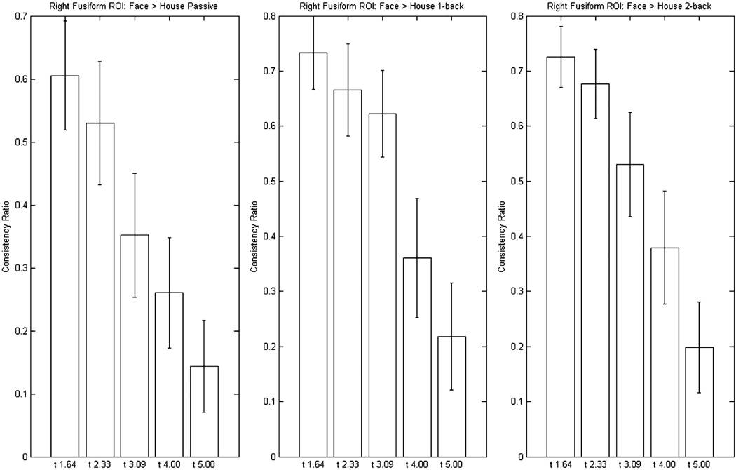
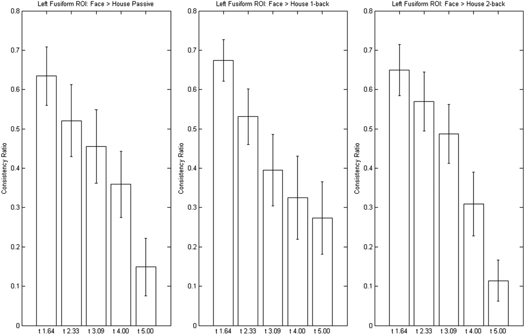
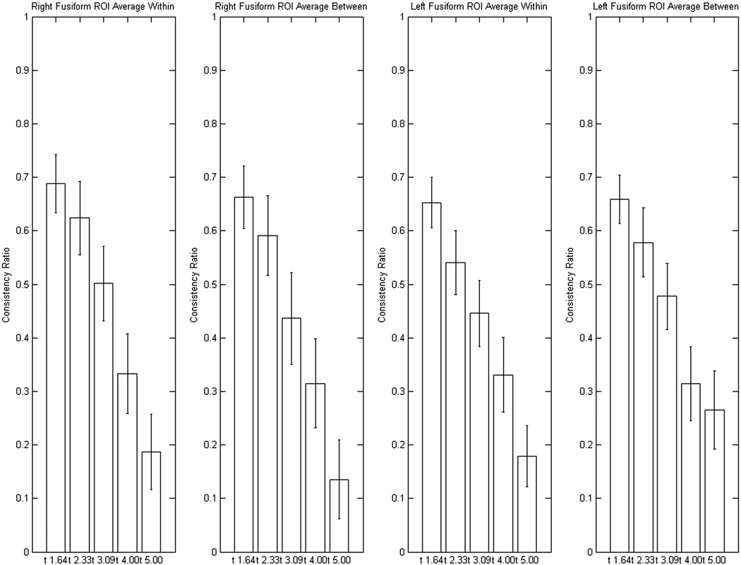
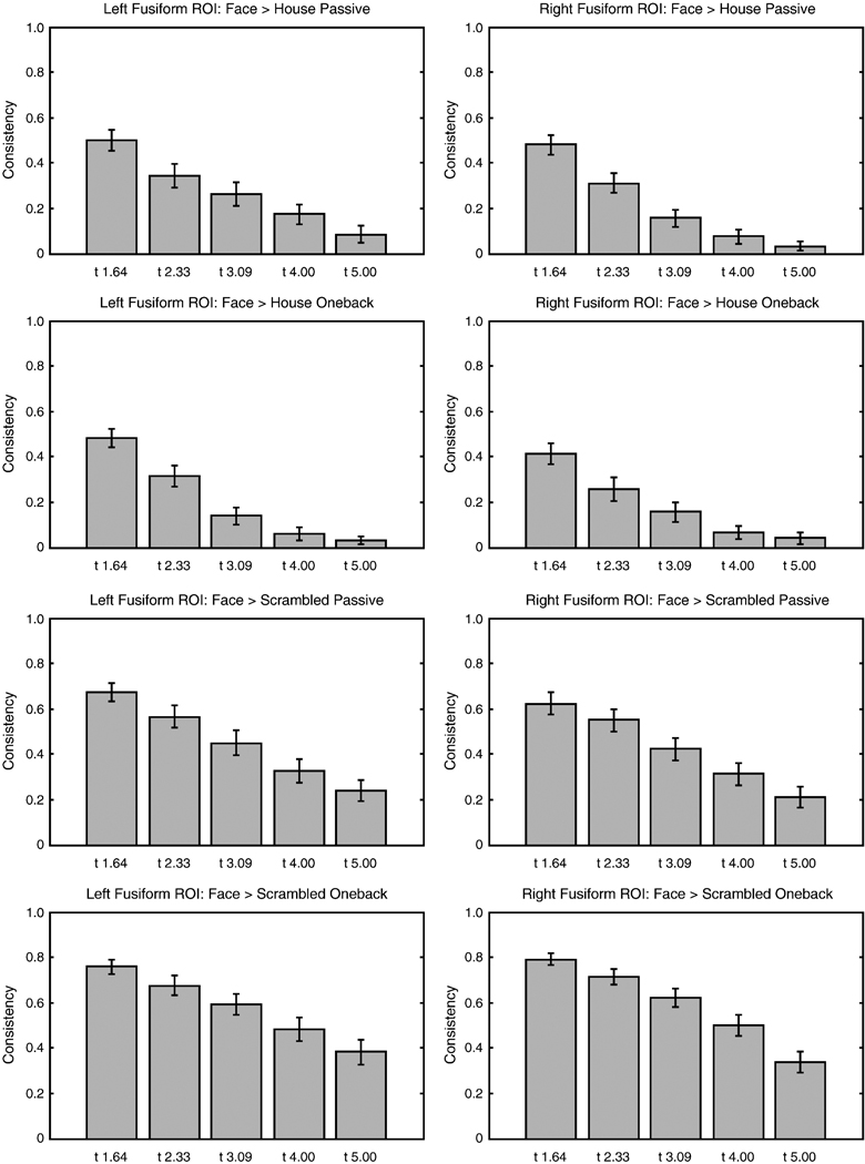
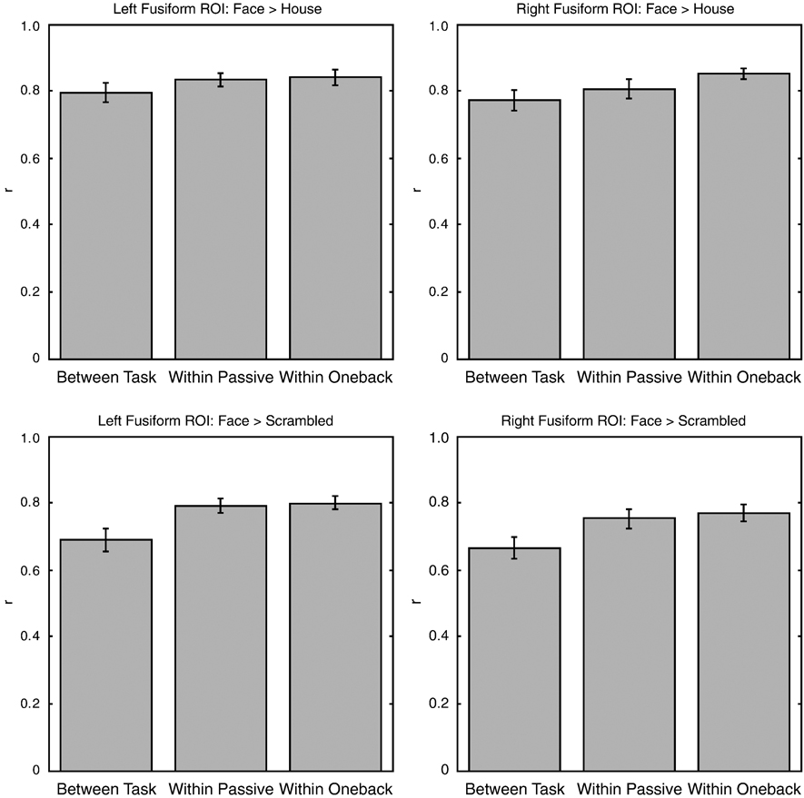
Functional localizers are routinely used in neuroimaging studies to test hypotheses about the function of specific brain areas. The specific tasks and stimuli used to localize particular regions vary widely from study to study even when the same cortical region is targeted. Thus, it is important to ask whether task and stimulus changes lead to differences in localization or whether localization procedures are largely immune to differences in tasks and contrasting stimuli. We present two experiments and a literature review that explore whether face localizer tasks yield differential localization in the fusiform gyrus as a function of task and contrasting stimuli. We tested standard localization tasks-passive viewing, 1-back, and 2-back memory tests--and did not find differences in localization based on task. We did, however, find differences in the extent, strength and patterns/reliabilities of the activation in the fusiform gyrus based on comparison stimuli (faces vs. houses compared to faces vs. scrambled stimuli).
Copyright (c) 2009 Elsevier Inc. All rights reserved.
Figures

Similar articles
-
Defining face perception areas in the human brain: a large-scale factorial fMRI face localizer analysis.Brain Cogn. 2012 Jul;79(2):138-57. doi: 10.1016/j.bandc.2012.01.001. Epub 2012 Feb 12. Brain Cogn. 2012. PMID: 22330606
-
Neuroanatomic correlates of the feature-salience hierarchy in face processing: an fMRI -adaptation study.Neuropsychologia. 2014 Jan;53:274-83. doi: 10.1016/j.neuropsychologia.2013.10.016. Epub 2013 Nov 1. Neuropsychologia. 2014. PMID: 24189157
-
Face categorization in visual scenes may start in a higher order area of the right fusiform gyrus: evidence from dynamic visual stimulation in neuroimaging.J Neurophysiol. 2011 Nov;106(5):2720-36. doi: 10.1152/jn.00672.2010. Epub 2011 Jul 6. J Neurophysiol. 2011. PMID: 21734108
-
The engagement of mid-ventrolateral prefrontal cortex and posterior brain regions in intentional cognitive activity.Hum Brain Mapp. 2008 Jan;29(1):107-19. doi: 10.1002/hbm.20378. Hum Brain Mapp. 2008. PMID: 17370344 Free PMC article.
-
Functional MRI evidence for distinctive binding and consolidation pathways for face-name associations: analysis of activation maps and BOLD response amplitudes.Top Magn Reson Imaging. 2009 Oct;20(5):271-8. doi: 10.1097/RMR.0b013e3181e8f1f9. Top Magn Reson Imaging. 2009. PMID: 20859188 Free PMC article. Review.
Cited by
-
Process and domain specificity in regions engaged for face processing: an fMRI study of perceptual differentiation.J Cogn Neurosci. 2012 Dec;24(12):2428-44. doi: 10.1162/jocn_a_00273. Epub 2012 Jul 31. J Cogn Neurosci. 2012. PMID: 22849402 Free PMC article.
-
Neural correlates of after-effects caused by adaptation to multiple face displays.Exp Brain Res. 2012 Aug;220(3-4):261-75. doi: 10.1007/s00221-012-3135-3. Epub 2012 Jun 7. Exp Brain Res. 2012. PMID: 22673875
-
Canonical template tracking: Measuring the activation state of specific neural representations.Front Neuroimaging. 2023 Jan 9;1:974927. doi: 10.3389/fnimg.2022.974927. eCollection 2022. Front Neuroimaging. 2023. PMID: 37555182 Free PMC article. Review.
-
New method for fMRI investigations of language: defining ROIs functionally in individual subjects.J Neurophysiol. 2010 Aug;104(2):1177-94. doi: 10.1152/jn.00032.2010. Epub 2010 Apr 21. J Neurophysiol. 2010. PMID: 20410363 Free PMC article.
-
Correlation and heritability in neuroimaging datasets: a spatial decomposition approach with application to an fMRI study of twins.Neuroimage. 2012 Jan 16;59(2):1132-42. doi: 10.1016/j.neuroimage.2011.06.066. Epub 2011 Jul 7. Neuroimage. 2012. PMID: 21763433 Free PMC article.
References
-
- Aguirre GK, Singh R, D'Esposito M. Stimulus inversion and the responses of face and object-sensitive cortical areas. Neuroreport. 1999;10(1):189–194. - PubMed
-
- Andrews TJ, Ewbank MP. Distinct representations for facial identity and changeable aspects of faces in the human temporal lobe. NeuroImage. 2004;23(3):905–913. - PubMed
-
- Andrews TJ, Schluppeck D. Neural responses to money images reveal a modular representation of faces in human visual cortex. NeuroImage. 2004;21(1):91–98. - PubMed
-
- Avidan G, Levy I, Hendler T, Zohary E, Malach R. Spatial vs. object specific attention in high-order visual areas. NeuroImage. 2003;19(2):308–318. - PubMed
-
- Caldara R, Seghier ML, Rossion B, Lazeyras F, Michel C, Hauert CA. The fusiform face area is tuned for curvilinear patterns with more high-contrasted elements in the upper part. NeuroImage. 2006;31(1):313–319. - PubMed
Publication types
MeSH terms
Grants and funding
LinkOut - more resources
Full Text Sources
