. 2010 Mar;222(1):42-50.
doi: 10.1016/j.expneurol.2009.12.006. Epub 2009 Dec 21.

Intracranial electrode implantation produces regional neuroinflammation and memory deficits in rats

Affiliations

Intracranial electrode implantation produces regional neuroinflammation and memory deficits in rats

Yafit Kuttner Hirshler et al. Exp Neurol. 2010 Mar.

Abstract

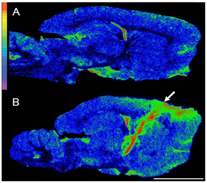
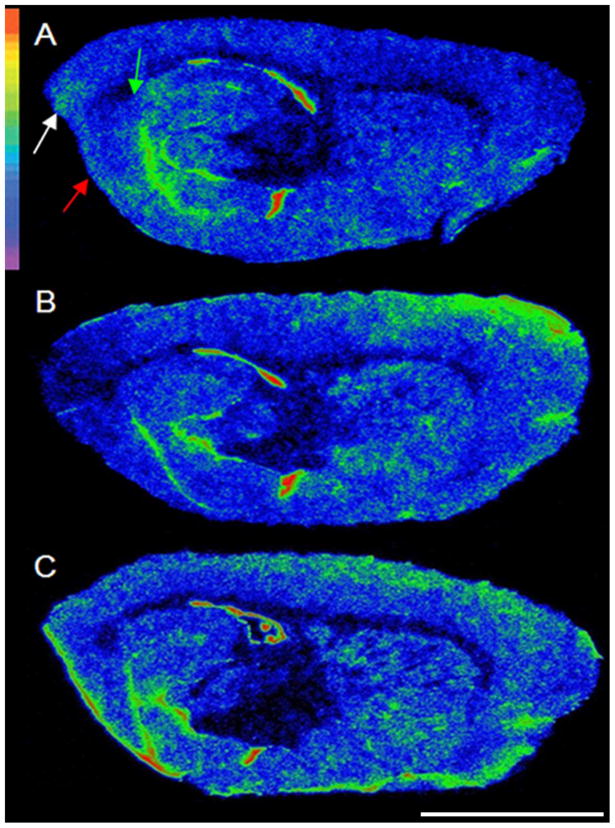
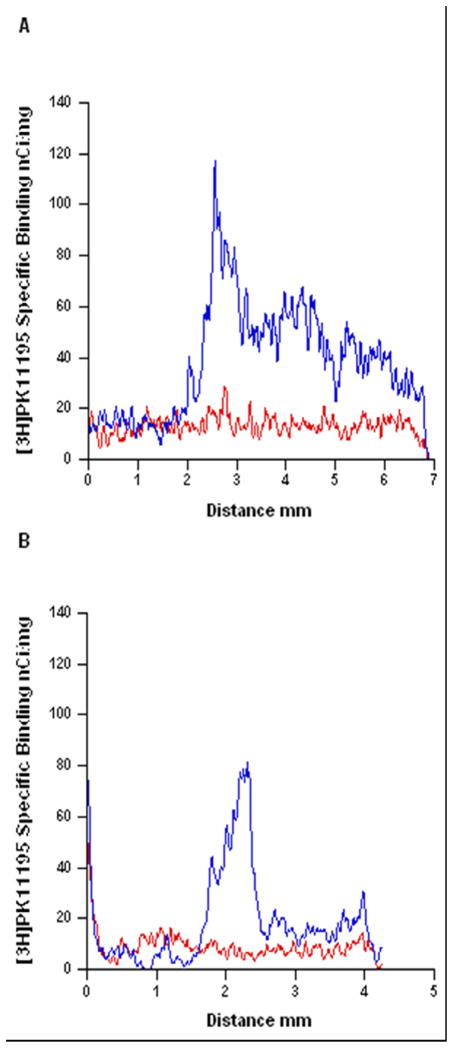
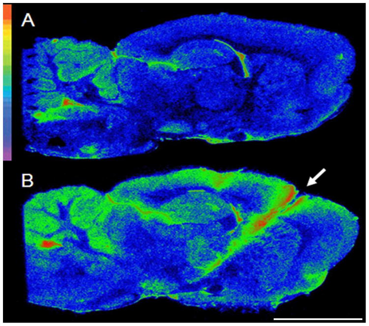
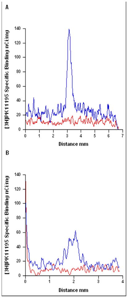
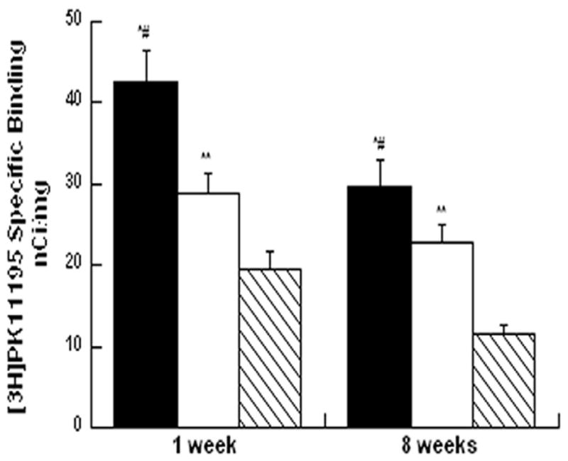
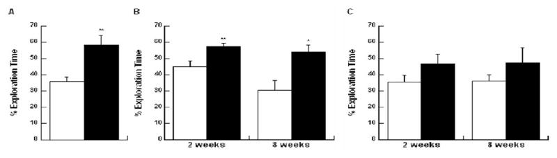
Deep brain stimulation (DBS) is an established treatment for advanced Parkinson's disease (PD). The procedure entails intracranial implantation of an electrode in a specific brain structure followed by chronic stimulation. Although the beneficial effects of DBS on motor symptoms in PD are well known, it is often accompanied by cognitive impairments, the origin of which is not fully understood. To explore the possible contribution of the surgical procedure itself, we studied the effect of electrode implantation in the subthalamic nucleus (STN) on regional neuroinflammation and memory function in rats implanted bilaterally with stainless steel electrodes. Age-matched sham and intact rats were used as controls. Brains were removed 1 or 8 weeks post-implantation and processed for in vitro autoradiography with [(3)H]PK11195, an established marker of microglial activation. Memory function was assessed by the novel object recognition test (ORT) before surgery and 2 and 8 weeks after surgery. Electrode implantation produced region-dependent changes in ligand binding density in the implanted brains at 1 as well as 8 weeks post-implantation. Cortical regions showed more intense and widespread neuroinflammation than striatal or thalamic structures. Furthermore, implanted animals showed deficits in ORT performance 2 and 8 weeks post-implantation. Thus, electrode implantation resulted in a widespread and persistent neuroinflammation and sustained memory impairment. These results suggest that the insertion and continued presence of electrodes in the brain, even without stimulation, may lead to inflammation-mediated cognitive deficits in susceptible individuals, as observed in patients treated with DBS.


Figures

Figure 1
Figure 1
Regional neuroinflammation at the level of the STN one week following electrode implantation. Autoradiograms of sagittal brain sections from: A= an intact animal, B= an animal implanted with thick electrodes. Autoradiograms were pseudocolored using the rainbow spectrum (top left) with red representing the highest value. The electrode insertion point is marked by an arrow. Scale bar: 0.5cm.
Figure 2
Figure 2
Regional neuroinflammation lateral to the electrode track one week following electrode implantation. Autoradiograms of sagittal brain sections from A= a sham animal B= an animal implanted with thick electrodes C=an animal implanted with thin electrodes. Autoradiograms were pseudocolored using the rainbow spectrum (top left). Note the decrease in PBR binding in the entorhinal cortex (red arrow), perirhinal cortex (white arrow) and subiculum (green arrow) with the thick, but not the thin, electrode. Scale bar: 0.5cm.
Figure 3
Figure 3
The spread of neuroinflammation within the cortex and striatum one week following electrode implantation. Line plot profile of [3H]PK11195 specific binding versus distance in the cortex (A) and striatum (B) of an intact animal (red line) and an implanted animal (blue line). The line in the cortex was drawn from the parietal cortex (above the ventricle), through the frontal cortex and prefrontal cortex and terminated at the edge of the section. The line in the striatum was drawn from the ventricle through the striatum and terminated at the corpus callosum.
Figure 4
Figure 4
Regional neuroinflammation at the level of the STN eight weeks following electrode implantation. Autoradiograms of sagittal brain sections from: A= an intact animal, B= an implanted animal. Autoradiograms were pseudocolored using the rainbow spectrum (top left) with red representing the highest value. Electrode insertion point is marked by a white arrow. Scale bar: 0.5cm.
Figure 5
Figure 5
The spread of neuroinflammation within the cortex and the striatum eight weeks following electrode implantation. Line plot profile of [3H]PK11195 specific binding versus distance in the cortex (A) and striatum (B) of an intact animal (red line) and an implanted animal (blue line).
Figure 6
Figure 6
Neuroinflammation in the peri-lesional areas one week and eight weeks following electrode implantation. Bars depict means±SEM of peri-lesional PBR density in cortex (N=11–16, black bars), striatum (N=10–16, white bars) and thalamus (6–15, hatched bars). ap<0.0001, cortex compared to striatum bp<0.0001 cortex compared to thalamus cp< 0.0002 striatum compared to thalamus, by one way ANOVA (by region).
Figure 7
Figure 7
ORT performance two weeks and eight weeks following electrodes implantation. Bars depict means±SEM of percentage of time exploring the familiar (white bars) or novel (black bars) object. A. Performance in the ORT pre-implantation (N=10) B. Performance of control animals two and eight weeks post sham procedure (N=9 at two weeks, N=8 at eight weeks) C. Performance of implanted animals two and eight weeks post implantation (N=7 at two weeks, N=9 at eight weeks). *p<0.05, **p<0.005 by paired t-test.

Similar articles

Cited by

References

    1. Alegret M, Junque C, Valldeoriola F, Vendrell P, Pilleri M, Rumia J, Tolosa E. Effects of bilateral subthalamic stimulation on cognitive function in Parkinson disease. Arch Neurol. 2001;58:1223–1227. - PubMed
    1. Ardouin C, Pillon B, Peiffer E, Bejjani P, Limousin P, Damier P, Arnulf I, Benabid AL, Agid Y, Pollak P. Bilateral subthalamic or pallidal stimulation for Parkinson’s disease affects neither memory nor executive functions: a consecutive series of 62 patients. Ann Neurol. 1999;46:217–223. - PubMed
    1. Balderas I, Rodriguez-Ortiz CJ, Salgado-Tonda P, Chavez-Hurtado J, McGaugh JL, Bermudez-Rattoni F. The consolidation of object and context recognition memory involve different regions of the temporal lobe. Learn Mem. 2008;15:618–624. - PMC - PubMed
    1. Benabid AL, Chabardes S, Mitrofanis J, Pollak P. Deep brain stimulation of the subthalamic nucleus for the treatment of Parkinson’s disease. Lancet Neurol. 2009;8:67–81. - PubMed
    1. Benabid AL, Chabardes S, Seigneuret E. Deep-brain stimulation in Parkinson’s disease: long-term efficacy and safety - What happened this year? Curr Opin Neurol. 2005;18:623–630. - PubMed

Publication types