. 2010 May;58(6):921-9.
doi: 10.1016/j.neuropharm.2009.12.005. Epub 2009 Dec 22.

Agonist-induced restoration of hippocampal neurogenesis and cognitive improvement in a model of cholinergic denervation

Affiliations

Agonist-induced restoration of hippocampal neurogenesis and cognitive improvement in a model of cholinergic denervation

Jackalina M Van Kampen et al. Neuropharmacology. 2010 May.

Abstract

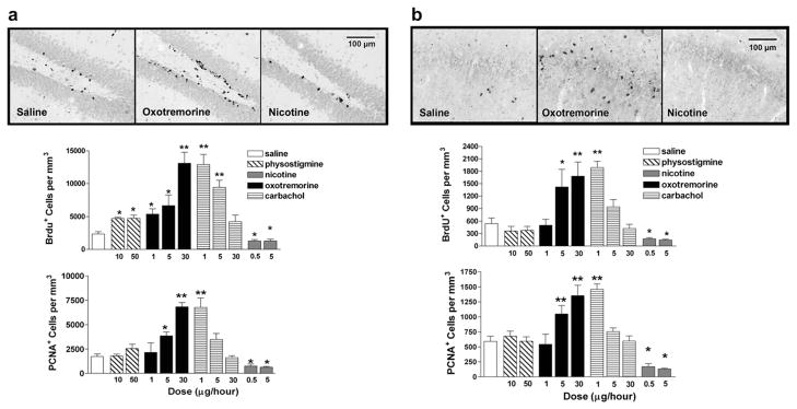
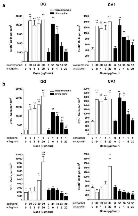
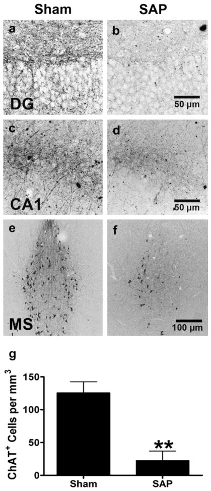
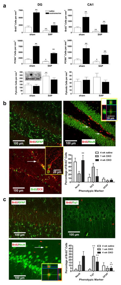
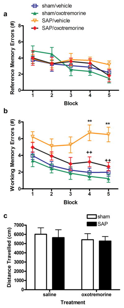
Loss of basal forebrain cholinergic innervation of the hippocampus and severe neuronal loss within the hippocampal CA1 region are early hallmarks of Alzheimer's disease, and are strongly correlated with cognitive status. Various therapeutic approaches involve attempts to enhance neurotransmission or to provide some level of neuroprotection for remaining cells. An alternative approach may involve the generation of new cells to replace those lost in AD. Indeed, a simple shift in the balance between cell generation and cell loss may slow disease progression and possibly even reverse existing cognitive deficits. One potential neurogenic regulator might be acetylcholine, itself, which has been shown to play a critical role in hippocampal development. Here, we report the effects of various cholinergic compounds on indices of hippocampal neurogenesis, demonstrating a significant induction following pharmacological activation of muscarinic M1 receptors, located on hippocampal progenitors in the adult brain. This is the first report that a small-molecule agonist may induce neurogenesis in the hippocampal CA1 region. Furthermore, such treatment reversed deficits in markers of neurogenesis and spatial working memory triggered by cholinergic denervation in a rodent model. This study suggests the use of small molecule, receptor agonists may represent a novel means to trigger the restoration of specific neuronal populations lost to a variety of neurodegenerative disorders, such as Parkinson's, Alzheimer's, Huntington's and Amyotrophic Lateral Sclerosis.


Figures

Fig. 1
Fig. 1
Cholinergic drugs differentially regulate indices of cell proliferation in the adult hippocampus. a, Both BrdU+ and PCNA+ cell counts were significantly reduced following chronic administration of nicotine in the DG, while the muscarinic agonists, oxotremorine and carbachol induced a dose-dependent and inversely dose-dependent elevation in immunolabeling, respectively, for both markers of cell proliferation. Slight elevations in BrdU labeling were also observed in response to the AChE inhibitor, phystostigmine. Representative photomicrographs depict BrdU immunolabeling in the DG following saline, nicotine or oxotremorine treatment. b, Similar elevations in both indices of cell proliferation were also evident in the CA1 region following treatment with oxotremorine or carbachol. By contrast, nicotine significantly reduced BrdU+ and PCNA+ cells in the CA1 region. Representative photomicrographs depict BrdU immunolabeling in the CA1 region following saline, nicotine or oxotremorine treatment. Each bar represents the mean (±S.E. M.) (n = 6) number of immunopositive cells per mm3. ** sig. diff. from saline-treated controls, p < 0.001; *p < 0.05. Scalebar = 100 μm.
Fig. 2
Fig. 2
Agonist-induced regulation of cell proliferation is blocked by receptor-selective antagonists indicating positive regulation mediated by the muscarinic M1 receptor. a, BrdU+ cell counts in the DG and CA1 regions of the hippocampus following oxotremorine. Chronic intraventricular administration of the M1 receptor antagonist, pirenzepine, but not the nicotinic antagonist, mecamylamine, dose-dependently blocked oxotremorine-induced elevations in BrdU immunolabeling in both the DG and CA1. b, Chronic intraventricular administration of the M1 receptor antagonist, pirenzepine, also dose-dependently blocked low dose carbachol-induced elevations in BrdU immunolabeling. By contrast, the nicotinic receptor antagonist, mecamylamine, had no effect. As described earlier, a higher dose of carbachol (30 μg/h) failed to affect cell proliferation. Co-administration with mecamylamine, however, dose-dependently elevated BrdU immunolabeling while co-administration with pirenzepine significantly reduced BrdU immunolabeling. Each bar represents the mean (±S.E.M.) (n = 6) number of BrdU+ cells per mm3. ** sig. diff. from saline-treated controls, p < 0.001, *p < 0.05; ++ sig. diff. from oxotremorine alone, p < 0.001, +p < 0.05.
Fig. 3
Fig. 3
Loss of Basal Forebrain Cholinergic Neurons Following Immunolesioning. Representative photomicrographs depicting choline acetyltransferase immunolabeling in the a,b dentate gyrus, c,d CA1 region and e,f medial septum of the adult rat brain following administration of a,c,e saline, or b,d,f 192IgG saporin. g, ChAT+ cell counts were significantly reduced in the medial septum four and eight weeks following saporin exposure. Each bar represents the mean (±S.E.M.) (n = 6) number of ChAT+ cells **per mm3. ** sig. diff. from saline-treated controls, p < 0.001. Scalebar = (a–d) 50 μm, (e,f) 100 μm.
Fig. 4
Fig. 4
Muscarinic receptor activation restores deficits in cell proliferation following immunolesioning with the majority of newly-generated cells expressing a neuronal phenotype. a, BrdU+ and PCNA+ cell counts were significantly reduced in both regions of the hippocampus following cholinergic depletion using 192IgG saporin. The muscarinic agonist, oxotremorine, induced a significant elevation in cell proliferation in sham animals and reversed the deficits in cell proliferation observed following immunolesioning without affecting apoptosis, as indicated by pyknotic cell counts. Each bar represents the mean (±S.E.M.) (n = 6) number of immunopositive cells per mm3. ** sig. diff. from saline-treated controls, p < 0.001; ++sig. diff. from sham controls, p < 0.001, +p < 0.05. Inset depicts an example of a pyknotic cell in the hippocampus. b,c, Representative fluorescent photomicrographs taken from the hippocampus of animals treated with oxotremorine for 4 weeks. b, In the DG, the majority of BrdU+ cells were also labeled with either the mature neuronal marker, NeuN, or doublecortin (DCX), a marker of immature migrating neurons. Significantly more BrdU+ cells expressed NeuN after 4 weeks as compared to 1 week, suggesting a time-dependent maturation. Few cells were co-labeled with the glial marker, GFAP. c, In the CA1 region, approximately 44% of BrdU+ cells were also labeled with NeuN or the immature neuronal marker, betaIII tubulin (Tuj1). Again, significantly more BrdU+ cells expressed the mature neuronal marker following 4 weeks, as compared to 1 week. Oxotremorine treatment significantly elevated the percentage of BrdU+ expressing a neuronal marker. Few cells were co-labeled with GFAP. Each bar represents the mean (±S.E.M.) (n = 6) percentage of BrdU+ cells also labeled for a phenotypic marker. ** sig. diff. from 1 week treatment, p < 0.001, *p < 0.05; ++ sig. diff. from saline-treated control, p < 0.001. Insets depict enlarged views, rotated along the x and y axes, scalebar = 20 μm. Arrows indicate representative examples of a double-labeled cell. Scalebar = 100 μm.
Fig. 5
Fig. 5
Muscarinic receptor activation reverses impairment in spatial working memory following immunolesioning. a, None of the groups differed in the number of reference errors committed. Reference memory errors were defined as entries into arms that were never baited. Each point represents the mean (±S.E.M.) (n = 6) number of reference memory errors. b, Significantly more working memory errors were committed by immunolesioned animals in the last two blocks of trials. These deficits in spatial working memory were significantly attenuated in animals treated with oxotremorine. Oxotremorine treatment was terminated 3 days prior to the final block of trials. In this final block, spatial working memory performance remained elevated. Working memory errors were defined as entries into previously visited arms. Each point represents the mean (±S.E.M.) (n = 6) number of working memory errors. ** sig. diff. from sham control, p < 0.001. c, No changes in locomotor activity were observed in any of the groups tested. Each bar represents the mean (±S.E.M.) (n = 6) total distance traveled (cm) in 1 h.

Similar articles

Cited by

References

    1. Abood LG, Saraswati M, Lerner-Marmarosh N, Hashmi M. Affinity ligands and related agents for brain muscarinic and nicotinic cholinergic receptors. Biochemical Pharmacology. 1993;45:2143–2148. - PubMed
    1. Ball MJ. Neuronal loss, neurofibrillary tangles and granulovacuolar degeneration in the hippocampus with ageing and dementia. Acta Neuropathologica. 1977;37:111–118. - PubMed
    1. Barker JL, Behar T, Li YX, Liu QY, Ma W, Maric D, Maric I, Schaffner AE, Serafini R, Smith SV, Somogyi R, Vautrin JY, Wen XL, Xian H. GABAergic cells and signals in CNS development. Perspectives on Developmental Neurobiology. 1998;5:305–322. - PubMed
    1. Bobinski M, de Leon MJ, Tarnawski M, Wegiel J, Bobinski M, Reisberg B, Miller DC, Wisniewski HM. Neuronal and volume loss in CA1 of the hippocampal formation uniquely predicts duration and severity of Alzheimer disease. Brain Research. 1998;805:267–269. - PubMed
    1. Borta A, Hoglinger GU. Dopamine and adult neurogenesis. Journal of Neurochemistry. 2007;100:587–595. - PubMed

MeSH terms