

6-Hz primed low-frequency rTMS to contralesional M1 in two cases with middle cerebral artery stroke
- PMID: 20026185
- PMCID: PMC2815205
- DOI: 10.1016/j.neulet.2009.12.023
6-Hz primed low-frequency rTMS to contralesional M1 in two cases with middle cerebral artery stroke
Abstract
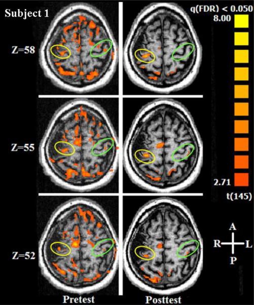
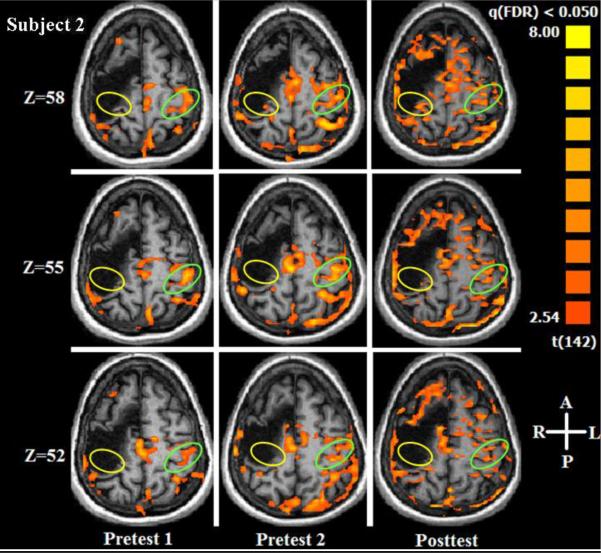
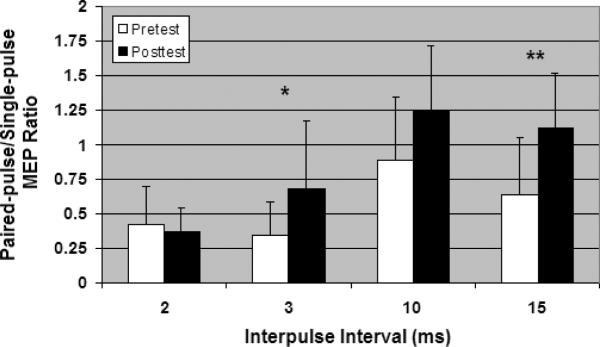
This case study contrasted two subjects with stroke who received 6-Hz primed low-frequency repetitive transcranial magnetic stimulation (rTMS) to the contralesional primary motor area (M1) to disinhibit ipsilesional M1. Functional magnetic resonance imaging (fMRI) showed that the intervention disrupted cortical activation at contralesional M1. Subject 1 showed decreased intracortical inhibition and increased intracortical facilitation following intervention during paired-pulse TMS testing of ipsilesional M1. Subject 2, whose precentral knob was totally obliterated and who did not show an ipsilesional motor evoked potential at pretest, still did not show any at posttest; however, her fMRI did show a large increase in peri-infarct zone cortical activation. Behavioral results were mixed, indicating the need for accompanying behavioral training to capitalize on the brain organization changes induced with rTMS.
(c) 2009 Elsevier Ireland Ltd. All rights reserved.
Figures
Similar articles
-
Differential effects of high-frequency repetitive transcranial magnetic stimulation over ipsilesional primary motor cortex in cortical and subcortical middle cerebral artery stroke.Ann Neurol. 2009 Sep;66(3):298-309. doi: 10.1002/ana.21725. Ann Neurol. 2009. PMID: 19798637
-
Effects of low-frequency repetitive transcranial magnetic stimulation of the contralesional primary motor cortex on movement kinematics and neural activity in subcortical stroke.Arch Neurol. 2008 Jun;65(6):741-7. doi: 10.1001/archneur.65.6.741. Arch Neurol. 2008. PMID: 18541794
-
Modulating cortical connectivity in stroke patients by rTMS assessed with fMRI and dynamic causal modeling.Neuroimage. 2010 Mar;50(1):233-42. doi: 10.1016/j.neuroimage.2009.12.029. Epub 2009 Dec 18. Neuroimage. 2010. PMID: 20005962 Free PMC article.
-
Changes in thresholds for intracortical excitability in chronic stroke: more than just altered intracortical inhibition.Restor Neurol Neurosci. 2013;31(6):693-705. doi: 10.3233/RNN-120300. Restor Neurol Neurosci. 2013. PMID: 23963339
-
Distinction of High- and Low-Frequency Repetitive Transcranial Magnetic Stimulation on the Functional Reorganization of the Motor Network in Stroke Patients.Neural Plast. 2021 Jan 20;2021:8873221. doi: 10.1155/2021/8873221. eCollection 2021. Neural Plast. 2021. PMID: 33542729 Free PMC article.
Cited by
-
5 Hz repetitive transcranial magnetic stimulation over the ipsilesional sensory cortex enhances motor learning after stroke.Front Hum Neurosci. 2014 Mar 21;8:143. doi: 10.3389/fnhum.2014.00143. eCollection 2014. Front Hum Neurosci. 2014. PMID: 24711790 Free PMC article.
-
Priming the brain to capitalize on metaplasticity in stroke rehabilitation.Phys Ther. 2014 Jan;94(1):139-50. doi: 10.2522/ptj.20130027. Epub 2013 Aug 15. Phys Ther. 2014. PMID: 23950225 Free PMC article. Review.
-
A Comparison of Primed Low-frequency Repetitive Transcranial Magnetic Stimulation Treatments in Chronic Stroke.Brain Stimul. 2015 Nov-Dec;8(6):1074-84. doi: 10.1016/j.brs.2015.06.007. Epub 2015 Jun 22. Brain Stimul. 2015. PMID: 26198365 Free PMC article.
-
Noninvasive brain stimulation in neurorehabilitation.Handb Clin Neurol. 2013;116:499-524. doi: 10.1016/B978-0-444-53497-2.00040-1. Handb Clin Neurol. 2013. PMID: 24112919 Free PMC article. Review.
-
New modalities of brain stimulation for stroke rehabilitation.Exp Brain Res. 2013 Feb;224(3):335-58. doi: 10.1007/s00221-012-3315-1. Epub 2012 Nov 29. Exp Brain Res. 2013. PMID: 23192336 Free PMC article. Review.
References
-
- Buonomano DV, Merzenich MM. Cortical plasticity: from synapses to maps. Annual Review of Neuroscience. 1998;21:149–186. - PubMed
-
- Butefisch CM, Khurana V, Kopylev L, Cohen LG. Enhancing encoding of a motor memory in the primary motor cortex by cortical stimulation. J. Neurophysiol. 2004;91:2110–2116. - PubMed
-
- Butefisch CM, Wessling M, Netz J, Seitz RJ, Homberg V. Relationship between interhemispheric inhibition and motor cortex excitability in subacute stroke patients. Neurorehabilitation & Neural Repair. 2008;22:4–21. - PubMed
-
- Carey JR. Manual Stretch: Effect on Finger Movement Control and Force Control in Stroke Subjects with Spastic Extrinsic Finger Flexor Muscles. Arch Phys Med Rehabil. 1990;71:888–894. - PubMed
Publication types
MeSH terms
Grants and funding
LinkOut - more resources
Full Text Sources
Medical
