. 2010 Feb;1803(2):275-87.
doi: 10.1016/j.bbamcr.2009.12.002. Epub 2009 Dec 21.

Protein kinase A regulation of P2X(4) receptors: requirement for a specific motif in the C-terminus

Affiliations

Protein kinase A regulation of P2X(4) receptors: requirement for a specific motif in the C-terminus

David A Brown et al. Biochim Biophys Acta. 2010 Feb.

Abstract

The P2X purinergic receptor sub-family of ligand-gated ion channels are subject to protein kinase modulation. We have previously demonstrated that P2X(4)R signaling can be positively regulated by increasing intracellular cAMP levels. The molecular mechanism underlying this effect was, however, unknown. The present study initially addressed whether protein kinase A (PKA) activation was required. Subsequently a mutational approach was utilized to determine which region of the receptor was required for this potentiation. In both DT-40 3KO and HEK-293 cells transiently expressing P2X(4)R, forskolin treatment enhanced ATP-mediated signaling. Specific PKA inhibitors prevented the forskolin-induced enhancement of ATP-mediated inward currents in P2X(4)R expressing HEK-293 cells. To define which region of the P2X(4)R was required for the potentiation, mutations were generated in the cytoplasmic C-terminal tail. It was determined that a limited region of the C-terminus, consisting of a non-canonical tyrosine based sorting motif, was required for the effects of PKA. Of note, this region does not harbor any recognizable PKA phosphorylation motifs, and no direct phosphorylation of P2X(4)R was detected, suggesting that PKA phosphorylation of an accessory protein interacts with the endocytosis motif in the C-terminus of the P2X(4)R. In support of this notion, using Total Internal Reflection Fluorescence Microscopy (TIRF)\ P2X(4)-EGFP was shown to accumulate at/near the plasma membrane following forskolin treatment. In addition, disrupting the endocytosis machinery using a dominant-negative dynamin construct also prevented the PKA-mediated enhancement of ATP-stimulated Ca(2+) signals. Our results are consistent with a novel mechanism of P2XR regulation, whereby PKA activity, without directly phosphorylating P2X(4)R, markedly enhances ATP-stimulated P2X(4)R currents and hence cytosolic Ca(2+) signals. This may occur at least in part, by altering the trafficking of a population of P2X(4)R present at the plasma membrane.


Figures
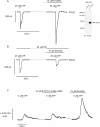
Fig 1
Fig 1
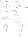
The forskolin-mediated enhancement of P2X4R signaling is blocked by PKA inhibition. An immunoblot of mock-transfected HEK-293 cells shows that they lack P2X4R, whereas P2X4R protein can readily be detected after transient overexpression of the receptor (Inset). Transfected HEK-293 cells were whole cell patch clamped at a holding potential of −30 mV. (A) Treatment of P2X4R-transfected HEK-293 cells with 25 μM ATP resulted in an inward current, which was enhanced by forskolin, consistent with our previous findings [19]. (B) Pretreatment with PKI, a cell permeable PKA inhibitor, abolished the enhanced inward current following forskolin treatment in P2X4R-transfected HEK-293 cells. The PKI/forskolin treated second current was not significantly different in magnitude to previously published control values (P2X4R control, 76 ± 10% of initial response [19] versus P2X4R PKI/forskolin treatment, 66 ± 16%, n = 8 and n = 4 respectively, p = 0.59). However, there was a significant difference between the magnitude of the second response between PKI/forskolin and previously published forskolin treated values (P2X4R forskolin treatment, 451 ± 101% of initial response [19] versus P2X4R PKI/forskolin treatment, 66 ± 16%, n = 9 and n = 4 respectively, p = 0.017 respectively). (C) [Ca2+]i signals mediated by ATP in DT-40 3KO cells stably expressing P2X4R were not modulated by the EPAC specific analog 8-CPT-2′-O-Me-cAMP. In the same cells, forskolin treatment resulted in a significant potentiation of Ca2+ signals. The data are presented as the mean ± S.E. Each trace is representative of three or more experiments.
Fig 2
Fig 2
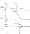
cAMP-mediated potentiation of ATP-mediated Ca2+ signals in DT-40 3KO cells and ATP-activated currents in HEK-293 cells is retained in cells overexpressing P2X4–EGFP. P2X4-EGFP was generated by tagging the C-terminus of P2X4R with EGFP. (A) Multiple responses to ATP. (B) Forskolin treatment enhanced ATP-mediated Ca2+ signaling in DT-40 3KO cells transiently overexpressing P2X4-EGFP receptors. (C) Forskolin enhanced ATP-activated current in HEK-293 cells overexpressing P2X4-EGFP receptors. Traces are representative of 4 or more experiments.
Fig 3
Fig 3
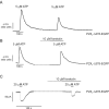
A chimeric P2X4R with a P2X1R C-terminus is not enhanced by raising cAMP. P2X4-C-P2X1-EGFP was generated using the N-terminal, first transmembrane domain, and extracellular regions from P2X4R and replacing the second transmembrane domain and C-terminus with the corresponding amino acids from P2X1R. (A) Transient transfection of P2X4-C-P2X1-EGFP receptors into DT-40 3KO cells elicited a Ca2+ spike when stimulated with ATP, which was no longer enhanced by forskolin treatment. (B) Forskolin did not enhance ATP-activated current in HEK-293 cells overexpressing chimeric P2X4-C-P2X1-EGFP receptors. Traces are representative of 4 or more experiments.
Fig 4
Fig 4
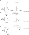
cAMP-mediated potentiation of ATP-mediated Ca2+ signals in DT-40 3KO cells and ATP-activated currents in HEK-293 cells is retained in cells overexpressing the truncation mutant receptor P2X4-Δ384-EGFP. P2X4-Δ384-EGFP was generated by removing the last 5 amino acids of the P2X4R C-terminus. (A) Multiple responses to ATP exposure in cells expressing this construct. (B) Forskolin treatment enhanced ATP-mediated Ca2+ signaling in DT-40 3KO cells overexpressing P2X4-Δ384-EGFP receptors. (C) Forskolin potentiated ATP-activated currents in HEK-293 cells overexpressing P2X4-Δ384-EGFP receptors. Traces are representative of 4 experiments.
Fig 5
Fig 5
Truncation of 11 amino acids in the P2X4R C-terminus prevents cAMP-mediated potentiation of ATP-mediated signaling. P2X4-Δ378-EGFP was generated by removing the last 11 amino acids of the P2X4R C-terminus. (A) Multiple responses to ATP exposure in cells expressing this construct. (B) Forskolin treatment did not enhance ATP-mediated Ca2+ signaling in DT-40 3KO cells overexpressing P2X4-Δ378-EGFP receptors. (C) Forskolin did not augment ATP-activated currents in HEK-293 cells overexpressing P2X4-Δ378-EGFP receptors. Traces are representative of 5 or more experiments.
Fig 6
Fig 6
Disruption of the YXXGL endocytosis motif in the full length P2X4R also prevents cAMP-mediated potentiation of ATP-mediated signaling. P2X4-AAA-EGFP was generated by three alanine point mutations in the YXXGL motif of the full length P2X4R. (A) Multiple responses to ATP exposure in cells expressing this construct. (B) Forskolin treatment did not enhance ATP-mediated Ca2+ signaling in DT-40 3KO cells overexpressing P2X4-AAA-EGFP receptors. (C) Forskolin did not enhance ATP-activated currents in HEK-293 cells overexpressing P2X4-AAA-EGFP receptors. Traces are representative of 7 or more experiments.
Fig 7
Fig 7
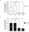
Data summary of ATP-mediated Ca2+ signals in DT-40 3KO cells and ATP-activated currents in HEK-293 cells overexpressing P2X4R mutants. (A) Data summary of ATP-mediated Ca2+ signaling in DT-40 3KO cells from paired experiments, where the forskolin-treated second response is expressed as a percentage of the control-treated first response (white bar graph). (B) Data summary of ATP-activated currents in HEK-293 cells from paired experiments, where the forskolin-treated second response is expressed as a percentage of the control-treated first response (black bar graph). The data are presented as the mean ± S.E. (*, p < 0.05).
Fig 8
Fig 8
PKA activation increases the number of P2X4-EGFP in the vicinity of the plasma membrane. Experiments were performed monitoring the movement of P2X4-EGFP using TIRF microscopy in cells stably expressing the receptor. (A) shows the fluorescence of P2X4-EGFP in wide field (WFM) and following establishing TIRF mode (TIRFM). (B) shows a series of images obtained at the time-points indicated in (C) for a group of HEK-293 cells stably expressing P2X4-EGFP. (C) shows kinetic plots from 3 regions of interest indicated in the grey scale image in (B). Following forskolin treatment fluorescence puncta grew larger and increased intensity. An exemplar experiment showing three regions of interest typical of data from 7 experimental runs. (D) shows similar experiments in which P2X4-AAA-EGFP was transiently expressed in cells. Forskolin treatment did not alter the distribution of this mutant receptor.
Fig 9
Fig 9
DT-40 3KO cells stably expressing P2X4R exhibit forskolin-mediated enhancement of ATP-evoked Ca2+ signals, which can be blocked by dominant negative dynamin K44A expression. (A) ATP-activated Ca2+ signals in mock-transfected DT-40 3KO cells stably expressing P2X4R are enhanced by forskolin treatment. (B) Forskolin treatment had no effect on ATP-mediated Ca2+ signals in DT-40 3KO cells stably expressing P2X4R and transiently overexpressing dominant negative dynamin K44A. Traces are representative of eight or more experiments. (C) Data summary of ATP-mediated Ca2+ signaling in DT-40 3KO cells stably expressing P2X4R from paired experiments after transient mock- or dynamin K44A-transfection (white and black bar graph, respectively), where the control- or forskolin-treated second response is expressed as a percentage of the control-treated first response.
Fig 10
Fig 10
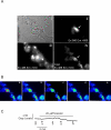
Expression of dominant negative K44A dynamin eliminates the forskolin-induced retention of P2X4-EGFP close to the PM. (Ai) bright field image of HEK-293 cells stably expressing P2X4-EGFP and transiently co-expressing HcRed and K44A dynamin. (Aii) Wide-field fluorescence image indicating HcRed expression (Ex 560 nm; Em > 600 nm). (Aiii) Wide-field fluorescence image of P2X4-EGFP (Ex 488 nm; Em 510 nm). (Aiv) TIRF fluorescence image of P2X4-EGFP. (B) Series of TIRF images captured at the time-points indicated in C in which the cells were exposed to forskolin. (C) Kinetic line plot of average fluorescence from the red box shown in B.

Similar articles

Cited by

References

    1. Schwiebert EM, Zsembery A. Extracellular ATP as a signaling molecule for epithelial cells. Biochim Biophys Acta. 2003;1615:7–32. - PubMed
    1. North RA. Molecular physiology of P2X receptors. Physiol Rev. 2002;82:1013–67. - PubMed
    1. Turner JT, Landon LA, Gibbons SJ, Talamo BR. Salivary gland P2 nucleotide receptors. Crit Rev Oral Biol Med. 1999;10:210–24. - PubMed
    1. Brake AJ, Wagenbach MJ, Julius D. New structural motif for ligand-gated ion channels defined by an ionotropic ATP receptor. Nature. 1994;371:519–23. - PubMed
    1. Khakh BS, North RA. P2X receptors as cell-surface ATP sensors in health and disease. Nature. 2006;442:527–32. - PubMed

Publication types

MeSH terms

LinkOut - more resources