

Review
doi: 10.1016/j.semcdb.2009.12.008.
Epub 2009 Dec 21.
Life and death of a BiP substrate
Affiliations
- PMID: 20026282
- PMCID: PMC2883687
- DOI: 10.1016/j.semcdb.2009.12.008
Item in Clipboard
Review
Life and death of a BiP substrate
Semin Cell Dev Biol.
2010 Jul.
Abstract
BiP is the mammalian endoplasmic reticulum (ER) Hsp70 orthologue that plays a major role in all functions of this organelle including the seemingly opposing functions of aiding the maturation of unfolded nascent proteins and identifying and targeting chronically unfolded proteins for degradation. The recent identification of mammalian BiP co-factors combined with delineation of the ER degradation machinery and data suggesting that the ER is subdivided into unique regions helps explain how these different functions can occur in the same organelle and raises some unresolved issues.
Figures
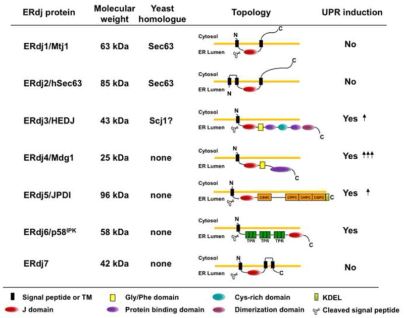
Seven DnaJ-like proteins have been identified in the mammalian ER. DnaJ proteins can be divided into three subgroups based on the similarities with E. coli DnaJ, the founding member of this family [77]. Type I proteins contain all three domains: an N-terminal J domain, a glycine/phenylalanine rich domain (yellow square), and a substrate binding domain, which is intersected with a cysteine-rich Zn2+ binding domain (light blue circle); type II proteins contain the first two domains and often bind to unfolded substrates, although this region is more poorly defined; and type III proteins have only a J domain, which can occur anywhere in the protein. In the mammalian ER most of the ERdjs are type III except for ERdj3 (type I) and ERdj4 (type II). ERdj5 contains four thioredoxin domains (orange squares) and ERdj6 has three tetratricopeptide domains (dark green squares). The detailed mechanism of their function can be found in the text.
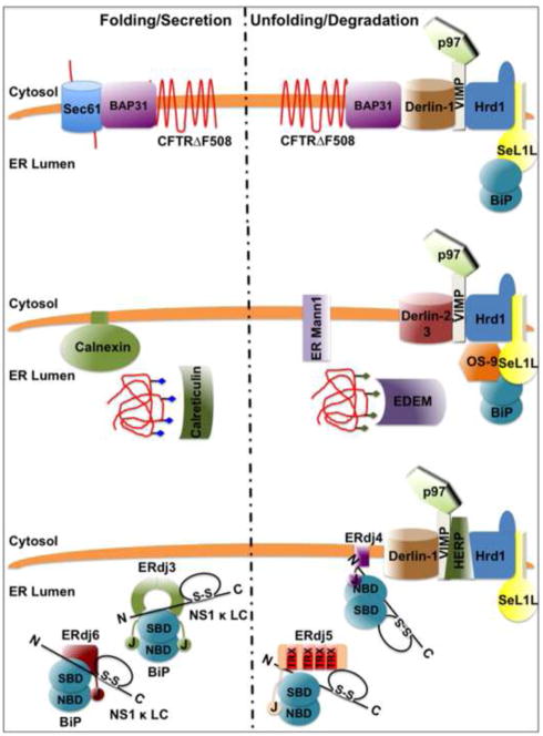
Top: Membrane proteins, like CFTR F508, bind BAP31, which cycles between sites of protein folding near the Sec61 translocon and the ERQC where it binds Derlin1 to promote retrotranslocation of ERAD substrates. Middle: Glycosylated proteins (blue diamonds) undergo cycles of binding and release from the lectins calnexin and calreticulin as they fold. If they fail to do so, their polysaccharide chains (green diamonds) are modified by ER mannosidase I, which prevents the unfolded protein from rebinding calnexin/calreticulin and allows it to bind EDEM, a soluble lectin. EDEM targets the protein to Sel1 and OS-9, which associate with a complex of Derlin 2/3, p97 and presumably the Hrd1 E3 ligase, promoting its retrotranslocation and degradation. Bottom: Non-glycosylated lumenal ERAD substrates, like the NS1 κ LC, bind BiP, which can promote their folding when the substrate is associated with ERdj3/ERdj6 or reduce and unfold the substrate when it is bound to ERdj4 or ERdj5. The fact that a number of the ERdjs are soluble and bind to both substrates and components of the folding machinery or degradation complexes may provide a mechanism for moving from one ER function to the other.
References
-
- Hammond C, Helenius A. Quality control in the secretory pathway. Curr Opin Cell Biol. 1995;7:523–9. - PubMed
-
- Jarosch E, Lenk U, Sommer T. Endoplasmic reticulum-associated protein degradation. Int Rev Cytol. 2003;223:39–81. - PubMed
-
- Hendershot LM. BiP is a master regulator of ER function. Mt Sinai J Med. 2004;71:289–97. - PubMed
-
- Molinari M, Helenius A. Chaperone selection during glycoprotein translocation into the endoplasmic reticulum. Science. 2000;288:331–3. - PubMed
Publication types
MeSH terms
Substances
Grants and funding
LinkOut - more resources
Full Text Sources
