. 2010 Apr;34(4):474-9.
doi: 10.1016/j.dci.2009.12.008. Epub 2009 Dec 28.

Isolation and characterization of chicken lung mesenchymal stromal cells and their susceptibility to avian influenza virus

Affiliations

Isolation and characterization of chicken lung mesenchymal stromal cells and their susceptibility to avian influenza virus

Mahesh Khatri et al. Dev Comp Immunol. 2010 Apr.

Abstract

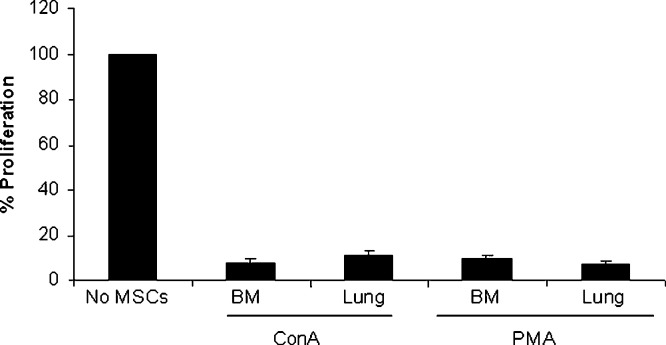
In this study, we isolated and characterized mesenchymal stromal cells (MSCs) from the lungs of 1- to 2-week-old chickens. Microscopically, the cultured cells showed fibroblast-like morphology. Phenotypically these cells expressed CD44, CD90, CD105 and the transcription factor PouV, which has been shown to be critical for stem cell self-renewal and pluripotency. The multipotency of chicken MSCs was demonstrated by their ability to undergo adipogenic and osteogenic differentiation. Like chicken bone marrow MSCs and mammalian MSCs, chicken lung MSCs had immunoregulatory activity and profoundly suppressed the proliferative capacity of T cells in response to a mitogenic stimulus. Next, we examined the susceptibility of these cells to H1N1 and H9N5 avian influenza (AI) viruses. The lung MSCs were shown to express known influenza virus alpha-2,3 and alpha-2,6 sialic acid receptors and to support replication of both the avian H1N1 and avian H9N5 influenza strains. Viral infection of MSCs resulted in cell lysis and cytokine and chemokine production. Further characterization of lung MSCs in chicken and other mammalian species may help in understanding the pathogenesis of infectious and non-infectious lung diseases and the mechanisms of lung injury repair.


Figures

Fig. 1
Fig. 1
Characterization of MSCs isolated from chicken lung. (A) Morphology of MSCs derived from lung. (B) Oil Red O staining for lipid-filled vesicles (red) after 21 days of adipogenic differentiation of MSCs derived from lung. (C) von Kossa staining for calcified mineral nodules (black) after 21 days of osteogenic differentiation of MSCs derived from lung. (D) Phenotype of chicken lung MSCs. Reverse transcription-polymerase chain reaction was performed using total RNA extracted from lung MSCs with specific primers. The center column shows the results with total RNA from lung MSCs. Total RNA prepared from normal chicken spleen cells was used as a positive control for CD44, CD90 and CD105 expression and for PouV, total RNA isolated from bone marrow MSCs was used as positive control (left column). (For interpretation of the references to color in this figure legend, the reader is referred to the web version of the article.)
Fig. 2
Fig. 2
MSCs inhibit T cells proliferation. Splenocytes (2 × 105) were stimulated with ConA (5 μg/ml) or PMA/ionomycin in the presence of MSCs (2 × 104) from BM and lung. Cultures were incubated for 48 h and for the final 6 h of culture [3H] TdR was added to the wells. Results are expressed as percentage of proliferation, with the positive control (in the absence of MSCs) at 100%. The values are averages ± S.D. of triplicate determinations. The data are representative of three separate experiments.
Fig. 3
Fig. 3
Replication of AI virus in lung MSCs. (A) MSCs express α-2,3 and α-2,6-linked sialic acid receptors. MSCs cultured on coverslips were fixed and stained with biotinylated Maackia amurensis lectin II (MAA) specific for α-2,3-linked sialic acid receptors and Sambucus niagra agglutinin (SNA), specific for α-2,6-linked sialic acid receptors, followed by the addition of ABC reagent. Brown color indicates positive reaction. (B) MSCs were infected with H1N1 or H9N5 at an MOI of 1. Virus production in infected culture supernatant as measured by titration in MDCK cells. (C) Virus-induced cell death was evaluated by counting the live and dead cells by trypan blue dye exclusion method. The values are averages ± S.D. of duplicate determinations. The data are representative of three separate experiments. (D) At 24 hpi, AI viral proteins were detected by IFA. (For interpretation of the references to color in this figure legend, the reader is referred to the web version of the article.)
Fig. 4
Fig. 4
Virus-induced apoptosis of MSCs. MSCs were infected with H1N1 or H9N5 viruses at an MOI of 1. At 24 hpi virus-induced apoptosis was examined by TUNEL assay. The arrows indicate apoptotic cells (brown). (For interpretation of the references to color in this figure legend, the reader is referred to the web version of the article.)
Fig. 5
Fig. 5
AI virus infection induces inflammatory cytokines in chicken lung MSCs. MSCs were infected with H1N1 or H9N5. After indicated times, total cellular RNA was extracted and subjected to qRT-PCRs by using specific primers to IL-6, IL-8 or 28S rRNA. Quantitation of the IL-6 and IL-8 mRNA was determined by the comparative cycle threshold (CT) method. RNA isolated from uninfected control cultures was used as the reference sample (calibrator) at each time point. The relative change in mRNA concentration (ΔCT) for each virus-infected sample was then determined from the difference between the calibrator CT and the CT of each virus-infected sample. Fold increase of IL-6 and IL-8 transcript expression over uninfected controls was calculated with the 2ΔΔCT method. The data are representative of two separate experiments.

Similar articles

Cited by

References

    1. Friedenstein A.J., Deriglasova U.F., Kulagina N.N., Panasuk A.F., Rudakowa S.F., Luria E.A. Precursors for fibroblasts in different populations of hematopoietic cells as detected by the in vitro colony assay method. Exp Hematol. 1974;2(2):83–92. - PubMed
    1. Iyer S.S., Rojas M. Anti-inflammatory effects of mesenchymal stem cells: novel concept for future therapies. Expert Opin Biol Ther. 2008;8(5):569–581. - PubMed
    1. Awad H.A., Boivin G.P., Dressler M.R., Smith F.N., Young R.G., Butler D.L. Repair of patellar tendon injuries using a cell-collagen composite. J Orthop Res. 2003;21(3):420–431. - PubMed
    1. Noel D., Djouad F., Jorgense C. Regenerative medicine through mesenchymal stem cells for bone and cartilage repair. Curr Opin Invest Drugs. 2002;3(7):1000–1004. - PubMed
    1. Pelttari K., Steck E., Richter W. The use of mesenchymal stem cells for chondrogenesis. Injury. 2008;39(Suppl. 1):S58–S65. - PubMed

MeSH terms