Review
. 2010 Aug;1800(8):834-45.
doi: 10.1016/j.bbagen.2009.12.005. Epub 2009 Dec 22.

The ferritin superfamily: Supramolecular templates for materials synthesis

Affiliations
Review

The ferritin superfamily: Supramolecular templates for materials synthesis

Masaki Uchida et al. Biochim Biophys Acta. 2010 Aug.

Abstract

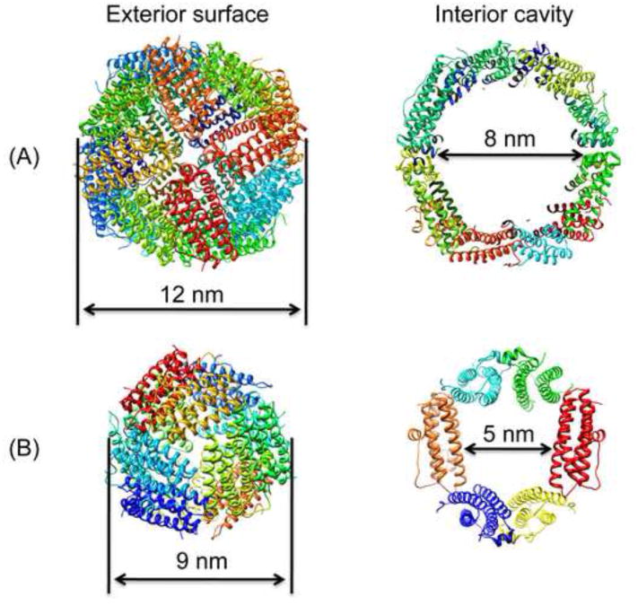
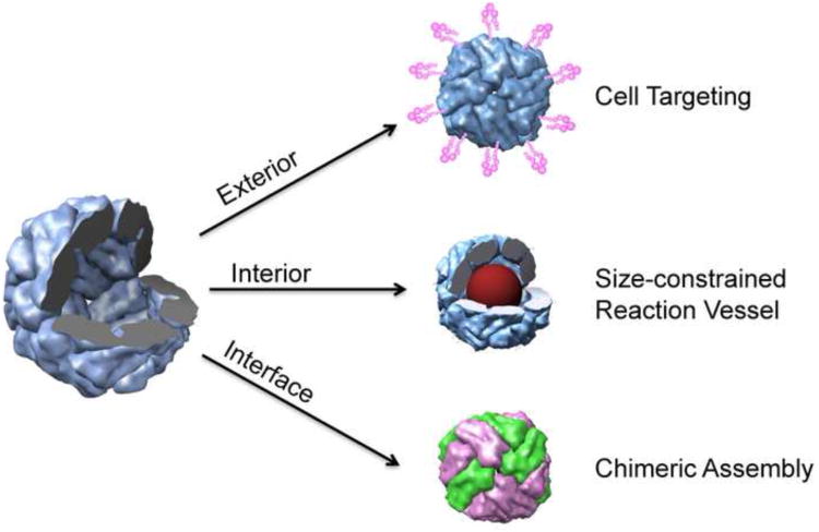
Members of the ferritin superfamily are multi-subunit cage-like proteins with a hollow interior cavity. These proteins possess three distinct surfaces, i.e. interior and exterior surfaces of the cages and interface between subunits. The interior cavity provides a unique reaction environment in which the interior reaction is separated from the external environment. In biology the cavity is utilized for sequestration of irons and biomineralization as a mechanism to render Fe inert and sequester it from the external environment. Material scientists have been inspired by this system and exploited a range of ferritin superfamily proteins as supramolecular templates to encapsulate nanoparticles and/or as well-defined building blocks for fabrication of higher order assembly. Besides the interior cavity, the exterior surface of the protein cages can be modified without altering the interior characteristics. This allows us to deliver the protein cages to a targeted tissue in vivo or to achieve controlled assembly on a solid substrate to fabricate higher order structures. Furthermore, the interface between subunits is utilized for manipulating chimeric self-assembly of the protein cages and in the generation of symmetry-broken Janus particles. Utilizing these ideas, the ferritin superfamily has been exploited for development of a broad range of materials with applications from biomedicine to electronics.


Figures

Fig. 1
Fig. 1
Ribbon diagrams of exterior surface view and interior cavity of (A) Human heavy-chain ferritin and (B) Listeria innocua Dps.
Fig. 2
Fig. 2
Schematic illustration of the three interfaces of a protein cages that can be exploited to impart designed functionalities.
Fig. 3
Fig. 3
Schematic illustration of the ion distribution at a charged surface according to Gouy-Chapman theory. The electronic double layer is formed nearby the charged surface through Coulombic interactions.
Fig. 4
Fig. 4
In vivo MRI of carotid arteries in (A) ligated mouse and (B) sham operated mouse; comparison of pre-injection, 24h and 48h post-injection of the mineralized HFn [80]. An atherosclerotic lesion was formed in left common carotid artery (LCCA) due to ligation of the artery but not formed either right common carotid artery (RCAA) of the same mouse or those arteries of the sham operated mouse. In the ligated mouse, concentric signal loss was observed around the LCCA lumen at 24 and 48 h post-injection of the mineralized HFn in comparison with pre-injection, but not the RCCA. No change was seen in either the LCCA or RCCA of sham operated mouse.
Fig. 5
Fig. 5
Fluorescence-activated cell sorting analysis of THP-1 cells incubated with fluorescently labeled RGD4C-Fn [86]. The data are plotted as histograms with their corresponding geometric (geo.) mean fluorescence values. Although the non-targeted protein cage (HFn) showed some interaction with the cells, the targeted cages (RGD4C-Fn) exhibit significantly increased interaction with the cells comparable to the positive control, anti-integrin αvβ3.
Fig. 6
Fig. 6
Schematic illustration of immobilization of [Ru(nbd)Cl]2 complex into apo-Fn cage followed by polymerization of phenylacetylene catalyzed by the Fn [88]. The polymerization reaction occurs site-specifically inside of the Fn cage.
Fig. 7
Fig. 7
(A) Schematic illustration of ferritin adsorption onto a positively charged 3-aminopropyltriethoxysilane (APTES) nanodisc formed on a negatively charged SiO2 substrate by an electrostatic interaction in solution under, (a) a short Debye length, λ, (b) a medium Debye length, and (c) a long Debye length. With a short λ, no selective adsorption onto the APTES area can be achieved because of short range interactions such as van der Waals force or hydrophobic interaction. With a long λ, ferritins can not reach to the APTES area due to the repulsive potential from the SiO2 substrate. With an optimized λ, ferritins can be adsorbed only on the APTES area. (B) Single ferritin placement on APTES nanodisc patterns formed on an oxidized Si substrate. APTES disks of 15 nm diameter are prepared at 100 nm intervals on the substrate. Each APTES disk generally has one ferritin molecule. [97].
Fig. 8
Fig. 8
Hysteresis loops measured at 2 K for field-cooled (8T) mixed oxide material with nominal composition of 66% Fe3O4 and 33% Co3O4 prepared by slow (30 min) or fast (5 min) synthesis [104]. The offset between the two hysteresis loops is the exchange bias. The fast synthesis leads to Co incorporation as ferrimagnetic CoxFe3−xO4 whereas slow synthesis results in a larger fraction of Co in antiferromagnetic Co3O4 which is available to bias the ferrimagnetic Fe3O4 present.
Fig. 9
Fig. 9
(A) Mass spectra of Fe-mineralized wild type LiDps at various loading factors of Fe(II). Charged peaks are indicated in the bottom spectrum [68]. (B) Fit (red line) of 22+ peaks of Fe-mineralized wt LiDps at lower loadings either with one or two Gaussians (green line). The peaks shift to higher m/z according to the increase of Fe2+ loading per cage indicates Fe2O3 particle growth in the cages. Fit of peaks demonstrates the presence of more than one mass distribution of the cages. The results allow us to establish a two-stage Fe2O3 particle growth process model[68].
Fig. 10
Fig. 10
Overlaid mass spectra of the Pt2+ ion bound (black) and Pt0 mineralized (red) phen-S138C LisDps cages at various loading ratios of Pt2+ per cage. The charged peaks of 23+ of the cages are indicated [70]. The peaks shifted to higher m/z in accordance with increasing the number of Pt2+ ions loaded per cage.
Fig. 11
Fig. 11
(A) Schematic representation of chimeric cage construction scheme. Two types of differentially modified S13C LiDps cages are disassembled to subunits at pH 2. Subsequently, the two types of subunits are mixed together with various ratios followed by reassembly to cages at pH 7. (B) Mass spectra of reassembled whole cages [110]. The peaks shift to higher m/z with increasing heavier subunit (blue subunits) ratio of the reassembled cages.
Fig. 12
Fig. 12
Schematic illustration of a Janus-like LiDps cage preparation scheme though a masking/unmasking technique on a solid bead support [138]. The protein cages are first immobilized on beads through disulfide bonds. The exposed sides of the protein cages are modified with maleimide-PEG2-Biotin, which can selectively bind with streptavidin. Subsequently, the modified LiDps cages are eluted from the beads by reduction and the free cysteine residues on the cages are labeled with fluorescein-5-maleimide.

Similar articles

Cited by

References

    1. Koeck PJB, Kagawa HK, Ellis MJ, Hebert H, Trent JD. Two-dimensional crystals of reconstituted .beta.-subunits of the chaperonin TF55 from Sulfolobus shibatae. Biochim Biophys Acta. 1998;1429:40–44. - PubMed
    1. Trent JD. A review of acquired thermotolerance, heat-shock proteins, and molecular chaperones in archaea. FEMS Microbiol Rev. 1996;18:249–258.
    1. Kim KK, Kim R, Kim SH. Crystal structure of a small heat-shock protein. Nature. 1998;394:595–599. - PubMed
    1. Bozzi M, Mignogna G, Stefanini S, Barra D, Longhi C, Valenti P, Chiancone E. A Novel Non-heme Iron-binding Ferritin Related to the DNA-binding Proteins of the Dps Family in Listeria innocua. J Biol Chem. 1997;272:3259–3265. - PubMed
    1. Grant RA, Filman DJ, Finkel SE, Kolter R, Hogle JM. The crystal structure of Dps, a ferritin homolog that binds and protects DNA. Nature Structural Biology. 1998;5:294–303. - PubMed

LinkOut - more resources