. 2010 Aug;20(8):1997-2006.
doi: 10.1093/cercor/bhp270. Epub 2009 Dec 21.

Correlated low-frequency BOLD fluctuations in the resting human brain are modulated by recent experience in category-preferential visual regions

Affiliations

Correlated low-frequency BOLD fluctuations in the resting human brain are modulated by recent experience in category-preferential visual regions

W Dale Stevens et al. Cereb Cortex. 2010 Aug.

Abstract

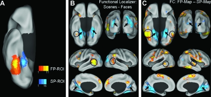
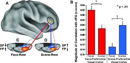
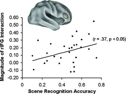
The resting brain is associated with significant intrinsic activity fluctuations, such as the correlated low-frequency (LF) blood oxygen level-dependent (BOLD) fluctuations measured by functional magnetic resonance imaging. Despite a recent expansion of studies investigating resting-state LF-BOLD correlations, their nature and function are poorly understood. A major constraint on LF-BOLD correlations appears to be stable properties of anatomic connectivity. There is also evidence that coupling can be modulated by recent or ongoing task performance, suggesting that certain components of correlated dynamics are malleable on short timescales. Here, we compared activity during extended periods of rest following performance of 2 distinct cognitive tasks using different categories of visual stimuli-faces and complex scenes. Prolonged exposure to these distinct categories of visual information caused frontal networks to couple differentially with posterior category-preferential visual regions during subsequent periods of rest. In addition, we report preliminary evidence suggesting that conditions exist in which the degree of modulation of LF-BOLD correlations predicts subsequent memory. The finding that resting-state LF-BOLD correlations are modulated by recent experience in functionally specific brain regions engaged during prior task performance clarifies their role as a dynamic phenomenon which may be involved in mnemonic processes.


Figures

Figure 1.
Figure 1.
Comparison of task-based functional localizer activity and FC maps. (A) Overlap of the FP- and SP-ROIs for all participants (n = 30) overlaid on the inflated right ventral cortical surface. (B) Group difference-maps for FP (yellow/orange scale) and SP (blue scale) regions defined by the contrast faces – scenes in the task-based functional localizer, overlaid on the inflated bilateral ventral (top left), posterior (top right), lateral (middle), and medial (bottom) cortical surfaces. (C) Group difference-maps for LF-BOLD correlations collapsed across all rest runs, specific to the FP-ROI (yellow/orange scale) and SP-ROI (blue scale) defined by the contrast FP-map – SP-map, overlaid on inflated cortical surfaces as in panel B. The functional localizer maps and FC difference-maps show similarities between the pattern of activation and correlated activity, respectively. Arrows indicate regions associated with scene processing: bilateral PHC, transverse occipital sulcus, and retrosplenial cortex, on the ventral, posterior, and medial surfaces, respectively. Circles indicate regions associated with face processing: FG and lateral temporal cortex, on the ventral and lateral surfaces, respectively.
Figure 2.
Figure 2.
LF-BOLD correlations during rest were modulated by previous task. (A) Region in the rIFG (circled) showing a task by ROI interaction for correlations with the FP- and SP-ROIs (t > 3.4, P < 0.001, 113 contiguous voxels; peak activation in MNI coordinates: x = 48, y = 36, z = 4; activation displayed at a threshold of: t > 2.76, P < 0.005). (B) Paired samples comparisons revealed a significant simple effect of task on correlations between rIFG and both the FP- and SP-ROIs (FP-ROI t = 3.0, P < 0.01, 2-tailed; SP-ROI t = −3.3, P < 0.01, 2-tailed). (C) Correlations with the FP-ROI increased during face–rest relative to scene–rest; (D) Correlations with the SP-ROI increased during scene-rest relative to face-rest. (C, D) The orange/yellow scale and blue-scale ROIs overlaid on the right ventral cortical surface images show the degree of overlap of all FP- and SP-ROIs, respectively, for all participants (n = 30); red and blue lines indicate correlation with FP-ROI and SP-ROI, respectively; solid lines and dotted lines indicate increased correlations and decreased correlations, respectively. Error bars represent standard error of the mean calculated for within-subjects design.
Figure 3.
Figure 3.
Both face and scene processing engage the rIFG during both task performance and subsequent rest. Region showing the task by ROI interaction in rIFG (A) overlapped peak activation for the task-based localizer contrast for faces (B: faces > fixation) and for scenes (C: scenes > fixation). (D) Conjunction of activation for all 3 contrasts, each contrast thresholded at: t > 2.76, P < 0.005. All activation maps displayed at a threshold of: t > 2.76, P < 0.005.
Figure 4.
Figure 4.
Coupling of LF-BOLD fluctuations in the rIFG with category-preferential visual regions during rest predicts subsequent memory. Across all participants (n = 30), the magnitude of the interaction in rIFG was significantly correlated with subsequent recognition accuracy for scenes (r = 0.37, P < 0.05, 2-tailed).

Similar articles

Cited by

References

    1. Albert NB, Robertson EM, Miall RC. The resting human brain and motor learning. Curr Biol. 2009;19:1023–1027. - PMC - PubMed
    1. Arfanakis K, Cordes D, Haughton VM, Moritz CH, Quigley MA, Meyerand ME. Combining independent component analysis and correlation analysis to probe interregional connectivity in fMRI task activation datasets. Magn Reson Imaging. 2000;18:921–930. - PubMed
    1. Bandettini PA. What's new in neuroimaging methods? Ann NY Acad Sci. 2009;1156:260–293. - PMC - PubMed
    1. Biswal B, Yetkin FZ, Haughton VM, Hyde JS. Functional connectivity in the motor cortex of resting human brain using echo-planar MRI. Magn Reson Med. 1995;34:537–541. - PubMed
    1. Boly M, Phillips C, Balteau E, Schnakers C, Degueldre C, Moonen G, Luxen A, Peigneux P, Faymonville ME, Maquet P, et al. Consciousness and cerebral baseline activity fluctuations. Hum Brain Mapp. 2008;29:868–874. - PMC - PubMed

Publication types