Review
. 2010 Apr;38(7):2119-33.
doi: 10.1093/nar/gkp1161. Epub 2009 Dec 21.

DNA supercoiling and its role in DNA decatenation and unknotting

Affiliations
Review

DNA supercoiling and its role in DNA decatenation and unknotting

Guillaume Witz et al. Nucleic Acids Res. 2010 Apr.

Abstract

Chromosomal and plasmid DNA molecules in bacterial cells are maintained under torsional tension and are therefore supercoiled. With the exception of extreme thermophiles, supercoiling has a negative sign, which means that the torsional tension diminishes the DNA helicity and facilitates strand separation. In consequence, negative supercoiling aids such processes as DNA replication or transcription that require global- or local-strand separation. In extreme thermophiles, DNA is positively supercoiled which protects it from thermal denaturation. While the role of DNA supercoiling connected to the control of DNA stability, is thoroughly researched and subject of many reviews, a less known role of DNA supercoiling emerges and consists of aiding DNA topoisomerases in DNA decatenation and unknotting. Although DNA catenanes are natural intermediates in the process of DNA replication of circular DNA molecules, it is necessary that they become very efficiently decatenated, as otherwise the segregation of freshly replicated DNA molecules would be blocked. DNA knots arise as by-products of topoisomerase-mediated intramolecular passages that are needed to facilitate general DNA metabolism, including DNA replication, transcription or recombination. The formed knots are, however, very harmful for cells if not removed efficiently. Here, we overview the role of DNA supercoiling in DNA unknotting and decatenation.


Figures

Figure 1.
Figure 1.
Atomic force microscopy (AFM) visualization of torsionally relaxed (A) and negatively supercoiled (B) bacterial plasmids pBR322. The deposition of the 4361 bp-long DNA chains is made on APTES modified mica (82).
Figure 2.
Figure 2.
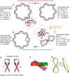
Topology and mechanics of DNA supercoiling. (A) Energy minimizing, torsionally relaxed, covalently closed DNA molecule with Lk = 8, Tw = 8, Wr = 0 and having the intrinsic DNA helicity of 10 bp/turn. The blow-up shows that interstrand crossings in the right-handed DNA helix are positive, since for topological considerations one assumes parallel orientation of DNA strands [see (E) for the topological convention of crossings’ signs]. (B) As a result of a combined action of DNA gyrase that decreased the linking number by two followed by the relaxation reaction of topo I, that increased the linking number by 1, the molecule presented in (A) has changed its linking number to Lk = 7. If that molecule were maintained in a planar configuration by ionic interaction with a charged surface, for example, its Tw would also change to 7, while Wr would remain unchanged. The change of DNA twist introduces a significant torsional tension into the elastic structure of the DNA as its helical repeat would need to change to 11.43 bp/turn, while its minimal torsional energy is achieved for a helix with 10 bp/turn. (C) The molecule presented in (B) has detached from a charged surface and minimized its elastic energy by adopting a supercoiled form with a Wr ≈−0.7 which permitted the molecule to greatly diminish its torsional tension as its twist has changed to ≈7.7, which is close to torsionally relaxed state. Due to the quadratic dependence of torsional and bending energies on the respective elastic deformations, it is energetically favourable to repartition the elastic stress due to the deficit of linking ΔLk into torsional and bending deformations. Usually ∼70% of the ΔLk are compensated by the acquired writhe (41,83). The blow-up shows that the inter-helix crossings between the two strands in negatively supercoiled DNA have negative sign as opposed to intra-helix crossings that have positive signs. (D) Opening of 10 bp by hybridization with nascent RNA, for example, is energetically more favourable in an unwound chain C than in the covalently closed, torsionally relaxed form A. The twist value is lower than in the torsionally relaxed DNA shown in (A). However, this causes no torsional stress as this twist is realized over a shorter region of pairing between the DNA strands, which re-establishes there the helicity of 10 bp/turn, while the open region is stabilized by the interaction with the hybridized RNA. (E) The topological sign convention. To determine the sign of individual crossings of two oriented curves, one checks in which sense one should turn the orientation vector of the overlying segment to have it pointing in the same direction as the vector of the underlying segment, while the rotation can not exceed 180°. If that rotation is clockwise, the crossing is negative and it is positive otherwise. (F) The concept of twist. Twist of DNA molecules is the sum of all the twists angles between the consecutive base pairs. The twist units are 360° rotations. (G) The concept of writhe. The same 3D curve, representing the axis of a given DNA molecule is observed from two different directions. The score provided by segment crossings can vary between the two cases, explaining why as writhe one takes the average value of crossings scores over all directions equisampling the sphere enclosing the 3D curve.
Figure 3.
Figure 3.
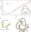
DNA knots, supercoiling and the geometric chirality. (A) Comparison of the elastic energies of simulated negatively supercoiled DNA molecules with 3000 bp that were either unknotted or formed left-handed trefoil knots [the energy graph is reproduced from ref. (40)]. ΔLk refers to the difference between the actual linking number of knotted or unknotted DNA molecules and that of torsionally relaxed unknotted DNA molecules. The light blue arrow indicates the energy difference between unknotted and knotted DNA molecules with the same ΔLke. The dark blue arrow indicates the energy difference between unknotted and knotted DNA molecules resulting from one round of a Topo II-like action. The black arrow indicates the energy difference between unknotted and knotted DNA molecules under the unrealistic assumption that topoisomerase II could mediate an intramolecular passage reaction without changing the linking number. Note that left-handed trefoil knots reach their minimal energy state for ΔLk ≈3.5 and that this closely corresponds to the average writhe of torsionally relaxed left-handed trefoil knots (43) (shown in the inset). For the convenience of the presentation the figure shows three different energetic consequences of formation of left-handed trefoil knot starting from unknotted DNA molecules that were kept at their supercoiling equilibrium at ΔLk ≈ −5. However, natural supercoiling of 3000 bp-long DNA would rather keep the unknotted molecules at ΔLk ≈−15. Therefore, at physiological level of negative supercoiling the passages leading to knot formation will be much stronger opposed by the free energy gradient than for the case analysed here. (B) The concept of geometrical chirality. To determine the geometrical chirality of a crossing one looks what is the smallest rotation of the overlying segment that brings it parallel with the underlying one. If that rotation is clockwise the crossing is left-handed and it is right-handed otherwise. (C) Knot 52L with indicated geometrical chirality of their crossings. An intersegmental passage at any of left-handed crossing will unknot the knot, while a passage at any of right-handed crossings will convert the 52L knot into 31L knot.
Figure 4.
Figure 4.
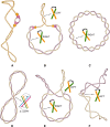
Topological transitions during replication of circular DNA. (A) Supercoiled DNA that just started its process of DNA replication. The molecule is still negatively supercoiled and shows right-handed interwinding. The negative supercoiling helps to initiate the process of DNA replication (7). (B) Partially replicated DNA molecule with positive torsional stress causing formation of left-handed interwinding in the unreplicated portion and of right-handed interwinding of precatenanes in the replicated portion. The left-handed interwinding can be easily relaxed by DNA gyrase or by topoisomerase IV. Right-handed interwinding is a poor substrate for the relaxation reaction by Topo IV. (C) Standard representation of freshly replicated molecules forming multiply interlinked DNA catenanes. All crossings in this representation have right-handed chirality but this representation is not the equilibrium form of multiply interlinked DNA catenanes. (D) Schematic presentation of the equilibrium form of multiply interlinked DNA catenanes. Minimization of the elastic energy of multiply interlinked catenanes leads to the formation of a left-handed folding of the entire catenanes (21,78). This folding leads to the apparition of left-handed DNA–DNA juxtapositions that are very good substrates for Topo IV-mediated passages that lead to decrease of DNA catenation. (E) Catenanes with decreasing number of interlinks. Individual rings get supercoiled by DNA gyrase, and left-handed crossings may form in the region deformed by the extrusion of supercoils. (F) Singly interlinked catenanes have a complete freedom to form left-handed juxtapositions. Supercoiling provides the necessary ‘pressure’ leading to unlinking. The representations of DNA molecules at different steps of DNA replication are not shown at the same scale but their size was chosen in order to make important points clear. However, all the drawings correspond to sequential stages of DNA replication of a molecule with 800 bp.

Similar articles

Cited by

References

    1. Du Q, Kotlyar A, Vologodskii A. Kinking the double helix by bending deformation. Nucleic Acids Res. 2008;36:1120–1128. - PMC - PubMed
    1. Cloutier TE, Widom J. Spontaneous sharp bending of double-stranded DNA. Mol. Cell. 2004;14:355–362. - PubMed
    1. Demurtas D, Amzallag A, Rawdon EJ, Maddocks JH, Dubochet J, Stasiak A. Bending modes of DNA directly addressed by cryo-electron microscopy of DNA minicircles. Nucleic Acids Res. 2009;37:2882–2893. - PMC - PubMed
    1. Vologodskii AV, Levene SD, Klenin KV, Frank-Kamenetskii M, Cozzarelli NR. Conformational and thermodynamic properties of supercoiled DNA. J. Mol. Biol. 1992;227:1224–1243. - PubMed
    1. Strick TR, Allemand JF, Bensimon D, Bensimon A, Croquette V. The elasticity of a single supercoiled DNA molecule. Science. 1996;271:1835–1837. - PubMed

Publication types

MeSH terms