. 2010 Mar 5;285(10):7405-16.
doi: 10.1074/jbc.M109.028365. Epub 2009 Dec 21.

Vesicle-associated membrane protein (VAMP) cleavage by a new metalloprotease from the Brazilian scorpion Tityus serrulatus

Affiliations

Vesicle-associated membrane protein (VAMP) cleavage by a new metalloprotease from the Brazilian scorpion Tityus serrulatus

Paul L Fletcher Jr et al. J Biol Chem. .

Abstract

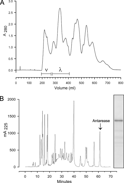
We present evidence that venom from the Brazilian scorpion Tityus serrulatus and a purified fraction selectively cleave essential SNARE proteins within exocrine pancreatic tissue. Western blotting for vesicle-associated membrane protein type v-SNARE proteins (or synaptobrevins) reveals characteristic alterations to venom-treated excised pancreatic lobules in vitro. Immunocytochemistry by electron microscopy confirms both the SNARE identity as VAMP2 and the proteolysis of VAMP2 as a marked decrease in secondary antibody-conjugated colloidal gold particles that are predominantly associated with mature zymogen granules. Studies with recombinant SNARE proteins were used to determine the specific cleavage site in VAMP2 and the susceptibility of VAMP8 (endobrevin). The VAMP2 cleavage site is between the transmembrane anchor and the SNARE motif that assembles into the ternary SNARE complex. Inclusion of divalent chelating agents (EDTA) with fraction nu, an otherwise active purified component from venom, eliminates SNARE proteolysis, suggesting the active protein is a metalloprotease. The unique cleavages of VAMP2 and VAMP8 may be linked to pancreatitis that develops following scorpion envenomation as both of these v-SNARE proteins are associated with zymogen granule membranes in pancreatic acinar cells. We have isolated antarease, a metalloprotease from fraction nu that cleaves VAMP2, and report its amino acid sequence.


Figures

FIGURE 1.
FIGURE 1.
TSV cleaves synaptobrevins in pancreatic lobules in vitro. A, dose-response curves for TSV, Fx ν, and Fx λ effects on secretion of radiolabeled (newly synthesized) proteins. Pancreatic lobules were pulsed with [3H]leucine for 10 min at 37 °C in KRB and then rinsed. Chase-incubation followed for 3 h, and then lobules were homogenized. Trichloroacetic acid-precipitable protein samples were processed for scintillation counting. Data for TSV are from unpublished experiments3 and previous publications (16, 20). Data for Fx ν and Fx λ are from one experiment performed with duplicate flasks (n = 2). Average resting secretion in untreated control lobules was 7.1%. B, Western blots (WB) for phosphotyrosine (PY20), VAMP2, and VAMP3 showing TSV dose effect on VAMP degradation. The TSV doses of 50 and 5 μg/ml are hyperstimulatory for secretion. The 1 μg/ml dose is optimal (see A). Control lobules (CON) are in KRB. C, time course for incubations at two temperatures. TSV is 50 μg/ml. D, Western blots for VAMP2 and VAMP8 in lobules incubated for 3 h in optimal stimulatory doses for carbachol (CARB) (10 μm) and caerulein (CAER) (1 nm) and hyperstimulatory toxin γ dose (100 nm). Lobules for Western blots were incubated in KRB in vitro at 37 °C under the conditions described and then homogenized. Homogenate samples (20 μg/lane) were electrophoresed by SDS-PAGE and then transblotted. For samples in B–D, n = 4 or 2 experiments with duplicate flasks.
FIGURE 2.
FIGURE 2.
Immunogold VAMP2 electron micrographs. Pancreatic lobules were incubated in vitro for 1 h at 37 °C. A, unstimulated control. Zymogen granules (Z) are positively immunostained for VAMP2 shown as 10-nm gold particles (arrowheads). B, hyperstimulation with 50 μg/ml TSV. The absence of gold particles signifies the loss of VAMP2. G, Golgi complex. The number of micrographs analyzed for each condition was 14 in A and 19 in B. The number of counted ZG was 159 in A and 194 in B. The number of counted gold particles was 2645 in A and 632 in B. Scale bars, 0.5 μm.
FIGURE 3.
FIGURE 3.
TSV and Fx ν cleave synaptobrevins in ZG and ZGM in vitro. After in vitro incubation at 37 °C, ZG, or ZGM (25 μg/lane) were analyzed by SDS-PAGE, followed by Western blot (WB). Shown is the effect of 50 μg/ml TSV on VAMP2 in isolated pancreatic ZG (A) and ZGM (B). C, effect of 10 μg/ml Fx ν on VAMP8. D, effects of 50 μg/ml TSV and 10 μg/ml Fx ν on VAMP2 and tyrosine phosphorylation (PY20) in isolated ZG in vitro. The number of experiments was as follows: ZG, n = 10; ZGM, n = 4.
FIGURE 4.
FIGURE 4.
Characterization of TSV and Fx ν cleavage of recombinant synaptobrevins. Transblots are shown. A, 10 μm VAMP2 cytoplasmic portion 1–94 proteins incubated in vitro at 37 °C for 30 min with 10 μg/ml TSV or 50 μg/ml Fx ν or Fx λ. B, inhibition of 10 μg/ml Fx ν proteolytic activity by a 60-min preincubation with 10 mm EDTA, pH 7, in vitro at 37 °C. 10 μm VAMP2-(1–94) proteins were then added for a further 60-min incubation. C, VAMP2 proteins incubated in vitro at 37 °C for 30 min with 10 μg/ml Fx ν or 10 μg/ml TSV. WT, wild type 10 μm VAMP2 cytoplasmic portion 1–96. K85A/R86S/K87A, mutant 10 μm VAMP2 cytoplasmic portion 1–96 with altered cleavage site. D, time course for 10 μm VAMP2-(1–94) cleavage by 10 μg/ml Fx ν at 37 °C. E, 10 μm VAMP2-(1–94) cleavage during incubation in vitro at 37 °C for 30 min with 50 μg/ml C. sculpturatus venom (CSV). The number of experiments under various conditions for each SNARE protein was as follows: VAMP2-(1–94), n = 20; WT VAMP2-(1–96), n = 9; VAMP2 K85A/R86S/K87A-(1–96), n = 3.
FIGURE 5.
FIGURE 5.
Isolation of cleaved VAMP2 peptides. C18 reverse phase chromatographic separation of WT 10 μm VAMP2-(1–94) peptides after in vitro incubation at 37 °C for 30 min. A, 10 μg/ml Fx ν-cleaved VAMP2 peptides with sequences. B, incubated control WT VAMP2.
FIGURE 6.
FIGURE 6.
Molecular model of VAMP2 and description of cleavage sites by clostridial toxins and Fx ν. Top, VAMP2 and VAMP8 FASTA homology alignment with cleavage sites (red) and transmembrane segments (violet). Center, VAMP2 amino acid sequence coordinated with a molecular model, where font color reflects sequence features: Fx ν cleavage (red) at broken ribbon, BoNT and TeNT cleavages (yellow and orange) at arrows, and SNARE motifs, X1 and X2 (blue). The intervening sequence ribbon is green. The model and FASTA homology were generated by InsightII (Accelrys).
FIGURE 7.
FIGURE 7.
TSV and Fx ν cleave other recombinant SNARE proteins. Transblots are shown. 90 μm WT endobrevin (VAMP8) cytoplasmic portion 1–74 (left) or 20 μm WT SNAP25 full-length 1–206 (right) was incubated with either 10 μg/ml TSV or 10 μg/ml Fx ν in vitro at 37 °C for 30 min. The number of experiments was as follows: VAMP8, n = 3; SNAP25, n = 7.
FIGURE 8.
FIGURE 8.
The assembled SNARE complex and Fx ν. A transblot is shown. SNARE complex (CPX) was incubated with 10 μg/ml Fx ν in vitro at 37 °C for 60 min. After mixing with Laemmli buffer, one sample was gel-loaded as intact complex (not boiled), and another sample was boiled for 5 min for disassembly prior to loading (boiled). The image is representative of 14 blots from three experiments using three sequentially purified SNARE complexes.
FIGURE 9.
FIGURE 9.
Chromatographic isolation and purification of Fx ν. A, gel filtration separation of T. serrulatus whole venom with Sephadex G-50. Pooled fractions Fx ν and Fx λ are indicated by the bars. B, reverse phase purification of Fx ν. The indicated peak (arrow) is the source of antarease. Inset, PAGE transblot of the antarease peak that was sequenced.
FIGURE 10.
FIGURE 10.
Ion exchange chromatographic purification of Fx ν. Anion exchange separation of Fx ν produced biologically active proteins. Pools I–IV were incubated with 20 μm WT VAMP2-(1–96) in vitro at 37 °C for 30 min to detect proteolytic activity. Insets, PAGE transblots of incubation samples. Fraction III-6 (arrowhead) cleaves VAMP2 and is a source for the antarease sequence determination.
FIGURE 11.
FIGURE 11.
Antarease primary structure. A minimum group of overlapping peptides was used to determine the amino acid sequence. Direct N-terminal sequence of N-isopropylcarboxyamidomethyl-antarease provided the initial 30 residues (Edmaneeee …). The peptides derived from subsequent cleavages are indicated as follows: trypsin peptides (Trypsintttttttt …), Asp-N peptides (AspNDnDnDnDnDn …), Arg-C peptides (ArgCRcRcRcRcRc …), CNBr peptides (CNBrBrBrBrBr …), Glu-C peptides (GluCEcEcEcEcEc …), and Lys-C peptides (LysCKcKcKcKcKc …). The putative zinc-binding site is shown underlined in boldface type. The arrows indicate the end of a peptide. The absence of an arrow indicates a sequence continuing on the next line. The antarease sequence has been included in the UniProt Knowledgebase with accession number P86392.

Similar articles

Cited by

References

    1. Ferro-Novick S., Jahn R. (1994) Nature 370, 191–193 - PubMed
    1. Rothman J. E. (1994) Nature 372, 55–63 - PubMed
    1. Südhof T. C. (1995) Nature 375, 645–653 - PubMed
    1. Jahn R., Scheller R. H. (2006) Nat. Rev. 7, 631–643 - PubMed
    1. Breidenbach M. A., Brunger A. T. (2005) Trends Mol. Med. 11, 377–381 - PubMed

Publication types

MeSH terms

Associated data