. 2010 Feb 26;285(9):6739-49.
doi: 10.1074/jbc.M109.058552. Epub 2009 Dec 21.

Dynamic changes in the subcellular distribution of Gpd1p in response to cell stress

Affiliations

Dynamic changes in the subcellular distribution of Gpd1p in response to cell stress

Sunhee Jung et al. J Biol Chem. .

Abstract

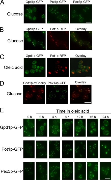
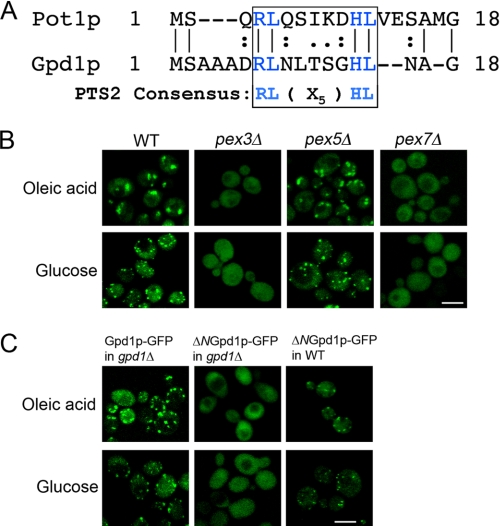
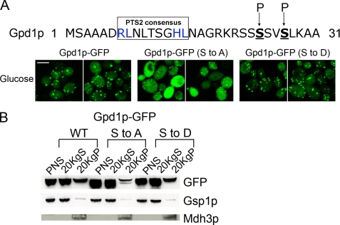
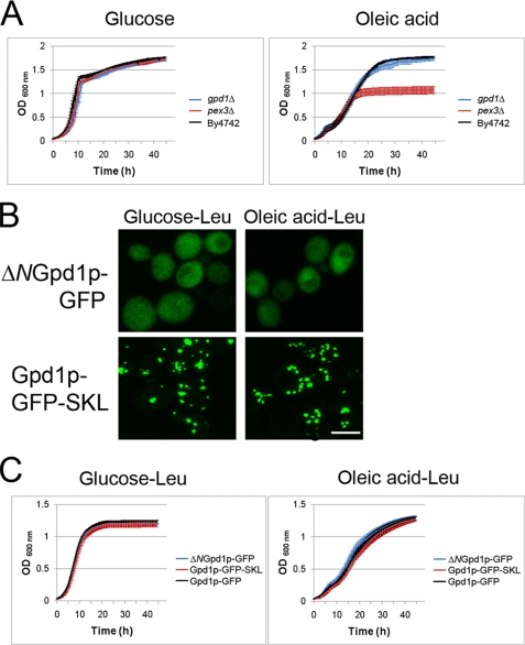
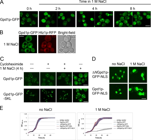
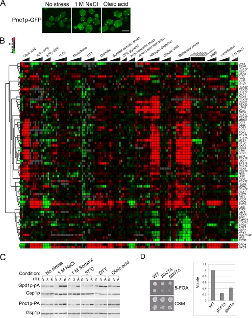
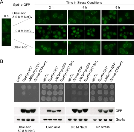
Gpd1p is a cytosolic NAD(+)-dependent glycerol 3-phosphate dehydrogenase that also localizes to peroxisomes and plays an essential role in the cellular response to osmotic stress and a role in redox balance. Here, we show that Gpd1p is directed to peroxisomes by virtue of an N-terminal type 2 peroxisomal targeting signal (PTS2) in a Pex7p-dependent manner. Significantly, localization of Gpd1p to peroxisomes is dependent on the metabolic status of cells and the phosphorylation of aminoacyl residues adjacent to the targeting signal. Exposure of cells to osmotic stress induces changes in the subcellular distribution of Gpd1p to the cytosol and nucleus. This behavior is similar to Pnc1p, which is coordinately expressed with Gpd1p, and under conditions of cell stress changes its subcellular distribution from peroxisomes to the nucleus where it mediates chromatin silencing. Although peroxisomes are necessary for the beta-oxidation of fatty acids in yeast, the localization of Gpd1p to peroxisomes is not. Rather, shifts in the distribution of Gpd1p to different cellular compartments in response to changing cellular status suggests a role for Gpd1p in the spatial regulation of redox potential, a process critical to cell survival, especially under the complex stress conditions expected to occur in the wild.


Figures

FIGURE 1.
FIGURE 1.
Cellular localization of Gpd1p. A, fluorescence micrographs depicting the subcellular localization of Gpd1p, Pot1p, and Pex3p genomically tagged with GFP in cells grown in glucose medium. B, double-labeling fluorescence confocal microscopy images of glucose-grown cells synthesizing genomically encoded Gpd1p-GFP and containing a plasmid coding for peroxisomal thiolase tagged with monomeric red fluorescent protein (Pot1p-RFP) showing co-localization of the two signals to punctate structures characteristic of peroxisomes (Overlay). C, fluorescence confocal microscopy images of doubly labeled cells as in B but incubated in oleic acid medium for 16 h. D, double-labeling fluorescence micrographs showing co-localization of Gpd1p-mCherry with Pex13p-GFP in glucose grown cells. E, localization of Gpd1p-GFP, Pot1p-GFP, and Pex3p-GFP in wild-type cells. The time in oleic acid medium is indicated. Bars, 10 μm.
FIGURE 2.
FIGURE 2.
PTS2-dependent targeting of Gpd1p to peroxisomes. A, sequence comparison of the N termini of Pot1p and Gpd1p showing they contain the PTS2 consensus sequence. B, peroxisomal targeting of Gpd1p is mediated by Pex7p. Wild-type BY4742 cells or the respective pex3Δ, pex5Δ, and pex7Δ mutants synthesizing genomically encoded Gpd1p-GFP were incubated in oleic acid medium for 8 h at 30 °C or in glucose medium and observed by confocal microscopy. The Gpd1p-GFP chimera fails to localize to peroxisomes in pex3Δ cells lacking peroxisomes and in pex7Δ cells lacking the PTS2 receptor but targets efficiently to peroxisomes in pex5Δ cells lacking the PTS1 receptor. Bar, 10 μm. C, Gpd1p-GFP import into peroxisomes is mediated by a functional PTS2 at the N terminus of Gpd1p. GFP chimeras of Gpd1p or Gpd1p lacking its N-terminal 17 amino acids (ΔNGpd1p-GFP) were localized in WT and gpd1Δ cells incubated in oleic acid or glucose medium for 8 h at 30 °C. ΔNGpd1p-GFP failed to localize to peroxisomes in gpd1Δ but not in wild-type cells. Bar, 10 μm.
FIGURE 3.
FIGURE 3.
Efficient peroxisomal import of Gpd1p by phosphorylation of two serine residues of Gpd1p. A, serines 24 and 27 (underlined and in boldface) of Gpd1p were changed to alanine (Ser to Ala) or aspartic acid (Ser to Asp), and the subcellular distributions of Gpd1p-GFP (Ser to Ala) and Gpd1p-GFP (Ser to Asp) were examined in glucose-grown cells by confocal microscopy. Bar, 10 μm. B, distribution of Gpd1p-GFP, Gpd1p-GFP (Ser to Ala), and Gpd1p-GFP (Ser to Asp) was analyzed by subcellular fractionation. Postnuclear supernatant (PNS), 20KgS fractions enriched for cytosol (loaded at 1 cell equivalent), and 20KgP fractions enriched for peroxisomes and mitochondria (loaded at 10 cell equivalents) were analyzed by Western blotting using anti-GFP antibodies. Gsp1p was used as a loading control for postnuclear supernatant and 20KgS. C-terminal SKL was used as a loading control for the 20KgP, and the molecular weight of the band corresponds to the molecular weight of Mdh3p.
FIGURE 4.
FIGURE 4.
Gpd1p is not required for β-oxidation of fatty acids. A, cell growth assay of gpd1Δ, pex3Δ, and WT cells in media with glucose or oleic acid as the carbon source. Growth was monitored using a liquid growth assay in a Bioscreen automatic reader and plotted as the average of three independent experiments and three technical replicates of each. gpd1Δ cells grew at the same rate as WT cells in oleic acid and glucose. Error bars indicate standard deviation. B, subcellular localization of ΔNGpd1p-GFP and Gpd1p-GFP-SKL in SM-Leu media containing glucose or oleic acid (8 h). C, growth assays as in A, but the strains shown are gpd1Δ cells transformed with pGPD1-GFP, pΔNGPD1-GFP, or pGPD1-GFP-SKL.
FIGURE 5.
FIGURE 5.
Gpd1p localizes to peroxisomes but redistributes to the nucleus under conditions of stress. A, localization of Gpd1p genomically tagged with GFP was analyzed by confocal microscopy at the different times upon exposure to 1 m NaCl. Bar, 10 μm. B, Gpd1p-GFP co-localizes in the nucleus with Htz1p-RFP as determined by confocal microscopy. Images were obtained after cells were exposed to 1 m NaCl for 4 h. Bar, 10 μm. C, peroxisomal Gpd1p does not exit peroxisomes upon exposure to 1 m NaCl. Cells expressing GFP chimeras of wild-type (pGPD1-GFP) or peroxisomal (pGPD1-GFP-SKL) Gpd1p were treated with 1 m NaCl for 4 h in the presence (+) or absence (−) of cycloheximide. D, subcellular localization of ΔNGpd1p-GFP-NLS and Gpd1p-GFP-NLS in glucose media in the absence or presence of 1 m NaCl. Cytoplasmic signals of both Gpd1p variants are increased by 1 m NaCl. E, cell growth assay of cells expressing GFP chimeras of wild-type (pGPD1-GFP), cytosolic (pΔNGPD1-GFP), peroxisomal (pGPD1-GFP-SKL), or nuclear (pΔNGPD1-GFP-NLS) Gpd1p. Cells were grown as in Fig. 4 but in the presence or absence of NaCl. All fusions were expressed from the plasmid pRS315 in gpd1Δ cells. Empty plasmid served as a control. The curves represent the average of three independent experiments and three technical replicates of each, and error bars represent standard deviation.
FIGURE 6.
FIGURE 6.
Similarities in expression and distribution of Pnc1p and Gpd1p. A, localization of Pnc1p-GFP chimera in glucose, 1 m NaCl (4 h), or oleic acid (4 h) as determined by confocal microscopy. Bar, 10 μm. B, hierarchical clustering of the transcriptional expression profiles of genes known to function in peroxisome biology over a broad range of stress conditions. GPD1 and PNC1 were among the most closely co-expressed genes within this dataset and within the whole yeast genome (correlation coefficient 0.85). C, co-synthesis of Gpd1p and Pnc1p across various stress conditions. Whole cell lysates of yeast strains subjected to various stress conditions were analyzed by Western blotting. Gsp1p was used as a loading control. D, strains lacking PNC1 or GPD1 failed to efficiently silence subtelomeric URA3. Wild-type, pnc1Δ, and gpd1Δ strains containing a subtelomeric URA3 gene were assayed for URA3 gene expression by spotting 10-fold dilutions onto CSM- and 5-fluoroorotic acid (5-FOA)-containing plates. The graph represents the silencing efficiency as the number of viable mutant colonies on 5-fluoroorotic acid normalized to wild type across nine independent experiments. Error bars represent the standard deviation.
FIGURE 7.
FIGURE 7.
Balance of subcellular distribution of Gpd1p is required for efficient growth in the combined oleic acid and 0.8 m NaCl stress. A, localization of Gpd1p-GFP chimera in cells exposed to the combined stress of NaCl and oleic acid. Wild-type cells expressing Gpd1p-GFP were incubated in oleic acid, 0.8 m NaCl, or oleic acid and 0.8 m NaCl. Fluorescence images were captured at the times indicated using confocal microscopy. Bar, 10 μm. B, cell growth on plates containing oleic acid, 0.8 m NaCl, or oleic acid plus 0.8 m NaCl. Strains were grown to exponential growth phase in minimal medium, and the culture was serially diluted and applied to plates containing no stress, oleic acid, 0.8 m NaCl, or oleic acid plus 0.8 m NaCl. The plates were photographed following 2 days of incubation for control (no stress), 3 days incubation for oleic acid or 0.8 m NaCl, and 5 days incubation for both oleic acid and 0.8 m NaCl at 30 °C. Whole cell lysates of the strains subjected to each of the different growth conditions for 16 h were analyzed by Western blotting with the indicated antibodies. Gsp1p serves as a loading control.

Similar articles

Cited by

References

    1. Larsson K., Ansell R., Eriksson P., Adler L. (1993) Mol. Microbiol. 10, 1101–1111 - PubMed
    1. Larsson C., Påhlman I. L., Ansell R., Rigoulet M., Adler L., Gustafsson L. (1998) Yeast 14, 347–357 - PubMed
    1. Aguilera J., Rodríguez-Vargas S., Prieto J. A. (2005) Mol. Microbiol. 56, 228–239 - PubMed
    1. Phillips S. A., Thornalley P. J. (1993) Eur. J. Biochem. 212, 101–105 - PubMed
    1. Vander Jagt D. L., Robinson B., Taylor K. K., Hunsaker L. A. (1992) J. Biol. Chem. 267, 4364–4369 - PubMed

Publication types

MeSH terms

Substances