. 2010 Jan 15;123(Pt 2):236-45.
doi: 10.1242/jcs.053264. Epub 2009 Dec 21.

Identification of beta-catenin as a target of the intracellular tyrosine kinase PTK6

Affiliations

Identification of beta-catenin as a target of the intracellular tyrosine kinase PTK6

Helena L Palka-Hamblin et al. J Cell Sci. .

Abstract

Disruption of the gene encoding protein tyrosine kinase 6 (PTK6) leads to increased growth, impaired enterocyte differentiation and higher levels of nuclear beta-catenin in the mouse small intestine. Here, we demonstrate that PTK6 associates with nuclear and cytoplasmic beta-catenin and inhibits beta-catenin- and T-cell factor (TCF)-mediated transcription. PTK6 directly phosphorylates beta-catenin on Tyr64, Tyr142, Tyr331 and/or Tyr333, with the predominant site being Tyr64. However, mutation of these sites does not abrogate the ability of PTK6 to inhibit beta-catenin transcriptional activity. Outcomes of PTK6-mediated regulation appear to be dependent on its intracellular localization. In the SW620 colorectal adenocarcinoma cell line, nuclear-targeted PTK6 negatively regulates endogenous beta-catenin/TCF transcriptional activity, whereas membrane-targeted PTK6 enhances beta-catenin/TCF regulated transcription. Levels of TCF4 and the transcriptional co-repressor TLE/Groucho increase in SW620 cells expressing nuclear-targeted PTK6. Knockdown of PTK6 in SW620 cells leads to increased beta-catenin/TCF transcriptional activity and increased expression of beta-catenin/TCF target genes Myc and Survivin. Ptk6-null BAT-GAL mice, containing a beta-catenin-activated LacZ reporter transgene, have increased levels of beta-galactosidase expression in the gastrointestinal tract. The ability of PTK6 to negatively regulate beta-catenin/TCF transcription by modulating levels of TCF4 and TLE/Groucho could contribute to its growth-inhibitory activities in vivo.


Figures

Fig. 1.
Fig. 1.
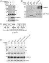
Tyrosine phosphorylation of β-catenin in cells expressing PTK6. HEK293 cells were co-transfected with wild-type PTK6, PTK6 YF or PTK6 KM and β-catenin. (A) Immunoblot analysis of Triton X-100 (TX-100)-soluble lysates using anti-phosphotyrosine (PY), β-catenin and PTK6 antibodies. α-tubulin was detected as a loading control. TX-100-soluble lysates from A were used to immunoprecipitate tyrosine-phosphorylated proteins using anti-PY antibodies (B) or β-catenin using a β-catenin-specific antibody (C). Immunoblot analysis was performed using anti-PY, β-catenin and PTK6 antibodies (B,C). Asterisk denotes the position of tyrosine-phosphorylated β-catenin (C). Size markers indicating molecular mass are shown to left of blots (kDa).
Fig. 2.
Fig. 2.
Downregulation of β-catenin transcriptional activity by PTK6. HEK293 cells were co-transfected with wild-type PTK6, PTK6 YF or PTK6 KM and β-catenin. (A) Immunoblot analysis of nuclear and cytoplasmic fractions using anti-PY, β-catenin and PTK6 antibodies. Immunoblotting for the nuclear marker Sp1 and the cytoplasmic marker α-tubulin served as controls. (B) β-catenin was immunoprecipitated from nuclear and cytoplasmic fractions using a β-catenin-specific antibody. Immunoblot analysis was performed using anti-PY antibodies and with the Myc epitope tag antibody to detect ectopically expressed β-catenin and PTK6. Asterisk denotes the position of tyrosine-phosphorylated β-catenin. Size markers indicating molecular mass are shown (kDa). (C) β-catenin/TCF reporter gene expression in HEK293 cells co-expressing PTK6 and β-catenin. HEK293 cells were transfected with the Super8XTOPFlash (TOPFlash) luciferase reporter construct containing eight TCF/Lef binding sites or the control Super8XFOPFlash (FOPFlash) construct containing eight mutated TCF/Lef binding sites, vector (−) PTK6 (WT), PTK6 YF (YF) or PTK6 KM (KM) DNA and β-catenin DNA. Luciferase activity was measured in cell lysates. The decrease in luciferase activity observed between vector and all forms of PTK6 as well as between wild-type PTK6 and PTK6 YF is statistically significant (means ± s.d.; *P<0.05, **P<0.005).
Fig. 3.
Fig. 3.
PTK6 directly associates with and phosphorylates several tyrosine residues in β-catenin in vitro. (A) Recombinant human PTK6 and wild-type β-catenin were incubated in kinase buffer with or without ATP. Immunoblot analysis of the in vitro kinase reaction was performed using anti-PY, β-catenin and PTK6 antibodies. (B) Recombinant human PTK6 and wild-type β-catenin were incubated in kinase buffer with or without ATP. Phosphorylated and unphosphorylated β-catenin was then incubated with PTK6 fusion proteins [full length (FL), SH2, SH3 and SH2-SH3 domains]. Immunoblot analysis of the GST pull-down was performed using β-catenin and GST tag antibodies. (C) Schematic representation of β-catenin that contains 12 armadillo repeats. Tyrosine residues 64 (Y64), Y142 and Y331 or Y333 were identified as sites phosphorylated by PTK6 using tandem mass spectrometry. (D) Recombinant human PTK6 and wild-type β-catenin or β-catenin YF point mutants were subjected to in vitro kinase assay and immunoblotting as described in A. β-catenin point mutants with a mutation at Y64 show no detectable tyrosine phosphorylation in vitro. Size markers indicating molecular mass are included in A, B and D (kDa). Asterisks denote the position of tyrosine-phosphorylated β-catenin and arrowheads indicate the position of activated PTK6 (A and D).
Fig. 4.
Fig. 4.
Phosphorylation of β-catenin on tyrosine is not essential for PTK6-mediated repression of β-catenin/TCF-regulated transcription. (A) HEK293 cells were transfected with PTK6 YF and Myc-tagged wild-type (WT) β-catenin or β-catenin YF point mutants. β-catenin was immunoprecipitated using a β-catenin antibody from TX-100-soluble cell lysates. Immunoblot analysis of immunoprecipitated wild-type β-catenin or β-catenin YF point mutants using anti-PY antibodies, the Myc epitope tag antibody to detect ectopically expressed β-catenin (β-catenin) and the PTK6 antibody. β-catenin point mutants with a mutation at Y64 show no detectable tyrosine phosphorylation in cells. Size markers indicating molecular mass are shown (kDa). Asterisk denotes the position of tyrosine phosphorylated β-catenin. (B) β-catenin/TCF reporter gene expression in HEK293 cells co-expressing PTK6 and β-catenin Y64F. HEK293 cells were transfected with Super8XTOPFlash (TOPFlash) or Super8XFOPFlash (FOPFlash), vector (−) PTK6 (WT), PTK6 YF (YF) or PTK6 KM (KM) DNA and β-catenin Y64F. The decrease in luciferase activity observed between vector and all forms of PTK6, as well as between wild-type PTK6 and PTK6 YF is statistically significant (*P<0.05, **P<0.005; mean ± s.d.). (C) β-catenin/TCF reporter gene expression in HEK293 cells co-expressing PTK6 YF and β-catenin point mutants (error bars indicate s.d.). HEK293 cells were transfected with Super8XTOPFlash, vector or PTK6 YF DNA and β-catenin Y142F, Y64F/Y142F or Y64F/Y142F/Y331,333F DNA. PTK6 YF was able to inhibit the transcriptional activity of all tested β-catenin mutants.
Fig. 5.
Fig. 5.
PTK6 regulates endogenous β-catenin transcriptional activity in SW620 cells. (A) β-catenin/TCF reporter gene expression in SW620 cells expressing nuclear-targeted PTK6. SW620 cells were transfected with Super8XTOPFlash (TOPFlash) or Super8XFOPFlash (FOPFlash), vector (−) NLS.PTK6 (WT), NLS.PTK6 YF (YF) or NLS.PTK6 KM (KM) DNA. Luciferase activity was measured in cell lysates. The decrease in luciferase activity observed between vector and PTK6 or PTK6 YF is statistically significant (*P<0.05; error bars indicate s.d.). (B) SW620 cells were transfected with nuclear-targeted PTK6, PTK6 YF or PTK6 KM. Immunoblot analysis was performed using β-catenin and PTK6 antibodies. (C) β-catenin/TCF reporter gene expression in SW620-expressing membrane-targeted PTK6. SW620 cells were transfected as in A with vector (−) Palm.PTK6 (WT), Palm.PTK6 YF (YF) or Palm.PTK6 KM (KM) DNA. Luciferase activity was measured in cell lysates. The increase in luciferase activity observed between vector and PTK6 or PTK6 YF is statistically significant (*P<0.005; error bars indicate s.d.). (D) SW620 cells were transfected with membrane-targeted PTK6, PTK6 YF or PTK6 KM. Immunoblot analysis was performed using β-catenin and PTK6 antibodies. Immunoblotting for Sp1 and E-cadherin served as controls. (E) Nuclear PTK6 expression upregulates TCF4 and TLE/Groucho protein levels. SW620 cells were transfected with nuclear-targeted PTK6, PTK6 YF or PTK6 KM. Nuclear fractions were resolved by SDS-PAGE. Immunoblot analysis was performed using anti-PY, β-catenin, TCF4, TLE/Groucho and PTK6 antibodies. Changes in protein expression levels were quantified using ImageJ and on average a two-fold increase was observed in TCF4 and TLE protein levels. Size markers indicating molecular mass are shown for B, D and E (kDa).
Fig. 6.
Fig. 6.
Endogenous PTK6 negatively regulates β-catenin in SW620 cells. (A) β-catenin/TCF reporter gene expression in PTK6-knockdown cells. Stable SW620 cell lines expressing two different PTK6 shRNAs (shRNA49, shRNA52) were transfected with Super8XTOPFlash (TOPFlash) or Super8XFOPFlash (FOPFlash) and the luciferase activity was measured in cell lysates. The increase in luciferase activity observed between vector and shRNA49 or shRNA52 is statistically significant (*P<0.005; error bars indicate s.d.) (B) Immunoblot analysis of PTK6-knockdown SW620 cells with β-catenin, PTK6, Myc and Survivin antibodies. Immunoblotting for β-actin served as a loading control. Size markers indicating molecular mass are shown (kDa).
Fig. 7.
Fig. 7.
Disruption of PTK6 gene expression leads to ectopic β-catenin/TCF transcriptional activity in the mouse intestine. (A) Enhanced reporter gene expression in distal colons of Ptk6−/− BAT-GAL mice. Colons from Ptk6+/+ (a,c,e) and Ptk6−/− (b,d,f) BAT-GAL animals were stained for β-galactosidase activity. Numerous positive crypt units could be detected in whole mount preparations at low magnification in Ptk6−/− BAT-GAL mice (b). Colons were sectioned and positive cells were found throughout crypt units in Ptk6 null colons (d,f). Colon sections were counterstained with Nuclear Fast Red. (B) Enhanced reporter gene expression in the small intestines of Ptk6−/− BAT-GAL mice. Small intestines from Ptk6+/+ (a) and Ptk6−/− (b-d) BAT-GAL animals were stained for β-galactosidase activity and sectioned. Single cells at the base of the crypts in PTK6-null intestines were positive for β-galactosidase activity (b, arrow). These cells contained granules characteristic of Paneth cells. (c) Magnification of boxed area in b. (d) Magnification of boxed area in c. Small intestine sections were counterstained with hematoxylin and eosin. Scale bars: 50 μm.

Similar articles

Cited by

References

    1. Andreu P., Colnot S., Godard C., Gad S., Chafey P., Niwa-Kawakita M., Laurent-Puig P., Kahn A., Robine S., Perret C., et al. (2005). Crypt-restricted proliferation and commitment to the Paneth cell lineage following Apc loss in the mouse intestine. Development 132, 1443-1451 - PubMed
    1. Batlle E., Henderson J. T., Beghtel H., van den Born M. M., Sancho E., Huls G., Meeldijk J., Robertson J., van de Wetering M., Pawson T., et al. (2002). Beta-catenin and TCF mediate cell positioning in the intestinal epithelium by controlling the expression of EphB/ephrinB. Cell 111, 251-263 - PubMed
    1. Behrens J., Vakaet L., Friis R., Winterhager E., Van Roy F., Mareel M. M., Birchmeier W. (1993). Loss of epithelial differentiation and gain of invasiveness correlates with tyrosine phosphorylation of the E-cadherin/beta-catenin complex in cells transformed with a temperature-sensitive v-SRC gene. J. Cell Biol. 120, 757-766 - PMC - PubMed
    1. Brauer P. M., Tyner A. L. (2009). RAKing in AKT: a tumor suppressor function for the intracellular tyrosine kinase FRK. Cell Cycle 8, 2728-2732 - PMC - PubMed
    1. Brembeck F. H., Schwarz-Romond T., Bakkers J., Wilhelm S., Hammerschmidt M., Birchmeier W. (2004). Essential role of BCL9-2 in the switch between beta-catenin's adhesive and transcriptional functions. Genes Dev. 18, 2225-2230 - PMC - PubMed

Publication types

MeSH terms