. 2009 Dec 28;187(7):1007-22.
doi: 10.1083/jcb.200905134. Epub 2009 Dec 21.

A novel intermembrane space-targeting signal docks cysteines onto Mia40 during mitochondrial oxidative folding

Affiliations

A novel intermembrane space-targeting signal docks cysteines onto Mia40 during mitochondrial oxidative folding

Dionisia P Sideris et al. J Cell Biol. .

Abstract

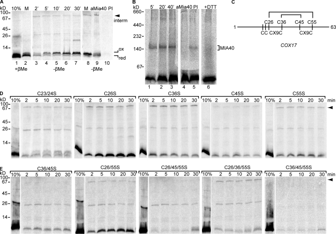
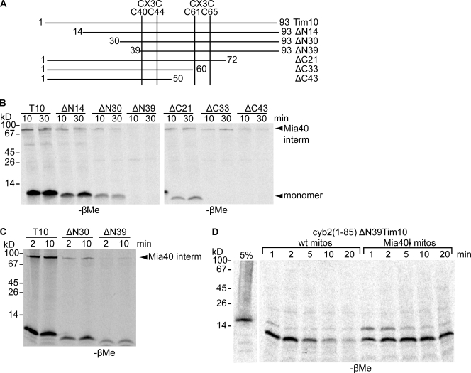
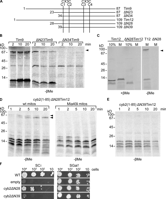
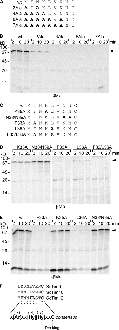
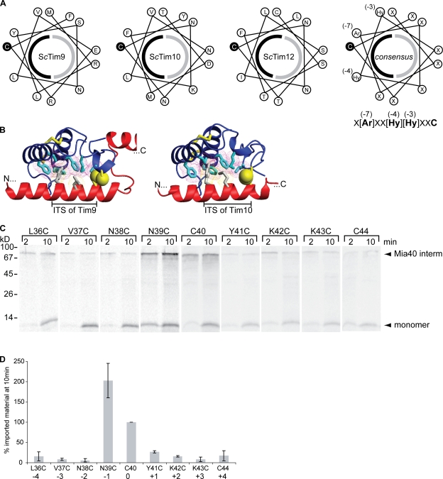
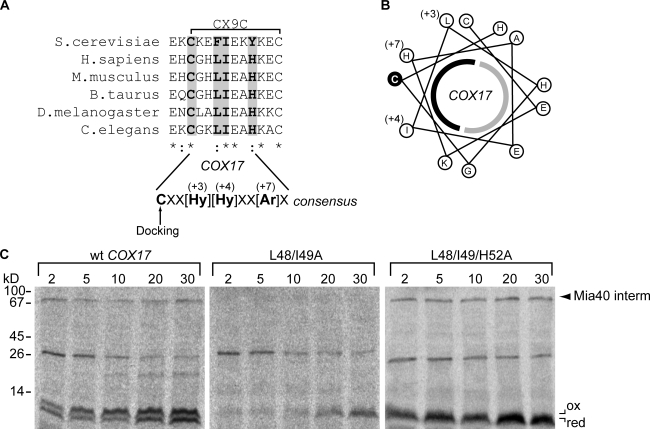
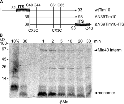
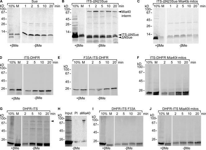
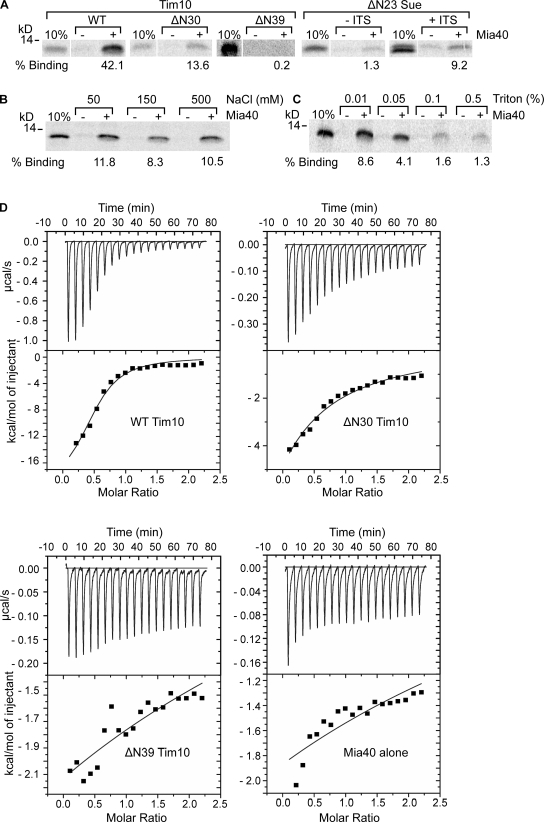
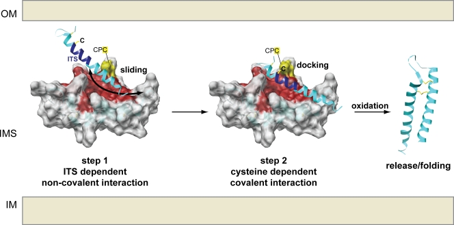
Mia40 imports Cys-containing proteins into the mitochondrial intermembrane space (IMS) by ensuring their Cys-dependent oxidative folding. In this study, we show that the specific Cys of the substrate involved in docking with Mia40 is substrate dependent, the process being guided by an IMS-targeting signal (ITS) present in Mia40 substrates. The ITS is a 9-aa internal peptide that (a) is upstream or downstream of the docking Cys, (b) is sufficient for crossing the outer membrane and for targeting nonmitochondrial proteins, (c) forms an amphipathic helix with crucial hydrophobic residues on the side of the docking Cys and dispensable charged residues on the other side, and (d) fits complementary to the substrate cleft of Mia40 via hydrophobic interactions of micromolar affinity. We rationalize the dual function of Mia40 as a receptor and an oxidase in a two step-specific mechanism: an ITS-guided sliding step orients the substrate noncovalently, followed by docking of the substrate Cys now juxtaposed to pair with the Mia40 active Cys.


Figures

Figure 1.
Figure 1.
Differential requirement of COX17 Cys residues for interaction with Mia40. (A) 35S-labeled COX17 import and immunoprecipitation as indicated, followed by SDS-PAGE. (B) As in A but using BN-PAGE. (C) Cys organization of COX17. (D) As in A for single Cys mutants. (E) As in D for multiply mutated Cys. (A, D, and E) Arrowheads indicate the Mia40 mixed intermediate (interm).
Figure 2.
Figure 2.
Deletion analysis of Tim10 reveals an internal targeting signal for the Mia40 pathway. (A) Tim10 truncations. (B) Import and analysis of Tim10 versions as in Fig. 1 A. (C) GST-Mia40 pull-downs of different Tim10 versions (as in Sideris and Tokatlidis, 2007). (D) Import of cyb2(1–85)ΔN39Tim10 in wild-type (wt) and Mia40-depleted (Mia40↓; Banci et al., 2009) mitochondria (mitos). interm, intermediate.
Figure 3.
Figure 3.
The Tim10 ITS is conserved among small Tims. (A) Tim9 and -12 truncations. (B) The intermediate with Mia40 is shown by an arrowhead. (C) As in B for Tim12 variants. 10% of in vitro translation mix was loaded as a standard. M, total mitochondria reisolated after import and protease treatment. (D) Import of cyb2(1–85)ΔN28 Tim12 fusion as in B. (C and D) Arrowheads indicate the Mia40 mixed intermediate. mitos, mitochondria; wt, wild type. (E) As in B but for the ΔN39Tim12 fragment. (F) In vivo complementation of Tim12 fusion variants using a GAL-Tim12 strain as described previously (Lionaki et al., 2008). SGal, GAL media; SC, glucose media; WT, wild type.
Figure 4.
Figure 4.
Ala mutagenesis identifies critical residues of the ITS. (A) The ITS of Tim10 with Ala substitutions (bold). (B) Imports of Tim10 Ala mutants performed as in Fig. 1. (C) Targeted Ala substitutions (bold) in the ITS of Tim10. (D) Import assays of the mutants of C. (E) As in D, but precursors were presented to mitoplasts. (B, D, and E) Arrowheads indicate the Mia40 mixed intermediate. (F) Alignment of the ITS sequence (residues 30–40 upstream of the N-terminal C40 in Tim10) in S. cerevisiae Tim9, -10, and -12. The consensus ITS sequence and the Cys docking to Mia40 are shown. The critical residues for the functionality of ITS are bolded, with their position relative to the docking Cys in parentheses. Colons indicate that conserved substitutions are observed, periods indicate that semiconserved substitutions are observed, and the asterisk indicates that the residues or nucleotides in the column are identical in all sequences in the alignment. wt, wild type.
Figure 5.
Figure 5.
Cys-scanning mutagenesis and conformational properties of the ITS. (A) Helical wheel representation of ITS in S. cerevisiae Tim9, -10, and -12. The hydrophobic (black) and hydrophilic (gray) faces of the helix are indicated as half circles. (B) Theoretical modeling of hTIM9 and hTIM10 docking to the hMIA40 substrate–binding cleft using HADDOCK. TIMS and MIA40 are shown as red and blue ribbons, respectively. Hydrophobic residues of MIA40 interacting with TIMS are in cyan. Conserved aromatic and hydrophobic residues in the first helix of the TIMS interacting with the MIA40 hydrophobic cleft are in gray. Cysteines are in yellow, and the docking cysteines of the TIMS and MIA40 are shown as yellow spheres. (C) Cys substitutions in Tim10 and import and Mia40 intermediate (interm) analysis. (D) Quantification of the Mia40 intermediate after 10-min import (three independent experiments). Error bars represent standard error of the mean.
Figure 6.
Figure 6.
An ITS is present in the C-terminal end of Cox17. (A) Alignment of Cox17 ITS sequences from different eukaryotes. Strictly conserved residues in the ITS consensus are shaded gray and bolded. Colons indicate that conserved substitutions are observed, and asterisks indicate that the residues or nucleotides in the column are identical in all sequences in the alignment. (B) Helical wheel projection for the ITS of COX17 as in Fig. 5 A. (C) COX17 mutants were imported and analyzed for mixed disulfide intermediate (interm) with Mia40 (arrowhead). wt, wild type.
Figure 7.
Figure 7.
Swapping the ITS from the N- to the C-terminal end of Tim10 does not affect targeting. (A) Schematic representation of the Tim10 ITS (zigzag line) and C-terminal end fusion to ΔN39Tim10. wt, wild type. (B) ΔN39Tim10-ITS was imported and analyzed for mixed disulfide intermediate (interm) formation with Mia40.
Figure 8.
Figure 8.
Fusion of the ITS can target to mitochondrial Mia40-independent substrates and nonmitochondrial proteins. (A) Import of Sue in wild-type mitochondria. (B) As in A for ITS-DHFR. interm, intermediate. (C) As in B but in Mia40-depleted mitochondria. (D) ITS-DHFR import as in A. (E) As in D but with the F33A Tim10 ITS mutant. (F) As in D but in Mia40-depleted mitochondria. (G) DHFR-ITS fusion and import as in D. The arrowhead indicates the Mia40 mixed intermediate. (H) Immunoprecipitation after the import of DHFR-ITS with anti-Mia40 or preimmune serum (PI). (I) As in G but with the F33A Tim10 ITS mutant. (J) As in G but in Mia40-depleted mitochondria.
Figure 9.
Figure 9.
The noncovalent ITS-dependent binding of substrates to Mia40 is mediated by hydrophobic interactions. (A) Purified His-tagged ΔN290Mia40SPS was bound to Ni2+-NTA beads. 35S-labeled proteins were incubated with Ni-NTA beads with or without ΔN290Mia40SPS at 15°C for 2 h. (B) 35S-labeled Tim10 was incubated with Ni-NTA bead–immobilized ΔN290Mia40SPS for 2 h at 30°C in the presence of 50, 150, or 500 mM NaCl. (C) As in B, but binding was performed in the presence of 0.01, 0.05, 0.1, or 0.5% Triton X-100. (D) ITC of 0.25 mM ΔN290Mia40SPS alone (bottom right) or with 0.025 mM of wild-type (WT) Tim10 (top left), Tim10 ΔN30 (top right), or Tim10 ΔN39 (bottom left).
Figure 10.
Figure 10.
Sliding–docking model for the interaction of substrates with Mia40. (step 1, sliding) The substrate slides onto Mia40, where it is oriented by the MISS/ITS through noncovalent, mainly hydrophobic interactions in the cleft of Mia40. The correct Cys of the substrate is thus primed to make the disulfide with Mia40. (step 2, docking) The substrate now docks onto Mia40 via the covalent mixed disulfide bond between the substrate-docking Cys and the juxtaposed active site Cys of Mia40. Finally, complete oxidation releases the substrate in a folded state. IM, inner membrane.

Similar articles

Cited by

References

    1. Abe Y., Shodai T., Muto T., Mihara K., Torii H., Nishikawa S., Endo T., Kohda D. 2000. Structural basis of presequence recognition by the mitochondrial protein import receptor Tom20. Cell. 100:551–560 10.1016/S0092-8674(00)80691-1 - DOI - PubMed
    1. Allen S., Lu H., Thornton D., Tokatlidis K. 2003. Juxtaposition of the two distal CX3C motifs via intrachain disulfide bonding is essential for the folding of Tim10. J. Biol. Chem. 278:38505–38513 10.1074/jbc.M306027200 - DOI - PubMed
    1. Arnesano F., Balatri E., Banci L., Bertini I., Winge D.R. 2005. Folding studies of Cox17 reveal an important interplay of cysteine oxidation and copper binding. Structure. 13:713–722 10.1016/j.str.2005.02.015 - DOI - PubMed
    1. Baker M.J., Webb C.T., Stroud D.A., Palmer C.S., Frazier A.E., Guiard B., Chacinska A., Gulbis J.M., Ryan M.T. 2009. Structural and functional requirements for activity of the Tim9-Tim10 complex in mitochondrial protein import. Mol. Biol. Cell. 20:769–779 10.1091/mbc.E08-09-0903 - DOI - PMC - PubMed
    1. Banci L., Bertini I., Ciofi-Baffoni S., Janicka A., Martinelli M., Kozlowski H., Palumaa P. 2008. A structural-dynamical characterization of human Cox17. J. Biol. Chem. 283:7912–7920 10.1074/jbc.M708016200 - DOI - PubMed

Publication types

MeSH terms

Substances