. 2009 Dec 28;187(7):1083-99.
doi: 10.1083/jcb.200909067. Epub 2009 Dec 21.

IKK phosphorylates Huntingtin and targets it for degradation by the proteasome and lysosome

Affiliations

IKK phosphorylates Huntingtin and targets it for degradation by the proteasome and lysosome

Leslie Michels Thompson et al. J Cell Biol. .

Abstract

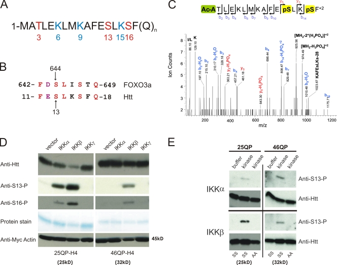
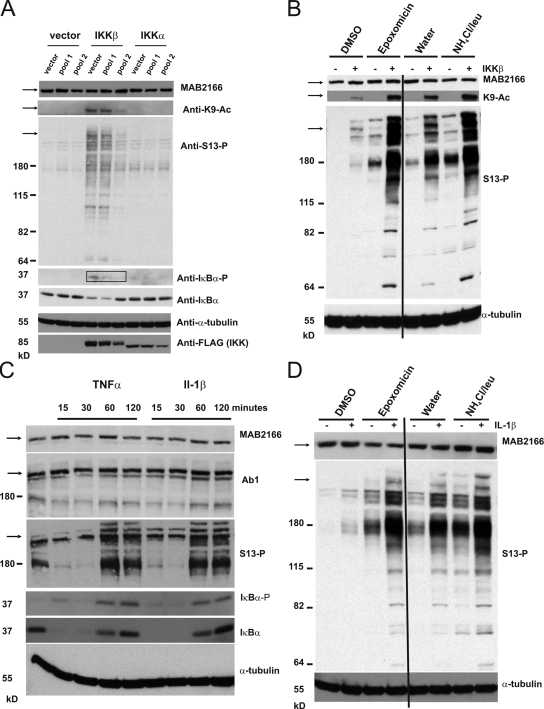
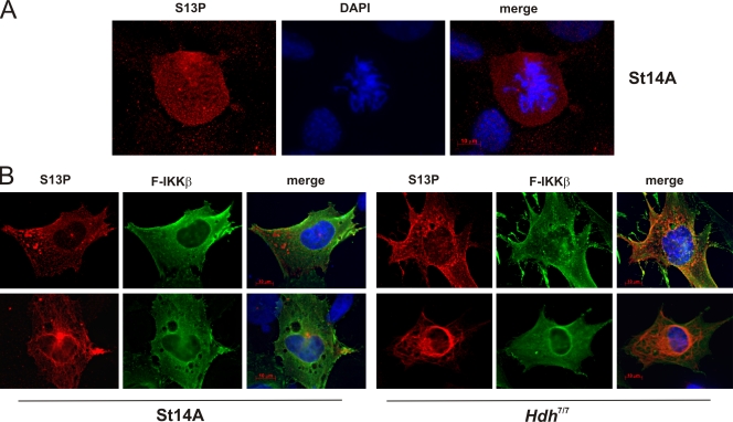
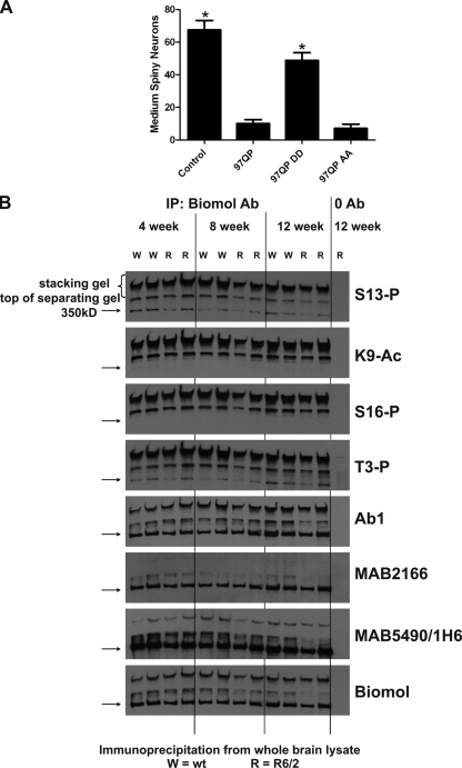
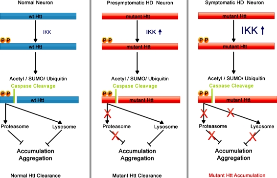
Expansion of the polyglutamine repeat within the protein Huntingtin (Htt) causes Huntington's disease, a neurodegenerative disease associated with aging and the accumulation of mutant Htt in diseased neurons. Understanding the mechanisms that influence Htt cellular degradation may target treatments designed to activate mutant Htt clearance pathways. We find that Htt is phosphorylated by the inflammatory kinase IKK, enhancing its normal clearance by the proteasome and lysosome. Phosphorylation of Htt regulates additional post-translational modifications, including Htt ubiquitination, SUMOylation, and acetylation, and increases Htt nuclear localization, cleavage, and clearance mediated by lysosomal-associated membrane protein 2A and Hsc70. We propose that IKK activates mutant Htt clearance until an age-related loss of proteasome/lysosome function promotes accumulation of toxic post-translationally modified mutant Htt. Thus, IKK activation may modulate mutant Htt neurotoxicity depending on the cell's ability to degrade the modified species.


Figures

Figure 1.
Figure 1.
IKK directly phosphorylates Htt. (A) The first 17 amino acids of the Htt protein contain three residues that may be phosphorylated (red) and three modifiable lysine residues (blue). (B) Htt S13 is within a domain similar to FOXO3a S644. (C) Mass spectrometry analysis shows that Htt serines 13 and 16 can be phosphorylated. 25QP-HBH was purified under denaturing conditions from St14A cells cotransfected with IKK-αβγ. ESI-MS/MS spectra were obtained after chymotryptic digestion and collision-induced dissociation (CID) for N-terminally acetylated and diphosphorylated peptide on S13 and S16 Ac-ATLEKLMKAFEpSLKpSF, with the parent ion, [MH2]+2, at m/z 1023.03+2 (M = 2044.06 D). (D) Htt phosphorylation of S13 and S16, detected with phospho-specific antibodies, is activated with coexpression of IKK-α or IKK-β. Httex1p was purified from St14A cells transiently transfected with 25QP-H4 or 46QP-H4 with vector control or plasmids encoding subunits of IKK. Total Htt was detected with CAG53b antibody, and myc-actin transfection control detected with anti-myc antibody. (E) IKK-α and IKK-β directly phosphorylate Htt S13 in vitro. An in vitro kinase assay was performed with 75 ng recombinant IKK-α or IKK-β protein, and wt (SS) or S13,16A (AA) mutant 25Q or 46Q purified Httex1p-H4. Htt was detected with CAG53b or anti-S13-P.
Figure 2.
Figure 2.
Phosphorylation of Httex1p regulates its ubiquitination, SUMOylation, acetylation, and nuclear localization. (A and B) Phosphorylation of serines 13 and 16 regulates mutant Httex1p ubiquitination and SUMOylation. St14A (A) or HeLa (B) cells were transiently transfected with vector or HIS-ubiquitin (A) or HIS-SUMO-1 (B) and control and mutant 97QP VL* Httex1p. Conjugated proteins were purified under denaturing conditions by Ni-NTA magnetic nickel beads and Htt detected with anti-Htt CAG53b by Western analysis. (C) Mass spectrometry analysis shows that Htt S13 phosphorylation can occur with K9 acetylation. 25QP-HBH was purified from St14A cells cotransfected with IKK-β and CBP and treated for 2 h with histone deacetylase inhibitors 200 mM Trichostatin A/5 mM Nicotinamide. ESI-MS/MS spectra were obtained after chymotryptic digestion and collision-induced dissociation (CID) for a peptide acetylated at K9 and phosphorylated at S13 MAcKAFEpSLKSF, [MH2]+2 at m/z 655.30+2 (M = 1308.60 D). (D) IKK-β overexpression increases phosphorylation of Htt S13 and acetylation of K9. St14A cells were transiently transfected with Httex1p-H4 with 25 or 46Qs, +/− IKK-β or with 46QP QEE-H4. Htt was purified and subjected to Western analysis with anti-K9-Ac, anti-S13-P, and CAG53b. (E) Mimicking phosphorylation significantly increases nuclear localization in primary neurons. Primary cortical neurons were cotransfected with pcDNA3.1-mRFP and 97QP-GFP or 97QP-DD-GFP plasmids. The subcellular distribution of these polypeptides was examined by measuring the fluorescence intensity of GFP, to which they are fused, in regions of the nucleus and cytoplasm for each cell. The extent to which these polypeptides localized preferentially to the nucleus or the cytoplasm was determined by calculating the ratio of nuclear/cytoplasmic GFP fluorescence intensity and comparing the distribution of the two polypeptides by t test. Error bars indicate SEM in arbitrary units.
Figure 3.
Figure 3.
Phosphorylation of unexpanded polyQ Httex1p and 586aa Htt is associated with its reduced abundance in cell culture. (A) Levels of unexpanded polyQ Httex1p are reduced with overexpression of IKK-β; this effect is inhibited with expansion of the polyQ repeat. 25QP-H4 or 46QP-H4 was cotransfected with myc-actin and with vector or IKK-β into St14A cells. Cells were treated for 16 h with DMSO, 100 nM epoxomicin in DMSO, or 20 mM ammonium chloride/100 µM leupeptin in DMSO. Lysates were subjected to filter-retardation assay and Western analysis using anti-myc to detect myc-actin, and anti-HA to detect Httex1p. (B) IKK-β overexpression reduces levels of unexpanded polyQ Httex1p in the presence of proteasome or lysosome inhibition. Scion software was used to quantitate triplicate levels of 25QP-H4 from the experiment represented in A, normalized to levels of myc-actin transfection control, within each treatment group: control, epoxomicin, or ammonium chloride/leupeptin. (C) Mimicking phosphorylation of unexpanded polyQ Httex1p reduces its abundance in cell culture; this effect is reduced with expansion of the polyQ repeat. 25QP-H4 or 46QP-H4, wt control or QEE, EE, AA, or 3R were cotransfected with myc-actin into St14A cells. Cells and lysates were treated as in A. (D) Levels of phosphorylated unexpanded polyQ 586aa Htt accumulate with inhibition of the proteasome or the lysosome; phosphorylation is reduced with expansion of the polyQ repeat. 15Q or 128Q 586aa Htt constructs were cotransfected into St14A cells with myc-actin and with vector or IKK-β. Cells were treated for 4 h with DMSO or 100 nM epoxomicin in DMSO (to eliminate any possible effect on the lysosome by epoxomicin), or for 16 h with water or 20 mM ammonium chloride/100 µM leupeptin in water. Lysates were subjected to filter-retardation assay and Western analysis using anti-myc, anti–S13-P, and anti-Htt 3B5H10.
Figure 4.
Figure 4.
Phosphorylated and acetylated endogenous wt Htt accumulates with inhibition of the proteasome and lysosome. (A) Overexpression of IKK-β increases phosphorylation of endogenous Htt and IκBα in cell culture. St14A cells were transiently transfected with vector, IKK-β, or IKK-α, together with vector or two different pools of anti–IKK-αβ shRNAs; a control pool that did not silence IKK well (pool 1) and one pool that was effective in silencing IKK (pool 2), as assessed by levels of FLAG-IKK-α and -β by Western. Lysates were subjected to Western analysis with anti-Htt MAB2166, anti–S13-P, anti–K9-Ac, anti-phosphoserine 32 IκBα (IκBα-P), anti–IκBα, anti–α-tubulin, and anti-FLAG to detect FLAG-tagged IKK-β and IKK-α. Bands the size of full-length endogenous Htt (350 kD) are shown by the arrow on the left. Boxed bands show the reduction in phosphorylated IκBα with IKK shRNA. (B) Phosphorylated and acetylated Htt accumulate with inhibition of the proteasome or the lysosome. St14A cells were transiently transfected with IKK-β or vector, and were treated as in Fig. 3 D. Lysates were subjected to Western analysis with anti–α-tubulin, and anti-Htt antibodies MAB2166, anti–S13-P or anti–K9-Ac. (C) Pharmacological activation of the IKK complex modulates levels of phosphorylated Htt and IκBα. St14A cells were incubated with 20 ng/ml TNF-α or IL-1β over a time course. Lysates were subjected to Western analysis as in A with additional detection by anti-Htt Ab1. (D) IL-1β–induced Htt S13-phosphorylated species accumulate with inhibition of the proteasome or lysosome. St14A cells were treated for 2 h with 20 ng/ml Il-1β before lysis. The proteasome and lysosome were inhibited, and Western analysis performed as in B.
Figure 5.
Figure 5.
Phosphorylated Htt can be detected by immunofluorescence in cell culture. (A) S13-phosphorylated Htt is present in untransfected mitotic rat S14A cells. (B) S13-phosphorylated Htt colocalizes with FLAG immunoreactivity in rat St14A cells lipofected with FLAG–IKK-β, and S13-phosphorylated Htt colocalizes with FLAG immunoreactivity in mouse Hdh7/7 cells nucleofected with FLAG–IKK-β.
Figure 6.
Figure 6.
LAMP-2A, Atg7, and Hsc70 may modulate Htt clearance and toxicity. (A) Reducing levels of LAMP-2A or Atg7 in cell culture increases abundance of S13-phosphorylated Htt. St14A cells were transiently transfected with shRNA for rat LAMP-2A or Atg7, or pSUPER vector control; antibodies used for Western analysis are shown to the left of the Western panels. (B) LAMP-2A levels modulate Httex1p abundance. St14A cells were transiently cotransfected with LAMP-2A shRNA, human HA-tagged LAMP-2A or vector control, and myc-actin transfection control. Lysates were subjected to Western analysis and filter-retardation assay, detected with anti-Htt CAG53b, anti–rat/mouse LAMP-2A, anti-HA to detect HA-hLAMP-2A, anti-myc, and MemCode protein stain. (C) Hsc70 interacts with phosphomimetic unexpanded polyQ Httex1p in vitro. Purified 25QP-H4 or 46QP-H4 wt and mutant proteins radiolabeled with 35S were incubated with isolated GST-Hsc70 or GST protein bound to glutathione-agarose beads. Where indicated, 5 mM ATP or ADP was added to the reaction. Bound proteins were washed, subjected to SDS-PAGE, and detected by phosphoimager autoradiography. (D) Hsc70 reduces Htt-mediated toxicity in Hdh111/111 cells. Hdh7/7 or Hdh111/111 cells were nucleofected with vector, Hsp70, or Hsc70 together with GFP. 47 h later, XTT cell viability assays were performed, and the percentage of relative survival was calculated correcting for transfection efficiency of the GFP control ± SEM from triplicates.
Figure 7.
Figure 7.
Phosphorylation may reduce toxicity but increase insolubility of Htt. (A) Mimicking 97QP Httex1p phosphorylation reduces its toxicity in acutely transfected rat cortico-striatal slice explants. Plasmids encoding YFP and 97QP-CFP, 97QP DD-CFP, 97QP AA-CFP, or CFP control were biolistically cotransfected into rat cortico-striatal brain slices. The number of healthy medium spiny neurons in the striatal region of each slice was scored 5 d after transfection. n = 9 for each condition. Error bars represent SEM. Asterisk = difference from 97QP at P < 0.0001. (B) Immunoprecipitated phosphorylated/acetylated wt Htt purified from mouse brain runs in an insoluble fraction in the SDS-PAGE stacking gel. 12 independent wt control (W) or R6/2 (R) mouse brains were collected at 4, 8, and 12 wk of age, snap frozen, and lysed. 500 µg of lysate was subjected to immunoprecipitation with PW0595 anti-Htt antibody or zero antibody control (lysate from lanes 12 and 13 are identical). Western analysis was performed with a series of antibodies listed to the right of the panel. Immunoprecipitated Htt is present as an insoluble species in the stacking gel and at the top of the separating gel, and as a soluble form at the standard 350-kD size, marked with an arrow. MAB2166 does not detect the insoluble species well.
Figure 8.
Figure 8.
Proposed molecular mechanism for the development of HD. Normal neuronal function: IKK phosphorylates wt Htt activating its post-translational modification, caspase cleavage, and clearance by the proteasome and lysosome. Presymptomatic HD neuronal function: With chronic expression of mutant Htt, proteasome activity is reduced, and lysosomal degradation of mutant Htt becomes essential. Mutant Htt triggers activation of the IKK complex; however, it is less efficiently phosphorylated than wt Htt. With the clearance mechanism activated, mutant and wt Htt in the presymptomatic cell are degraded by the lysosome. Symptomatic HD neuronal function: Lysosomal degradation of Htt is impaired through reduction of LAMP-2A levels or other loss of lysosomal function caused by aging and by mutant Htt expression. Uncleared mutant Htt and Htt fragments accumulate and take on toxic functions, enhancing HD pathogenesis.

Similar articles

Cited by

References

    1. Aiken C.T., Steffan J.S., Guerrero C.M., Khashwji H., Lukacsovich T., Simmons D., Purcell J.M., Menhaji K., Zhu Y.Z., Green K., et al. 2009. Phosphorylation of threonine 3: implications for Huntingtin aggregation and neurotoxicity. J. Biol. Chem. 284:29427–29436 10.1074/jbc.M109.013193 - DOI - PMC - PubMed
    1. Anne S.L., Saudou F., Humbert S. 2007. Phosphorylation of huntingtin by cyclin-dependent kinase 5 is induced by DNA damage and regulates wild-type and mutant huntingtin toxicity in neurons. J. Neurosci. 27:7318–7328 10.1523/JNEUROSCI.1831-07.2007 - DOI - PMC - PubMed
    1. Apostol B.L., Simmons D.A., Zuccato C., Illes K., Pallos J., Casale M., Conforti P., Ramos C., Roarke M., Kathuria S., et al. 2008. CEP-1347 reduces mutant huntingtin-associated neurotoxicity and restores BDNF levels in R6/2 mice. Mol. Cell. Neurosci. 39:8–20 10.1016/j.mcn.2008.04.007 - DOI - PubMed
    1. Arrasate M., Finkbeiner S. 2005. Automated microscope system for determining factors that predict neuronal fate. Proc. Natl. Acad. Sci. USA. 102:3840–3845 10.1073/pnas.0409777102 - DOI - PMC - PubMed
    1. Arrasate M., Mitra S., Schweitzer E.S., Segal M.R., Finkbeiner S. 2004. Inclusion body formation reduces levels of mutant huntingtin and the risk of neuronal death. Nature. 431:805–810 10.1038/nature02998 - DOI - PubMed

Publication types

MeSH terms