. 2010 Jun;51(6):1535-45.
doi: 10.1194/jlr.M002717. Epub 2009 Dec 21.

Mutation of conserved cysteines in the Ly6 domain of GPIHBP1 in familial chylomicronemia

Affiliations

Mutation of conserved cysteines in the Ly6 domain of GPIHBP1 in familial chylomicronemia

Gunilla Olivecrona et al. J Lipid Res. 2010 Jun.

Abstract

We investigated a family from northern Sweden in which three of four siblings have congenital chylomicronemia. LPL activity and mass in pre- and postheparin plasma were low, and LPL release into plasma after heparin injection was delayed. LPL activity and mass in adipose tissue biopsies appeared normal. [(35)S]Methionine incorporation studies on adipose tissue showed that newly synthesized LPL was normal in size and normally glycosylated. Breast milk from the affected female subjects contained normal to elevated LPL mass and activity levels. The milk had a lower than normal milk lipid content, and the fatty acid composition was compatible with the milk lipids being derived from de novo lipogenesis, rather than from the plasma lipoproteins. Given the delayed release of LPL into the plasma after heparin, we suspected that the chylomicronemia might be caused by mutations in GPIHBP1. Indeed, all three affected siblings were compound heterozygotes for missense mutations involving highly conserved cysteines in the Ly6 domain of GPIHBP1 (C65S and C68G). The mutant GPIHBP1 proteins reached the surface of transfected Chinese hamster ovary cells but were defective in their ability to bind LPL (as judged by both cell-based and cell-free LPL binding assays). Thus, the conserved cysteines in the Ly6 domain are crucial for GPIHBP1 function.


Figures

Fig. 1.
Fig. 1.
Pedigree in three generations of the family. Filled symbols denote family members with chylomicronemia. Birth years for the second generation are shown.
Fig. 2.
Fig. 2.
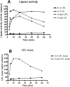
Release of LPL activity and mass into plasma after heparin injection in patient II-2 and a healthy, age-matched control subject. A single intravenous injection of heparin (100 U/ kg body weight) was given at time 0. Values are means of triplicate measurements. A: LPL activity measured after immunoinhibition of HL activity in the samples and HL activity measured in an assay containing 1 M NaCl to inactivate LPL. B: LPL mass in patient II-2 and a control subject measured with bovine LPL as standard for the ELISA.
Fig. 3.
Fig. 3.
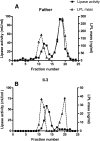
Separation of lipases in postheparin plasma by chromatography on heparin-Sepharose. Fresh postheparin plasma (9 ml) was applied to columns with 3 ml of heparin-Sepharose. A: Postheparin plasma from the father (I-1). B: Postheparin plasma from the affected son (II-3). HL displayed activity in this assay (about 25% of the activity in the assay optimized for measurements of HL with 1 M NaCl to inactivate LPL). Values for activity are means of duplicate determinations. In this experiment, human LPL was used as standard for the ELISA.
Fig. 4.
Fig. 4.
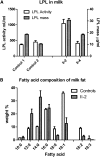
LPL activity and mass in milk samples. A: LPL activity and mass in milk from subjects II-2 and II-4 were compared with two healthy mothers. From patient II-2, three milk samples were analyzed from days 3, 10, and 18 postpartum. From patient II-4, eight milk samples were taken during days 14–24. From control subject 1, a total of 37 milk samples were taken during days 18–22 postpartum, and from control subject 2, a total of 42 milk samples were taken from a similar time period. Human LPL was used as standard for the ELISA, and data are mean values ± SD. B: Milk fatty acid composition. Values are mean ± SD from seven samples taken at days 3–60 postpartum from subject II-2 and of samples from five healthy mothers taken <1 month postpartum (control subjects, mean of three samples from each mother ± SD).
Fig. 5.
Fig. 5.
Identification of mutations in GPIHBP1. Filled symbols indicate family members with chylomicronemia. Black circles within the symbols indicate heterozygous carriers of GPIHBP1 mutations. Question mark indicates the lack of DNA sequencing in the son (II-1). Electropherograms including the two mutations, C65S and C68G, in exon 3 of GPIHBP1 are shown below the symbols to the right. Arrows indicate heterozygous changes.
Fig. 6.
Fig. 6.
Expression of wild-type and mutant forms of the GPIHBP1 protein in CHO pgsA-745 cells. The cells were electroporated with an empty vector, a construct encoding an S-tagged human GPIHBP1, or the mutants C65S and C68G. GPIHBP1 in nonpermeabilized (A) and permeabilized (B) cells was assessed by immunofluorescence microscopy with an antiserum against the S-protein tag (green). Cell nuclei were visualized with 4’,6-diamidino-2-phenylindole (blue).
Fig. 7.
Fig. 7.
Release of wild-type and mutant GPIHBP1 proteins from the cell surface with PIPLC. CHO pgsA-745 cells were electroporated with expression vectors encoding wild-type mouse GPIHBP1, a mutant mouse GPIHBP1 (N76Q) that eliminates the sole N-linked glycosylation site (38), wild-type human GPIHBP1, and mutant human GPIHBP1-C65S or GPIHBP1-C68G proteins. All of the GPIHBP1 constructs contained an S-protein tag. The amounts of GPIHBP1 released into the medium by PIPLC (5 U/ml for 1 h at 37°C) were assessed by Western blotting.
Fig. 8.
Fig. 8.
Binding of LPL to cells expressing wild-type and mutant GPIHBP1. CHO pgsA-745 cells were electroporated with constructs encoding S-protein-tagged wild-type GPIHBP1 or the mutant forms C65S, C68G, or Q115P. Twenty-four hours later, the cells were incubated with V5-tagged human LPL in the presence or absence of heparin (500 U/ml). The cells were washed six times, cell extracts were prepared, and the level of LPL bound to the cells was assessed by Western blotting with a V5-specific antibody. Simultaneously, the level of GPIHBP1 in cell extracts was assessed by Western blotting with an antibody against the S-protein tag. Actin was used as a loading control.
Fig. 9.
Fig. 9.
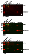
Elution of human LPL together with soluble wild-type GPIHBP1 or the soluble forms of the mutants C65S and C68G from agarose beads coated with anti-GPIHBP1 monoclonal antibody (11A12). V5-tagged human LPL was mixed with the different forms of soluble mouse GPIHBP1, along with antibody 11A12-coated beads. After three washes, the bound proteins were eluted with 0.1 M glycine, pH 2.5. GPIHBP1 and LPL in the starting material, the unbound fraction, the washes, and elution fractions were detected by Western blotting. In all cases, most of the GPIHBP1 protein was captured by the antibody 11A12-coated beads, but LPL was coeluted only in the presence of wild-type GPIHBP1 (A). With the GPIHBP1 mutants (B and C), no LPL was detectable in the eluates.

Similar articles

Cited by

References

    1. Wang H., Eckel R. H. 2009. Lipoprotein lipase: from gene to obesity. Am. J. Physiol. Endocrinol. Metab. 297: E271–E288. - PubMed
    1. Olivecrona T., Olivecrona G. 2009. The ins and outs of adipose tissue. In Cellular Lipid Mmetabolism Ehnholm C., editor Springer, Heidelberg, Germany: 315–369.
    1. Krieger M., Herz J. 1994. Structures and functions of multiligand lipoprotein receptors: macrophage scavenger receptors and LDL receptor-related protein (LRP). Annu. Rev. Biochem. 63: 601–637. - PubMed
    1. Beisiegel U. 1995. Receptors for triglyceride-rich lipoproteins and their role in lipoprotein metabolism. Curr. Opin. Lipidol. 6: 117–122. - PubMed
    1. Gliemann J. 1998. Receptors of the low density lipoprotein (LDL) receptor family in man. Multiple functions of the large family members via interaction with complex ligands. Biol. Chem. 379: 951–964. - PubMed

Publication types

MeSH terms