

Immunocytochemical localization of sex steroid hormone receptors in normal human mammary gland
- PMID: 20026671
- PMCID: PMC2874183
- DOI: 10.1369/jhc.2009.954644
Immunocytochemical localization of sex steroid hormone receptors in normal human mammary gland
Abstract
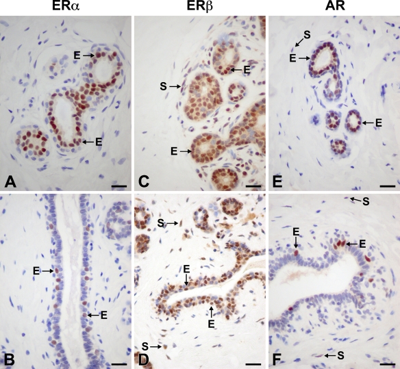
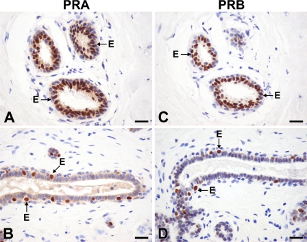
The sex steroids, estrogens, progesterone, and androgens, all play a role in mammary development and function. To precisely identify the sites of action of these steroids, we studied the localization of the estrogen receptor alpha (ERalpha) and ERbeta, the progesterone receptor A (PRA) and PRB, and androgen receptors (AR) in the normal human mammary gland. Immunocytochemical localization of ERalpha, ERbeta, PRA, PRB, and AR was performed with reduction mammoplasty specimens from premenopausal women. ERalpha, PRA, PRB, and AR were localized mostly to the inner layer of epithelial cells lining acini and intralobular ducts, as well as to myoepithelial cells scattered in the external layer of interlobular ducts. AR was also found in some stromal cells. ERbeta staining was more widespread, resulting in epithelial and myoepithelial cells being labeled in acini and ducts as well as stromal cells. These results suggest that all sex steroids can directly act on epithelial cells to modulate development and function of the human mammary gland. Estrogens and androgens can also indirectly influence epithelial cell activity by an action on stromal cells.
Figures
Similar articles
-
Sex steroid hormone receptors in human skin appendage and its neoplasms.Endocr J. 2005 Jun;52(3):317-25. doi: 10.1507/endocrj.52.317. Endocr J. 2005. PMID: 16006726
-
Steroid hormone receptor expression in male breast cancer.Eur J Surg Oncol. 2006 Feb;32(1):44-7. doi: 10.1016/j.ejso.2005.09.013. Epub 2005 Nov 2. Eur J Surg Oncol. 2006. PMID: 16260112
-
Profiling estrogen, progesterone, and androgen receptors in colorectal cancer in relation to gender, menopausal status, clinical stage, and tumour sidedness.Front Endocrinol (Lausanne). 2023 May 3;14:1187259. doi: 10.3389/fendo.2023.1187259. eCollection 2023. Front Endocrinol (Lausanne). 2023. PMID: 37206439 Free PMC article.
-
Localization of androgen and estrogen receptors in rat and primate tissues.Histol Histopathol. 2000 Oct;15(4):1261-70. doi: 10.14670/HH-15.1261. Histol Histopathol. 2000. PMID: 11005250 Review.
-
Localization of sex steroid receptors in human skin.Histol Histopathol. 2004 Apr;19(2):629-36. doi: 10.14670/HH-19.629. Histol Histopathol. 2004. PMID: 15024720 Review.
Cited by
-
The Tissue-Selective Estrogen Complex (Bazedoxifene/Conjugated Estrogens) for the Treatment of Menopause.Int J Endocrinol. 2017;2017:5064725. doi: 10.1155/2017/5064725. Epub 2017 Dec 5. Int J Endocrinol. 2017. PMID: 29358948 Free PMC article. Review.
-
Importance of Breast Cancer Subtype in the Development of Androgen Receptor Directed Therapy.Curr Breast Cancer Rep. 2014 Jun 1;6(2):71-78. doi: 10.1007/s12609-014-0140-5. Curr Breast Cancer Rep. 2014. PMID: 24860642 Free PMC article.
-
Oestrogen receptors in breast cancer: basic mechanisms and clinical implications.Ecancermedicalscience. 2013 Nov 5;7:370. doi: 10.3332/ecancer.2013.370. Ecancermedicalscience. 2013. PMID: 24222786 Free PMC article. Review.
-
Effects of the Overexpression of Progesterone Receptors on a Precancer p53 and Rb-Defective Human Fallopian Tube Epithelial Cell Line.Int J Mol Sci. 2023 Jul 23;24(14):11823. doi: 10.3390/ijms241411823. Int J Mol Sci. 2023. PMID: 37511582 Free PMC article.
-
Relationship between morphological development and sex hormone receptor expression of mammary glands with age in male rats.Exp Anim. 2018 Jul 30;67(3):361-371. doi: 10.1538/expanim.17-0134. Epub 2018 Mar 9. Exp Anim. 2018. PMID: 29526866 Free PMC article.
References
-
- Anderson E, Clarke RB (2004) Steroid receptors and cell cycle in normal mammary epithelium. J Mammary Gland Biol Neoplasia 9:3–13 - PubMed
-
- Aupperlee M, Kariagina A, Osuch J, Haslama SZ (2005a) Progestins and breast cancer. Breast Dis 24:37–57 - PubMed
-
- Aupperlee MD, Haslam SZ (2007) Differential hormonal regulation and function of progesterone receptor isoforms in normal adult mouse mammary gland. Endocrinology 148:2290–2300 - PubMed
-
- Aupperlee MD, Smith KT, Kariagina A, Haslam SZ (2005b) Progesterone receptor isoforms A and B: temporal and spatial differences in expression during murine mammary gland development. Endocrinology 146:3577–3588 - PubMed
-
- Bocchinfuso WP, Korach KS (1997) Mammary gland development and tumorigenesis in estrogen receptor knockout mice. J Mammary Gland Biol Neoplasia 2:323–334 - PubMed
MeSH terms
Substances
LinkOut - more resources
Full Text Sources
Research Materials
