Comparative Study
. 2010 Feb 1;184(3):1379-91.
doi: 10.4049/jimmunol.0903016. Epub 2009 Dec 21.

A small, variable, and irregular killer cell Ig-like receptor locus accompanies the absence of MHC-C and MHC-G in gibbons

Affiliations
Comparative Study

A small, variable, and irregular killer cell Ig-like receptor locus accompanies the absence of MHC-C and MHC-G in gibbons

Laurent Abi-Rached et al. J Immunol. .

Abstract

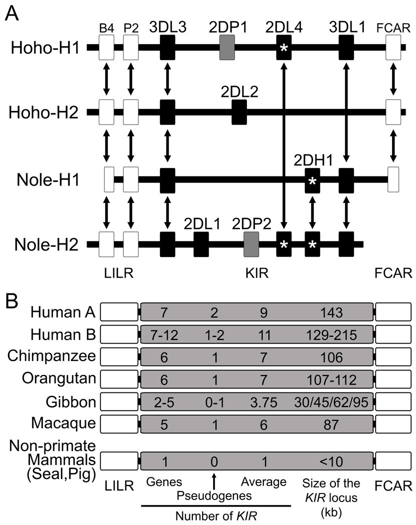
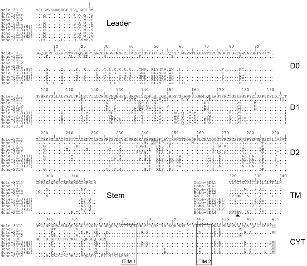
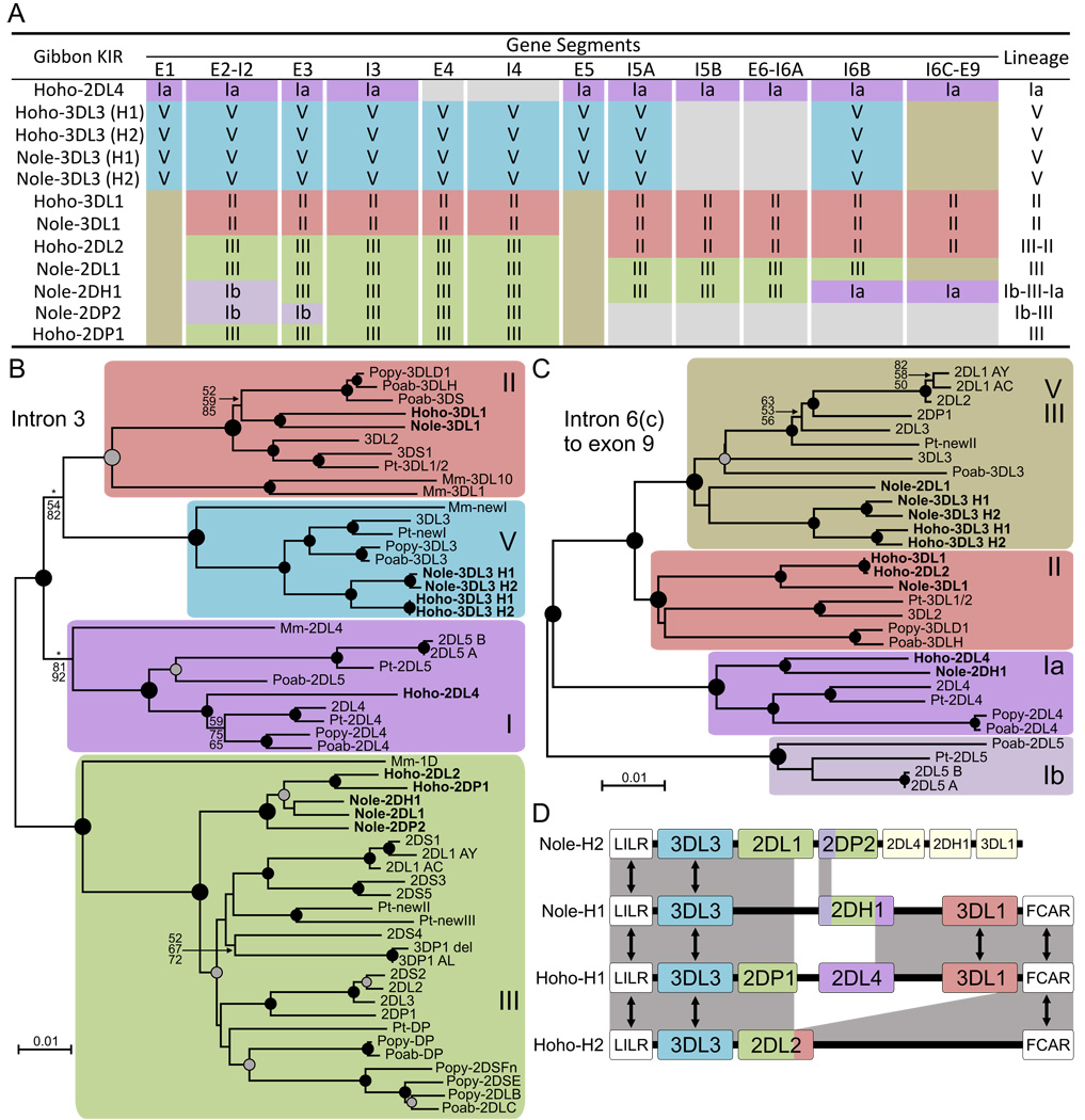
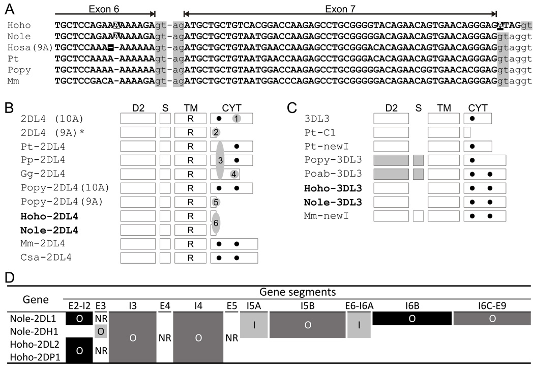
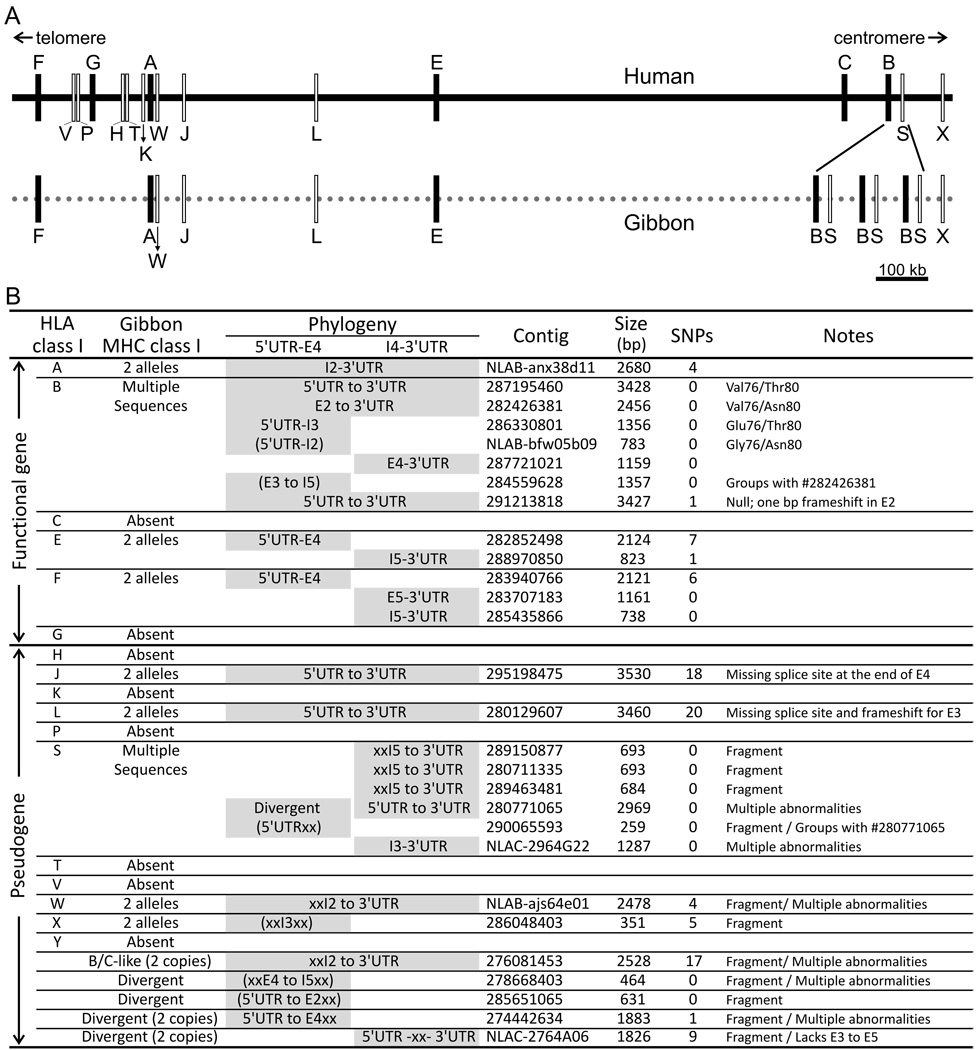
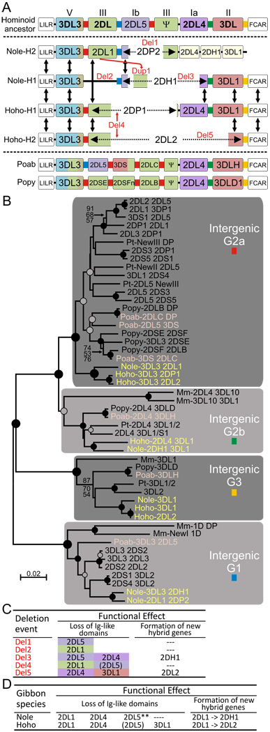
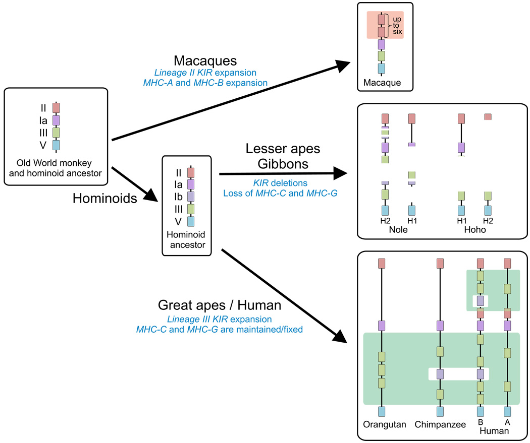
The killer cell Ig-like receptors (KIRs) of NK cells recognize MHC class I ligands and function in placental reproduction and immune defense against pathogens. During the evolution of monkeys, great apes, and humans, an ancestral KIR3DL gene expanded to become a diverse and rapidly evolving gene family of four KIR lineages. Characterizing the KIR locus are three framework regions, defining two intervals of variable gene content. By analysis of four KIR haplotypes from two species of gibbon, we find that the smaller apes do not conform to these rules. Although diverse and irregular in structure, the gibbon haplotypes are unusually small, containing only two to five functional genes. Comparison with the predicted ancestral hominoid KIR haplotype indicates that modern gibbon KIR haplotypes were formed by a series of deletion events, which created new hybrid genes as well as eliminating ancestral genes. Of the three framework regions, only KIR3DL3 (lineage V), defining the 5' end of the KIR locus, is present and intact on all gibbon KIR haplotypes. KIR2DL4 (lineage I) defining the central framework region has been a major target for elimination or inactivation, correlating with the absence of its putative ligand, MHC-G, in gibbons. Similarly, the MHC-C-driven expansion of lineage III KIR genes in great apes has not occurred in gibbons because they lack MHC-C. Our results indicate that the selective forces shaping the size and organization of the gibbon KIR locus differed from those acting upon the KIR of other hominoid species.


Figures

FIGURE 1
FIGURE 1
Gibbons have small, irregular and diverse KIR haplotypes. (A) Schematic representation of the four gibbon KIR haplotypes. Haplotypes Hoho-H1 and Hoho-H2 are from Hoolock hoolock leuconedys, haplotypes Nole-H1 and Nole-H2 are from Nomascus leucogenys leucogenys. Double arrows indicate genes that are either equivalent within species (alleles) or between species (orthologs). Black boxes represent genes encoding functional KIR, whereas gray boxes represent KIR pseudogenes, and white boxes represent FCAR and LILR genes flanking the KIR locus.*, activating KIR. For Nole-H2, KIR3DL3, 2DL1 and 2DP2 were fully characterized through sequencing of a BAC; 2DL4, 2DH1 and 3DL1 were characterized by analysis of whole genome shotgun sequences. (B) Shown are the numbers of KIR genes, KIR pseudogenes, and the average number of KIR genes and pseudogenes; the size of the KIR locus in the KIR haplotypes of several primate and non-primate species is also given. For gibbons, the size of each of the four haplotypes is given.
FIGURE 2
FIGURE 2
Structural diversity of gibbon KIR. Shown is an alignment of the deduced amino acid sequences of the ten functional gibbon KIR encoded by genes in the four KIR haplotypes of Fig. 1. The alignment is organised according to the seven functional domains of the protein: D0, D1 and D2 are the three Ig-like domains, the STEM separates the ligand-binding Ig-like domains from the transmembrane region (TM) and the cytoplasmic tail (CYT). Sequence identity with Nole-KIR3DL1 is indicated by a dot, an asterisk (*) denotes a stop codon. Immunoreceptor tyrosine-based inhibition motifs (ITIM) are boxed. Other functionally significant residues are gray shaded. The characteristic arginine residue at position 323 in the transmembrane region of activating KIR is also indicated by an arrowhead. Nole-KIR are from Nomascus leucogenys leucogenys; Hoho-KIR are from Hoolock hoolock leuconedys. Nole-H2 KIR2DL4, 2DH1 and 3DL1 genes characterized by analysis of whole genome shotgun sequences were not included in this alignment.
FIGURE 3
FIGURE 3
All four primate KIR lineages are represented in gibbons. To assess the lineage affinities of gibbon KIR with other hominoid KIR, the genes were divided into 12 segments that were independently subjected to phylogenetic analyses. Panel (A) summarizes the results of the phylogenetic analyses. Lineage assignment for each segment is shown, and the boxes are colour-coded according to lineage: Ia (dark purple), Ib (light purple), II (pink), III (green) and V (blue). Segments for which lineage affinity could not be narrowed down to a single lineage are coloured in light brown: exon1 (lineages Ib, II and III), exon 5 (lineages II and III) and intron 6c to exon 9 (lineages III and V). Light gray boxes indicate gene segments absent in some gibbon KIR. (B–C) Representative phylogenetic trees for two of the 12 KIR gene segments are shown in panel (B), for intron 3, and in panel (C), for the segment from intron 6c to exon 9; analyses for the other ten segments are in Supplemental Fig. S1. Phylogenetic reconstruction was performed using neighbor-joining (NJ), maximum-likelihood (ML) and maximum-parsimony (MP) approaches. The NJ tree is shown (midpoint rooting), with support from all three methods being indicated at the nodes where the bootstrap proportion (BP) ≥50 for two of the three methods (from top to bottom: NJ, MP and ML). At the nodes, black circles denote strong phylogenetic support (BP≥80 with the three methods), and gray circles denote moderate support (BP≥60). *, BP<50. Mm, Macaca mulatta; Poab, Pongo abelii; Popy, Pongo pygmaeus; Pt, Pan troglodytes. (D) Relationships between the four gibbon KIR haplotypes. Regions that are equivalent are shaded in gray. Black arrows indicate equivalent genes. (A–D) The KIR2DL4, 2DH1 and 3DL1 genes of Nole-H2 were not included in the phylogenetic analysis because they are incomplete and contain segments of draft quality.
FIGURE 4
FIGURE 4
Unique patterns of evolution for gibbon KIR. (A) Sequence alignment of exon 7 and the 3’ part of exon 6 for KIR2DL4 from six primate species: gibbon (Hole and Nole), human (Hosa), chimpanzee (Pt), orangutan (Popy) and rhesus macaque (Mm). A common dimorphism in KIR2DL4 is the run of either nine (9A) or ten (10A) adenosines in exon 6. Positions of frameshift are marked by a black square. Intronic and exonic nucleotides are indicated by lower and upper case letters, respectively. Splice sites are shaded gray. The Nole-2DL4 sequence was obtained by analysis of the N. leucogenys whole genome shotgun sequences (see methods); each base pair of the segment presented here has a quality >80 (1 error/ 100,000,000 bp). (B–C) Schematic representation of the potential for signal transduction by primate KIR2DL4 (B) and KIR3DL3 (C): The letter 'R' in the transmembrane domain (TM) indicates the presence of a charged residue (arginine). Dots in the cytoplasmic domain (CYT) indicate canonical ITIM motifs. (B) The numbers (1 to 6) in the gray circles denote six independent evolutionary events that affected KIR2DL4 ITIMs. *, Human KIR2DL4 with 9A does not encode a cell surface receptor (71, 72). (C) In orangutan KIR3DL3, the exons encoding D2 and Stem (S) originate from lineage III and are shaded gray. (D) Summary of the relationships between the gibbon and hominid (great apes and human) lineage III KIR sequences. The KIR genes were divided into 11 segments which are shown to scale. For each of the four gibbon KIR with lineage III gene segments, the phylogenetic relationships with hominid lineage III sequences are indicated as follows: ‘O’ for outgroup (orthologous to all hominid lineage III KIR), ‘I’ for ingroup, or NR for not resolved. For the ‘outgroup’ and ‘ingroup’ categories the strength of the phylogenetic support is indicated by the shading underlying the segment: black for strong support (BP≥80 with the three methods used), dark gray for moderate support (BP≥60 with three methods), light gray for weak support (BP≥50 with two or three methods). (A–D) Species abbreviations are: eastern hoolock gibbon (Hoho), northern white-cheeked gibbon (Nole), human (Hosa), chimpanzee (Pt), orangutans (Popy and Poab), rhesus macaque (Mm), green monkey (Csa).
FIGURE 5
FIGURE 5
Gibbon MHC haplotypes lack MHC-C and MHC-G. The MHC class I gene sequences generated by the whole-genome shotgun sequencing project for N. leucogenys were assembled into 28 contiguous sequences (contigs) and compared to human HLA class I by phylogenetic analysis (Supplemental Fig. S2). Although the majority of gibbon MHC class I have orthologs in the HLA class I region, notably absent are gibbon counterparts to HLA-G, and closely linked pseudogenes, and HLA-C. (A) In this summary diagram, the inferred MHC class I gene content of the gibbon MHC is compared to the established map of the human HLA class I genomic region. Functional genes are indicated by black boxes, pseudogenes by white boxes. The terms centromeric and telomeric refer to the orientation of the HLA complex on the short arm of human chromosome 6. Results from a study that combined paired-end sequence analysis and molecular cytogenetics suggest that in N. leucogenys the MHC is on chromosome 1b, with an opposite centromeric/telomeric orientation comparing to human (73). (B) This shows the characteristics of the 28 gibbon MHC class I-containing contigs and their relationships to human HLA class I genes and pseudogenes. The first column on the left lists the six HLA class I genes and the eleven largest HLA class I pseudogenes; the second column indicates presence or absence of orthologs in the gibbon genome. Under ‘Phylogeny’, gray boxes indicate whether phylogenetic analysis was performed on the 5’UTR-E4 part of the class I gene and/or the I4-3’UTR part, and they also show the part of the MHC-class I gene region covered by each of the 28 gibbon MHC class I contigs. Parentheses indicate segments that were analyzed separately (see methods) and 'xx' indicates a sequence that cannot be aligned with the other MHC class I sequences, suggestive of a class I gene fragment. Under 'Notes', the sequence motif at positions 76 and 80 is given for MHC-B, because valine 76, asparagine 80 defines the C1 epitope, a ligand for lineage III KIR; also given is the diagnosis of certain sequences as pseudogenes or null genes. To demonstrate the coverage of analysis, the single nucleotide polymorphism (SNP) content of each contig is given, excluding those representing the duplicated MHC-B and MHC-S, for which the allelic relationships are uncertain.
FIGURE 6
FIGURE 6
Successive deletions diversified gibbon KIR haplotypes. (A) Shows the relationships between the genes and intergenic segments of the four gibbon KIR haplotypes. Vertical black arrows indicate equivalent genes or gene segments. Red arrows indicate deletion (Del) or duplication (Dup) events. Dotted arrows denote hybrid genes with segments originating from different ancestral genes. The KIR intergenic regions are color-coded based on the phylogenetic analysis presented in panel B. At the top is shown the predicted ancestral hominoid KIR haplotype, with framework genes in bold. At the bottom are shown two orangutan KIR haplotypes. KIR genes are color-coded according to lineage: Ia (dark purple), Ib (light purple), II (pink), III (green) and V (blue). (B) Shows phylogenetic analysis of KIR intergenic segments. Analysis and display are as described for Fig. 3B–C. Sequences characterized in this study are coloured yellow (gibbon) or pink (orangutan). (C–D) Summarizes the functional effects of the five deletions shown in panel A. All five deletions led to the loss of Ig-like domains; two of them also formed new hybrid genes (C). Both species of gibbon experienced deletions that caused loss of Ig-like domains and formation of new genes (D). Because KIR2DL5 is not a framework gene, its Ig domains were only considered ‘lost’ when a KIR2DL5 remnant was present on the haplotype; other potential KIR2DL5 ‘losses’ were are indicated by parentheses. Deletions were numbered as in panel A. **, two independent events. (A–D) The KIR2DL4, 2DH1 and 3DL1 genes of Nole-H2 were not included in the analysis because they are incomplete and contain segments of draft quality.
FIGURE 7
FIGURE 7
Model for the diversification of KIR in hominoids and macaques. KIR genes are color-coded according to lineage: Ia (dark purple), Ib (light purple), II (pink), III (green) and V (blue). In great apes and human, the lineage III genomic regions are highlighted by green boxes; in macaques, the lineage II region is highlighted by a pink box. To simplify the display, KIR pseudogenes were not included in the model, except for gibbons.

Similar articles

Cited by

References

    1. Vilches C, Parham P. KIR: diverse, rapidly evolving receptors of innate and adaptive immunity. Annu. Rev. Immunol. 2002;20:217–251. - PubMed
    1. Bashirova AA, Martin MP, McVicar DW, Carrington M. The killer immunoglobulin-like receptor gene cluster: tuning the genome for defense. Annu. Rev. Genomics Hum. Genet. 2006;7:277–300. - PubMed
    1. Khakoo SI, Thio CL, Martin MP, Brooks CR, Gao X, Astemborski J, Cheng J, Goedert JJ, Vlahov D, Hilgartner M, Cox S, Little AM, Alexander GJ, Cramp ME, O'Brien SJ, Rosenberg WM, Thomas DL, Carrington M. HLA and NK cell inhibitory receptor genes in resolving hepatitis C virus infection. Science. 2004;305:872–874. - PubMed
    1. Alter G, Martin MP, Teigen N, Carr WH, Suscovich TJ, Schneidewind A, Streeck H, Waring M, Meier A, Brander C, Lifson JD, Allen TM, Carrington M, Altfeld M. Differential natural killer cell-mediated inhibition of HIV-1 replication based on distinct KIR/HLA subtypes. J. Exp. Med. 2007;204:3027–3036. - PMC - PubMed
    1. Martin M, Qi Y, Gao X, Yamada E, Martin J, Pereyra F, Colombo S, Brown E, Shupert W, Phair J, Goedert J, Buchbinder S, Kirk G, Telenti A, Connors M, O'brien S, Walker B, Parham P, Deeks S, McVicar D, Carrington M. Innate partnership of HLA-B and KIR3DL1 subtypes against HIV-1. Nat. Genet. 2007;39:733–740. - PMC - PubMed

Publication types

MeSH terms

Substances