. 2010 Jan 15;184(2):869-76.
doi: 10.4049/jimmunol.0901804. Epub 2009 Dec 21.

Naturally occurring altered peptide ligands control Salmonella-specific CD4+ T cell proliferation, IFN-gamma production, and protective potency

Affiliations

Naturally occurring altered peptide ligands control Salmonella-specific CD4+ T cell proliferation, IFN-gamma production, and protective potency

Tanner M Johanns et al. J Immunol. .

Abstract

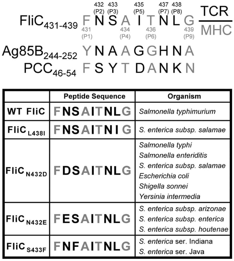
T cell activation required for host defense against infection is an intricately regulated and precisely controlled process. Although in vitro studies indicate that three distinct stimulatory signals are required for T cell activation, the precise contribution of each signal in regulating T cell proliferation and differentiation after in vivo infection is unknown. In this study, altered peptide ligands (APLs) derived from the protective Salmonella-specific FliC Ag and CD4+ T cells specific for the immune-dominant FliC(431-439) peptide within this Ag were used to determine how changes in TCR stimulation impact CD4+ T cell proliferation, differentiation, and protective potency. To explore the prevalence and potential use of altered TCR stimulation by bacterial pathogens, naturally occurring APLs containing single amino acid substitutions in putative TCR contact residues within the FliC(431-439) peptide were identified and used for stimulation under both noninfection and infection conditions. On the basis of this analysis, naturally-occurring APLs that prime proliferation of FliC-specific CD4+ T cells either more potently or less potently compared with the wild-type FliC(431-439) peptide were identified. Remarkably, despite these differences in proliferation, all of the APLs primed reduced IFN-gamma production by FliC(431-439)-specific CD4+ T cells after stimulation in vivo. Moreover, after expression of the parental FliC(431-439) peptide or each APL in recombinant Listeria monocytogenes, only CD4+ T cells stimulated with the wild-type FliC(431-439) peptide conferred significant protection against challenge with virulent Salmonella. These results reveal important and unanticipated roles for TCR stimulation in controlling pathogen-specific CD4+ T cell proliferation, differentiation, and protective potency.


Conflict of interest statement

DISCLOSURES

The authors have no financial conflicts of interest.

Figures

Figure 1
Figure 1
Alignment of FliC431–439 with other I-Ab peptides. Putative TCR contact or MHC anchor residues within each peptide are indicated by black and gray font, respectively. Numbers in parentheses indicate the amino acid residues that correspond to each position for the FliC431–439 peptide compared with the indicated residues from antigen-85B (Ag85B) or pigeon cytochrome C (PCC) (top). Alignment of each naturally-occurring FliC431–439 –derived APL compared with the parental FliC431–439 peptide (bottom).
Figure 2
Figure 2
FliC-specific CD4+ T cell proliferation and activation after in vitro stimulation. A. CFSE dilution in SM1 CD4+ T cells after stimulation with WT FliC peptide or each APL (line histogram) at the indicated concentration, or after stimulation with Control peptide (shaded histogram, 10 μM) for 5 days. Numbers in each plot indicate the percent CFSElo cells. B. CD25, CD44, CD62L and Annexin V expression by FliC-specific CD4+ T cells after stimulation with 10 μM WT FliC or each APL (line histogram) or Control peptide (shaded histogram) for 5 days. These results are representative of three independent experiments each with similar results.
Figure 3
Figure 3
Reduced IFN-γ and T-bet expression after stimulation with each APL compared with WT FliC peptide. A. Percent IFN-γ producing CD4+ T cells after stimulation with each peptide at the indicated concentration. Numbers in each plot represent percent IFN-γ+ cells among FliC-specific CD4+ T cells. B. T-bet expression after stimulation with WT FliC or each APL (line histogram) and Control peptide (shaded histogram) at the indicated peptide concentration. Numbers in each plot indicate mean fluorescent intensity of T-bet staining. These results are representative of at least three independent experiments each with similar results.
Figure 4
Figure 4
Pathogen-specific CD4+ T cell expansion and cytokine production after in vivo stimulation. A. Percent FliC-specific (CD45.1+) T cells among CD4+ splenocytes. Number in each plot indicates percent CD45.1+ cells among CD4+ T cells. B. Total number of CD45.1+CD4+ T cells day 5 after intravenous injection of each peptide (50 μg). C. Total number of IFN-γ producing CD45.1+CD4+ T cells for mice described in A after ex vivo peptide stimulation (10 μM; shaded bar) with either WT FliC (top left), FliCL438I (top right), FliCN432D (bottom left), or Control peptide (bottom right) compared to no stimulation control (open bar). These results are representative of two independent experiments each with similar results containing six mice per group. Bar, standard error.
Figure 5
Figure 5
FliC-specific CD4+ T cell expansion and cytokine production after recombinant Lm infection. A. Construct map indicating placement of coding sequences for WT FliC and each APL within the pAM401-based expression vector (Phly, Lm hly promoter; SS, signal sequence of hly; HA, hemagglutinin tag; cat, chloramphenicol acetlytransferase) (left). B. Western blot of supernatant protein from rLM-WT (lane 1), rLM-L438I (lane 2), rLM-N432D (lane 3), and rLM-Control (lane 4) (right). C. Total number of FliC-specific CD45.1+CD4+ T cells at day 3 (left) and day 30 (right) after infection with each recombinant Lm strain. D. Percent IFN-γ producing CD45.1+CD4+ T cells after ex vivo peptide stimulation with WT FliC peptide (10 μM) among splenocytes day 3 after infection with each recombinant Lm strain. Number in each plot indicates percent IFN-γ+ cells among CD45.1+CD4+ T cells. E. Total number of IFN-γ producing CD45.1+CD4+ T cells among splenocytes after ex vivo stimulation with WT FliC peptide (10 μM; shaded bar) or no peptide (open bar) day 3 (left) or day 30 (right) following recombinant Lm infection. These results are combined from at least two independent experiments each with similar results containing four to six mice per group. Bar, standard error.
Figure 6
Figure 6
WT FliC peptide expressed in recombinant Lm confers protection to Salmonella. Number of recoverable Salmonella CFUs in the spleen (top) and liver (bottom) of mice day 5 after challenge with virulent Salmonella typhimurium. Each group of mice were adoptively transferred with FliC-specific CD4+ T cells and initially infected with the indicated recombinant Lm or no Lm infection 30 days prior to Salmonella challenge. These data represent eight to fourteen mice per group combined from four independent experiments each with similar results. Bar, standard error. *, p < 0.05.

References

    1. Curtsinger JM, Lins DC, Mescher MF. Signal 3 determines tolerance versus full activation of naive CD8 T cells: dissociating proliferation and development of effector function. J Exp Med. 2003;197:1141–1151. - PMC - PubMed
    1. Janeway CA, Jr, Bottomly K. Signals and signs for lymphocyte responses. Cell. 1994;76:275–285. - PubMed
    1. Gett AV, Sallusto F, Lanzavecchia A, Geginat J. T cell fitness determined by signal strength. Nat Immunol. 2003;4:355–360. - PubMed
    1. Lafferty KJ, Prowse SJ, Simeonovic CJ, Warren HS. Immunobiology of tissue transplantation: a return to the passenger leukocyte concept. Annu Rev Immunol. 1983;1:143–173. - PubMed
    1. Jenkins MK, Johnson JG. Molecules involved in T-cell costimulation. Curr Opin Immunol. 1993;5:361–367. - PubMed

Publication types

LinkOut - more resources