. 2010 Feb 25;463(7284):1042-7.
doi: 10.1038/nature08752.

Reprogramming towards pluripotency requires AID-dependent DNA demethylation

Affiliations

Reprogramming towards pluripotency requires AID-dependent DNA demethylation

Nidhi Bhutani et al. Nature. .

Abstract

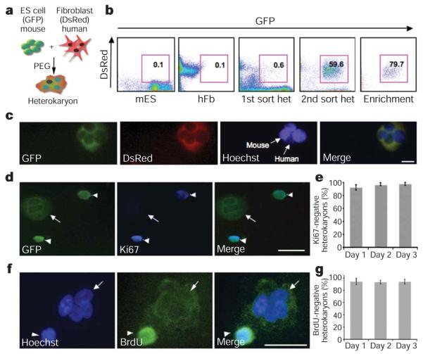
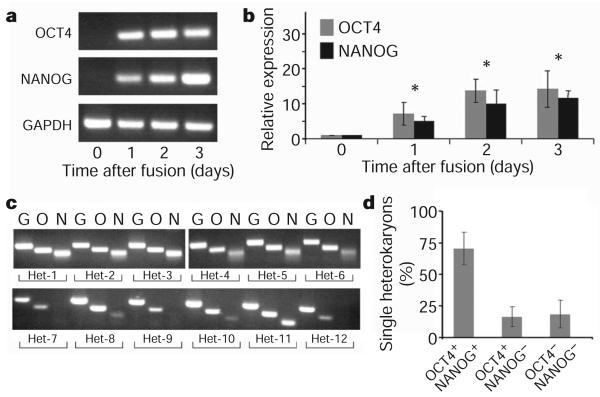
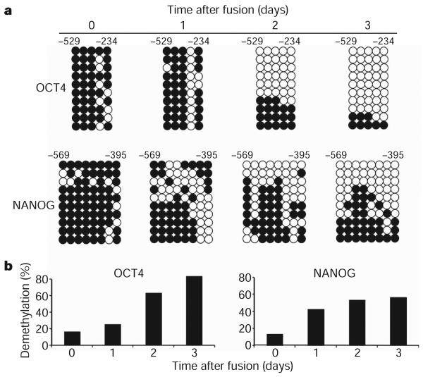
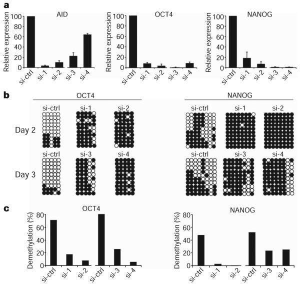
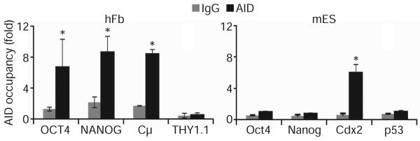
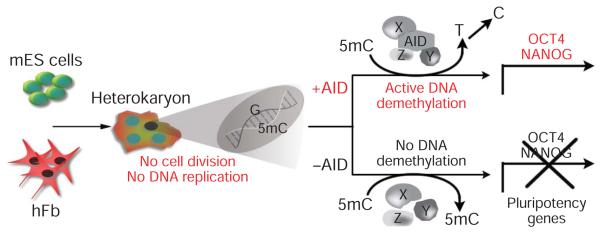
Reprogramming of somatic cell nuclei to yield induced pluripotent stem (iPS) cells makes possible derivation of patient-specific stem cells for regenerative medicine. However, iPS cell generation is asynchronous and slow (2-3 weeks), the frequency is low (<0.1%), and DNA demethylation constitutes a bottleneck. To determine regulatory mechanisms involved in reprogramming, we generated interspecies heterokaryons (fused mouse embryonic stem (ES) cells and human fibroblasts) that induce reprogramming synchronously, frequently and fast. Here we show that reprogramming towards pluripotency in single heterokaryons is initiated without cell division or DNA replication, rapidly (1 day) and efficiently (70%). Short interfering RNA (siRNA)-mediated knockdown showed that activation-induced cytidine deaminase (AID, also known as AICDA) is required for promoter demethylation and induction of OCT4 (also known as POU5F1) and NANOG gene expression. AID protein bound silent methylated OCT4 and NANOG promoters in fibroblasts, but not active demethylated promoters in ES cells. These data provide new evidence that mammalian AID is required for active DNA demethylation and initiation of nuclear reprogramming towards pluripotency in human somatic cells.


Figures

Figure 1
Figure 1. Absence of cell division and DNA replication in heterokaryons
a, Heterokaryon fusion scheme. GFP+ mouse ES (mES) cells were co-cultured with DsRed+ primary human fibroblasts (hFb) and then fused using PEG. b, FACS profiles of GFP+ mES, DsRed+ hFb, and GFP+ DsRed+ day 2 heterokaryons (het). c, Representative image of GFP+ DsRed+ day 2 heterokaryons. Hoechst 33342 (blue) denotes the nuclei. The heterokaryon shown has three distinct, unfused bright mouse nuclei, and one uniformly stained human nucleus. Scale bar, 50 μm. d, GFP+ DsRed+ day 2 heterokaryons were cytospun and stained for Ki67 (blue) to assess cell division. The GFP+ heterokaryon has two distinct nuclei (arrow) that are negative for Ki67 (blue) in contrast to the mononuclear cells (arrowheads) that stain positive for Ki67. Scale bar, 50 μm. e, Heterokaryons on days 1, 2 and 3 after fusion were scored on the basis of Ki67 staining, and 98±2% heterokaryons were non-dividing (mean±s.e.m., P, 0.05). f, Heterokaryons, generated using GFP2 ES cells, were enriched using a human fibroblast marker THY1.1 (see Methods) on day 1 post fusion, and stained for BrdU (green) and nuclei (blue) using Hoechst 33258. The heterokaryon (arrow) has three uniformly stained human nuclei and one bright, punctate mouse nucleus, and is negative for BrdU staining. In contrast, the indicated human mononuclear cell (arrowhead) stains positive for BrdU. Scale bar, 50 μm. g, Day 1, 2 and 3 heterokaryons were scored on the basis of nuclear and BrdU staining. DNA replication did not occur in 94±3% heterokaryons (mean±s.e.m., P, 0.05).
Figure 2
Figure 2. Time course of human fibroblast pluripotency gene expression in heterokaryons at the single-cell level
a, Human-specific primers against OCT4, NANOG and GAPDH were used for transcript analysis by RT–PCR of unfused co-cultures on day 0 and heterokaryons (mES 3 hFb) isolated on days 1, 2 and 3 after fusion. b, Real-time PCR to assess the upregulation of OCT4 (grey) and NANOG (black) in day 1, 2 and 3 heterokaryons using human-specific primers (mean ± s.e.m.; *P, 0.03). Unfused co-cultures served as day 0 controls and the expression of OCT4 and NANOG was normalized to GAPDH expression. Data shown are from three independent fusion experiments. c, Single heterokaryon nested PCR was used to assess the efficiency of reprogramming in the heterokaryon population. Direct reverse transcription and nested PCR were performed simultaneously on day 3 single heterokaryons, using human-specific primers for GAPDH (G), OCT4 (O) and NANOG (N) as indicated. Twelve heterokaryons analysed from a single fusion experiment are shown. Supplementary Fig. 3 shows 41 heterokaryons analysed from two additional fusion experiments. d, The frequency of heterokaryons expressing both OCT4 and NANOG is 70 ± 13%, showing that a high proportion of heterokaryons initiate reprogramming towards pluripotency. Data shown are a summary of three independent fusion experiments (mean ± s.e.m.).
Figure 3
Figure 3. Time course of DNA demethylation at pluripotency gene promoters in heterokaryons
a, Bisulphite sequencing analysis of methylation status of the human OCT4 and NANOG promoter in heterokaryons. Both human OCT4 and NANOG promoters in heterokaryons show rapid and progressive DNA demethylation on days 1, 2 and 3 after fusion compared to the co-culture control. White circles indicate unmethylated, black circles indicate methylated CpG dinucleotides. b, The percentage of demethylation at the human OCT4 and NANOG promoters in heterokaryons after fusion shows a progressive increase in demethylation. Thirty clones were analysed at each time point in two to three independent experiments; ten representative clones are shown.
Figure 4
Figure 4. Requirement of AID-dependent DNA demethylation for initiation of reprogramming towards pluripotency in heterokaryons
a, AID and human pluripotency gene expression in heterokaryons subjected to siRNA treatment, as assessed by real time PCR. Day 2 heterokaryons were treated with siRNA-1 and -2 (si-1 and si-2), and day 3 heterokaryons were treated with si-3 and si-4. Total levels of mouse and human AID transcripts were assessed using a set of degenerate primers, whereas human-specific primers were used for human OCT4 and NANOG. Gene expression was normalized internally to GAPDH (degenerate primers) for AID expression, and to human GAPDH for human OCT4 and NANOG expression. The samples were then normalized to the corresponding day 2 or 3 sample treated with the control siRNA (si-ctrl), represented as 100%. b, Human OCT4 and NANOG promoters on days 2 and 3 post fusion after AID knockdown remain methylated. c, The percentage of demethylation at the human OCT4 and NANOG promoters after AID knockdown shows a block in demethylation compared to their respective day 2 and day 3 control samples treated with control siRNA.
Figure 5
Figure 5. AID binding to pluripotency gene promoters by chromatin immunoprecipitation
ChIP with anti-AID antibody was performed in human fibroblasts (left) and mouse ES cells (right). AID occupancy is shown relative to background IgG signal (mean±s.e.m.). Significant AID binding was detected in human fibroblasts for the methylated NANOG and OCT4 promoters as well as for the positive control Cμ (P<0.002). In mouse ES cells, AID binding was detected only in the positive control, Cdx2 promoter region (P<0.007), whereas no significant binding was observed for the unmethylated Oct4 and Nanog promoters.
Figure 6
Figure 6. Model for AID-dependent active DNA demethylation in reprogramming
AID-mediated reprogramming and DNA demethylation in heterokaryons takes place in the absence of cell division or DNA replication, showing that AID is a component of an active DNA demethylation complex in mammals. The other putative components of this mammalian DNA demethylase complex (X, Y and Z) that may act together with the deaminase, AID, remain to be identified. 5mC, 5-methyl-cytosine.

Similar articles

Cited by

References

    1. Gurdon JB. Adult frogs derived from the nuclei of single somatic cells. Dev. Biol. 1962;4:256–273. - PubMed
    1. Briggs R, King TJ. Transplantation of living nuclei from blastula cells into enucleated frogs' eggs. Proc. Natl Acad. Sci. USA. 1952;38:455–463. - PMC - PubMed
    1. Takahashi K, et al. Induction of pluripotent stem cells from adult human fibroblasts by defined factors. Cell. 2007;131:861–872. - PubMed
    1. Takahashi K, Yamanaka S. Induction of pluripotent stem cells from mouse embryonic and adult fibroblast cultures by defined factors. Cell. 2006;126:663–676. - PubMed
    1. Wernig M, et al. In vitro reprogramming of fibroblasts into a pluripotent ES-cell-like state. Nature. 2007;448:318–324. - PubMed

Publication types

MeSH terms